Cell Ultrastructure, Prokaryotes, Cytoskeleton Questions OCR A
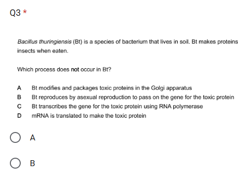
Self-marking multiple choice quiz for Cell Ultrastructure, Ions, and Elements. Using OCR A past paper exam questions.
Key Concept: Surface Area to Volume ratio (SA:V)
Some concepts turn up again and again in A-level biology. Taking a little time to ensure you really understand these key concepts from the start can save a lot of effort overall.
Surface area to volume ratio (SA:V) is vital for understanding a wide range of topics including transport across cell membranes, gas exchange, digestion, heat exchange, and mass transport. SA:V explains why the inner membrane of a mitochondrion is folded, why elephants have big ears, and why jellyfish don’t need blood vessels.
Some concepts turn up again and again in A-level biology. Taking a little time to ensure you really understand these key concepts from the start can save a lot of effort overall.
Surface area to volume ratio (SA:V) is vital for understanding a wide range of topics including transport across cell membranes, gas exchange, digestion, heat exchange, and mass transport. SA:V explains why the inner membrane of a mitochondrion is folded, why elephants have big ears, and why jellyfish don’t need blood vessels.
How confident are you in calculating this value and understanding its significance?
What are surface-area-to-volume ratios?
"Microvilli-Duodenum" by Wbensmith is licensed under CC BY-SA 3.0.
The ratio tells you about how changing the shape or volume of something affects its surface area. It’s not as simple as many people think: doubling the volume of ice-cream in a choc-ice doesn’t double the area of chocolate needed to cover it.
Surface areas are really important in biology. And so the shapes and volumes of things including organelles, cells, and organs are often optimised with respect to surface area. Whenever you see a weirdly folded or worm-shaped structure, think about SA:V.
Surface area to volume ratio (SA:V) is a value calculated by dividing a thing’s surface area by its volume. It is a single number that tells you about the relationship between the two input values.
To be precise: SA:V tells you how much surface area there is per unit of volume.
How to calculate SA:V (it’s not difficult, you just divide)
Quick maths refresher
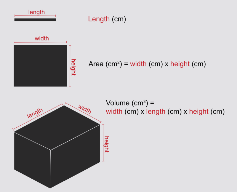
You will need to think about length, area, and volume and understand their units. Here’s a reminder for cuboids:
Simple example
Imagine a cube where every edge is 1 cm long. (Note: this blog doesn’t let me write little superscript numbers so I’ll use ^ to indicate that the following number should be written superscript, so 1^2 would mean 1-squared.)
Each square face of the cube has a surface area of 1 cm x 1 cm = 1 cm^2
Cubes have six faces, so the total surface area is 6 x 1 cm^2 = 6 cm^2
The volume of the cube is 1 cm x 1 cm x 1 cm = 1 cm^3
SA:V = surface area / volume. In this example that’s 6 cm^2 / 1 cm^3 = 6 cm^-1
Notice the funny unit there. You’ve divided cm^2 by cm^3 so they don’t cancel out perfectly.
But does it matter what this unit is? Let’s try it using the same cube but measuring it in millimetres.
Each square face of the 1 cm cube has a surface area of 10 mm x 10 mm = 100 mm^2
Cubes have six faces, so the total surface area is 6 x 100 mm^2 = 600 mm^2
The volume of the cube is 10 mm x 10 mm x 10 mm = 1000 mm^3
SA:V = surface area / volume. Using millimetres that’s 600 mm^2 / 1000 mm^3 = 0.6 mm^-1
1 mm is 1/10 of 1 cm and the SA:V calculated in mm is 1/10 that of the value calculated in cm. If you are comparing SA:V values, make sure they have the same unit.
Of course you don’t find a lot of cubes in nature. In exams they might simplify cells or other bodies to be imagined as spheres of a known radius, and give you equations for you to calculate the surface area and volume. And there are much weirder shapes to think about too. But before we get to all that …
How does SA:V change as things increase in scale?
We know the 1 cm cube has a SA:V of 6 cm^-1.
So, what if we get a handful of these 1 cm cubes and stick them together to make a scaled-up cube where every edge is 2 cm long? (See picture). How will the SA:V of this new structure compare? It’s still a cube, after all; it’s just a bit bigger.
2-cm cube:
Each square face of the assembled cube has a surface area of 2 cm x 2 cm = 4 cm^2
Cubes have six faces, so the total surface area is 6 x 4 cm^2 = 24 cm^2
The volume of the assembled cube is 2 cm x 2 cm x 2 cm = 8 cm^3
SA:V = surface area / volume. In this example that’s 24 cm^2 / 8 cm^3 = 3 cm^-1
The larger cube’s SA:V is only half that of the unit cube!
SA:V is surface area divided by volume. Higher values of SA:V mean there is more surface area for each unit of volume, and lower values of SA:V mean there is less surface area for each unit of volume.
So our 2 cm cube has less surface area per unit volume than the 1 cm cube did.
The larger cube has a lower SA:V. And if you work out the numbers for an even bigger one, you’ll get an even lower SA:V.
This makes sense. To build the 2 cm cube, you stack eight 1 cm cubes together. By doing this, you bury some of their faces within the structure. You end up with the same volume as eight individual 1-cm cubes, but you have reduced the surface area that is exposed to the outside world.
Also, if you look carefully at the picture, you’ll see that each cube in the assembled structure has half of its faces buried. So there’s only half the surface area per unit volume compared to the single cube. And so it makes sense that the SA:V is half what it was for the single cube.
Each small cube is only showing three of its six faces. So for each 1 cm^3 volume (ie for each small cube) there is 3 cm^2 surface area exposed, giving us an overall SA:V of 3 cm^-1.
Key concept: as size increases, SA:V decreases. This is a good thing to explicitly state in any question about SA:V and size, where the shape stays the same.
Try it: calculate SA:V for a 3 cm x 3 cm cube. What value do you guess you might get? And what is the actual value?
Why size matters
"Zygote" by Nina Sesina is licensed under CC BY-SA 4.0.
Imagine you are a single cell (well, you were once, don’t you remember?).
Your cell membrane is your connection with the outside world. Through it you take in nutrients, water and other necessary things. And you expel your waste through it.
This membrane is your surface; you have as much surface area as you have cell membrane.
Meanwhile, inside the cell membrane you have cytoplasm, organelles, etc. In this region you are busily metabolising molecules. You can measure this region as your volume.
Question: can you take in enough nutrients, and expel enough waste, to keep up with your metabolism?
More-important question: if you grow bigger, will this still be true?
Valonia ventricosa aka Sailors eyeball - a single cell that can grow up to 5 cm in diameter. But … how???
As a cell grows larger its SA:V falls. This is just like we saw with scaling up the cubes above. The growth of the cell’s surface area (cell membrane) just can’t keep up with the growth of its volume (where the metabolism is). This means there will be a point at which there just isn’t enough cell membrane (surface area) to transport everything that needs to be transported, limiting the cells ability to function.
This is one reason why cells tend to be pretty small!
But … wait a minute, what about the freakish cell in this next photo? A shiny, bright green, single-celled alga that can get up to 5 cm in diameter. A single cell! Charmingly known as ‘sailors’ eyeballs’.
But how can one cell possibly be so large? Isn’t its SA:V ridiculously low? (Spoiler: yes it is). How can it transport everything it needs with such a low SA:V? Well, it has a trick …
Shape matters
This giant cell is cheating. Inside, all the metabolic activity is limited to an area very close to its cell membrane. Nearly all of the cell’s interior is filled up by one giant vacuole that pushes all the interesting stuff out to the region next to the membrane. So there isn’t nearly as much metabolism going on in there as you might imagine from its total volume. If you re-calculated its SA:V ignoring the volume inside its vacuole, you’d get a much more sensible number. (It also has multiple nuclei spread throughout its cytoplasm.)
In exams, questions are usually about solid spheres or cuboids. We’ve looked at cuboids, so let’s look at spheres.
Spheres
If you have to calculate for spheres, they will give you the equations you need to calculate volume and surface area. So don’t worry about remembering these.
Volume of a sphere = 4/3 x π x r^3
Surface area of a sphere = 4 x π x r^2
So, if you have a sphere of radius 1 cm (remember this is just the distance from the centre to the outside, so the whole thing is 2 cm across):
The surface area is 4 x π x r^2. If r = 1, r^2 = 1. So the surface area is 4 x 3.14 x 1 = 12.6 cm^2
The volume is 4/3 x π x r^3. If r = 1, r^3 = 1. So the volume is 4/3 x 3.14 x 1 = 4.19 cm^3
SA:V = surface area / volume. In this example that’s 12.6 cm^2 / 4.19 cm^3 = 3 cm^-1
This is the same SA:V as the 2-cm cube!
That important phrase “as size increases, SA:V decreases”, is talking about increasing the size of something while keeping the same shape overall. Shape is also really important!
Spheres have the lowest possible surface area for their volume. This is one of the reasons that nature likes them. For example, bubbles are spheres because surface tension pulls their surface in to minimise its area.
Stretching out
If a cell needs to grow to large dimensions but also needs to keep a high SA:V, it needs to avoid being a sphere. More-complex shapes will always have higher SA:V.
Consider neurons. You have a single neuron that reaches from your big toe to your spine. A single cell over a metre long! But these neurons are definitely not spheres. They are very skinny.
Question: what happens to SA:V when you make long/thin shapes?
Let’s go back to the 1 cm cubes. The 1 cm cube had a SA:V of 6. And the 2 cm cube had a SA:V of 3.
The 2 cm cube had a volume of 8 cm^3 : it was made out of eight small cubes. Let’s take those eight small cubes and rearrange them into the shape of a flatworm: a rectangular block of 2 cm x 4 cm x 1 cm (see picture above).
The upper and lower faces of this shape each have a surface area of 8 cm^2
The two long sides of this shape each have a surface area of 4 cm^2
The two ends of this shape each have a surface area of 2 cm^2
The total surface area is (2 x 8 cm^2) + (2 x 4 cm^2) + (2 x 2 cm^2) = 28 cm^2 (the 2 cm cube had 24 cm^2)
The volume of the assembled cube is 2 cm x 4 cm x 1 cm = 8 cm^3 (ok that isn’t a surprise)
SA:V = surface area / volume. In this example that’s 28 cm^2 / 8 cm^3 = 3.5 cm^-1 (the 2 cm cube was 3 cm^-1)
Rearranging it from a 2 cm cube to a flatworm of the same volume has increased its SA:V.
Ok so let’s push it even further and turn it into a long worm.
The upper and lower faces of this shape each have a surface area of 8 cm^2
The two long sides of this shape each have a surface area of 8 cm^2
The two ends of this shape each have a surface area of 1 cm^2
The total surface area is (2 x 8 cm^2) + (2 x 8 cm^2) + (2 x 1 cm^2) = 34 cm^2
The volume of the assembled cube is 1 cm x 8 cm x 1 cm = 8 cm^3 (no surprise)
SA:V = surface area / volume. In this example that’s 28 cm^2 / 8 cm^3 = 4.25 cm^-1 (the 2 cm cube was 3 cm^-1)
Your neurons can be really, really long because they’re also skinny, because that means they still have a really high SA:V.
And of course this works for whole animals too. Many small animals lack blood vessels and rely on molecules simply diffusing through their bodies. They need high surface area for transport and they also need every part of their interior to be not-to-far from that surface. This is (one of) the reasons we don’t have truly-giant insects: they are limited by their ability fo transport things to/from the outside world. Sponges and jellyfish also lack blood vessels and rely on diffusion.
Nemotode by Bob Goldstein, UNC Chapel Hill http://bio.unc.edu/people/faculty/goldstein/ Licenced under a Creative Commons Attribution-Share Alike 3.0 Unported licene
Tardigrade by Alexander Klepnev licenced under CC BY-SA 4.0.
Humans, alongside other larger animals, have much lower SA:V and suffer from low diffusion rates. So we need specialised structures to aid exchange and transport.
Maximising SA:V
Structures that are involved in transport and exchange rely upon having very high surface areas of membrane across which transport can take place. To maximise exposed surface area, structures are folded, or shaped into fingers, or branches.
In the human body, you find extremely high SA:V ratio in specialised structures including:
Gut - the gut wall has microvilli, finger-like structures that extend into the lumen. These increase surface area for absorption of water and nutrients
Lungs - the lungs have many branches terminating in tiny alveoli offering a very high surface area for gas exchange.
Capillaries - capillaries have much higher SA:V than arteries or veins. This offers a higher surface area for transporting things between the blood and tissue fluids. It also increases fluid resistance, which is important to keep the blood moving forward.
Red blood cells have a biconcave shape that increases their SA:V allowing better gas exchange with the blood plasma.
In other organisms, the shapes of structures like leaves, chloroplasts, gills, root hairs, and fungal hyphae, among many other examples, are similarly optimised to maximise surface area to volume ratios.
Folded membranes
You also find folded membranes used as a way to squeeze a load of membrane into a small space. Eg:
Mitochondria have a highly folded inner membrane that offers more surface area to embed the enzymes and proteins involved in oxidative phosphorylation, so that more ATP can be produced.
Golgi apparatus and endoplasmic reticulum are similarly folded to maximise their surface area.
Thermoregulation
Who is is feeling cold, and who is feeling hot?
Body surfaces also exchange heat with the environment.
We naturally change our SA:V when we get hot or cold by changing the position of our limbs and body. Which of the people in the image has the highest SA:V? Which person is feeling cold, and which is feeling hot?
Dogs increase their surface area by opening their mouths when hot and panting (passing air over their wet surfaces to lose heat through evaporation). Cats form neat loafs when cold but sprawl dramatically across the floor when hot.
Organisms’ entire body shape will also be related to their thermoregulatory needs.
Animals that need to lose heat from their bodies usually have a higher SA:V, and animals that need to conserve heat usually have a lower SA:V. This is why elephants have such huge ears (more surface area from which to lose heat) and why animals from cold environments tend to be more spherical than those from hot climates.
"Emperor Penguins" by Christopher.Michel is licensed under CC BY 2.0.
"Black Necked Stork" by AntoGros is licensed under CC BY 2.0.
Keep it in mind
If you can get an intuitive feeling for Surface Area to Volume ratios, and keep them in mind whenever you see a question about folded structures, or about similar shapes of different sizes, then this will help you understand what questions are really asking. A level Biology is about the why and how much more than it is about remembering facts, and SA:V goes a long way to help you understand things on this deeper level.
This article was written by Dr Jenny Shipway with guidance from Tom Whitburn
Introduction to Mitosis (with Cell Division and the Cell Cycle)
This article is for anyone who is totally confused by mitosis, or who hasn’t yet started the topic.
Mitosis is part of cell division. It explains how multicellular organisms grow and is vital for understanding the challenging topic of autosomal linkage. Mitosis is also similar to the first half of meiosis; learning mitosis first will make meiosis much easier to understand.
Remember that for A level biology, you always need to understand why, as well as how. So, get a strong grip of the fundamental concept of mitosis before you try to memorise the details. Understanding the purpose of mitosis will also make it a lot easier to remember the individual stages of the process.
This article is for anyone who is confused by mitosis, or who hasn’t yet started the topic.
Mitosis is part of cell division. It is key to understanding how multicellular organisms develop from a single cell. Mitosis is also similar to the second half of meiosis; learning mitosis first will make meiosis much easier to understand.
Remember that for A level biology, you always need to understand why, as well as how. So, get a strong grip of the fundamental concept of mitosis before you try to memorise the details. Understanding the purpose of mitosis will also make it a lot easier to remember the individual stages of the process.
Being a Cell
Most of the time, cells hang out just being cells. Doing cell stuff. In the cell cycle, this is called G1 phase. It’s just the normal state of a cell.
The other phases (S, G2, M) only happen when a cell is preparing to divide, and actually dividing. Some cell types will do this more often than others.
Dividing cells, Kuan-Chung Su, London Research Institute, Cancer Research UK. Source: Wellcome Collection. This Image is licensed under CC BY 4.0. DNA is shown in red. Read it like a cartoon strip, starting in the middle and spiraling out. The central image is of the mother cell, and the final image shows the separated daughter cells.
Mitosis is a part of Cell Division
Cells come from cells. New cells are created when existing cells divide into two by cell division.
The cell that is going to divide is called the “mother cell”, and the two resulting cells are called “daughter cells”.
But - this is not like a mother birthing two daughters to send out into the world. As these daughters arrive, the mother cell ceases to exist. Because it has become the daughter cells.
Mitosis is just one part of the cell division process (alternatively meiosis can be used instead, but most cells only do mitosis).
In cell division with mitosis, the daughter cells are both genetically identical to the mother cell.
How to Divide a Cell: why DNA matters
So, what does a cell need to be able to divide in two? If you divide a human in two you get … a mess. Cells are much easier to split in half.
For a start, most intracellular molecules and organelles have multiple copies. For example, human cells contain thousands of mitochondria. These can be shared out between the two daughter cells. And large organelles like the endoplasmic reticulum can be divided easily enough because their membranes are fluid. But there’s a big problem …
The mother cell has only one copy of the organism’s DNA.
Before it can divide to form two identical daughter cells, the mother cell will need to synthesise the extra DNA required to provide two identical sets (one for each daughter cell), and then separate the two sets of DNA ready for cell division. This DNA synthesis step is S phase in the cell cycle. It is followed by G2 phase where the cell prepares itself for mitosis.
Mitosis is the process of separating the two sets of DNA. Mitosis and the process of actually splitting the cell into two both happen during M phase.
Vocabulary check
Cell division is the process by which a cell divides in two, creating two separate cells.
The Cell Cycle describes different phases of a cell’s activity. All except G1 are related to cell division.
G1 phase = normal cell stuff
S phase = replication of DNA ready for cell division
G2 phase = preparation for M phase
M phase = organisation of the DNA into two different sets, and cytokinesis (the splitting of the cell into two)
Mitosis is the process by which a cell separates its DNA into two identical sets before and during cell division.
Meiosis is an alternative process to mitosis, used to make gametes (sperm, eggs, pollen). Meiosis is not discussed in this article.
What is a Chromosome
The very long DNA molecules inside cells are not just big messy tangles - they are organised with the help of proteins. These include proteins called histones. Sections of a DNA molecule that are not in use are coiled around histones to organise it into neat packages. Together, one molecule of DNA plus the proteins that help organise its structure is called a chromosome.
When a cell is doing its normal cell behaviours, just doing its job, it will be actively using its DNA as a template to make RNA. For this reason, large parts of its DNA will be uncoiled and exposed within the nucleus. A light microscope can’t see the DNA when it’s like this because the DNA molecule is extremely thin. And if you could see it, it’d look pretty messy.
But during cell division, the DNA is packaged up very neatly and densely. During this time, the chromosome can be seen as a dark body. In fact, this is where the word comes from (“chromo” - dark, “some”= body).
Just like cotton thread, DNA is much easier to see when it’s neatly organised for transport/storage, than when it is uncoiled and exposed for use. (Note: DNA does not coil around a single spool like thread does, it has a more complex but similarly dense structure.)
Normally, a human cell contains two sets of chromosomes. The DNA code in one set comes from the biological mother of the person, and that in the other set comes from the biological father. The two sets are very similar, but have small differences due to them having different versions of genes.
What do Chromosomes look like (top tip: don’t think of an X)
One normal chromosome, with its DNA neatly packaged up so we can see it. Notice that it’s not an X shape!
A chromosome is one molecule of DNA with the proteins that help manage its structure. Its structure varies a lot depending on how tightly it is package. When a chromosome is packaged up very densely, it looks like a long sausage shape - see the picture to the right. You can imagine this structure as the equivalent of a spool of thread as shown above (although the DNA is rolled up in a different structure from the thread).
But wait - if you google an image of a chromosome, you see images of a blobby X shape!
OK so yes, that’s a chromosome too, but they only look like that at one specific time during mitosis, after DNA replication! That image of an X-shape is NOT a good place to start your understanding! It’s a bit like looking at a woman half-way through giving birth and saying “that’s what a human looks like”.
Sister Chromatids
Chromosomes only look like X-shapes after the chromosome’s DNA has been replicated but before it separates.
The Chromosome on the left is a double stranded molecule of DNA. The chromosome on the right has been replicated and now has twice as much DNA as before. Each sister chromatid is a double stranded molecule of DNA.
Confusingly it’s still called “a” chromosome at this point, but it now has twice as much DNA and comprises two identical “sister chromatids”. The chromatids are joined together at their centromeres, creating the classic X shape (see diagram to right).
During cell division, the sister chromatids will separate and move to the two separate cells. After cell division has finished, they will just be called chromosomes.
To be clear, these are not the pairs of slightly-different chromosomes where one carries code from each parent. These sister chromatids are identical copies of the code from just one parent. There will be another pair of sister chromatids carrying the code from the other parent.
Semi-conservative Replication
The original chromosome, on the left, has two strands. These are separated and used to template new strands. This means that both of the sister chromatids have one of the original strands of DNA, now pair-bonded to a new strand that has been freshly synthesised using it as a template.
An important note:
Neither of the sister chromatids is “the original one”. The original DNA has separated into two strands, and each of the sister chromatids contains one of these original strands, now pair-bonded to a freshly synthesised complementary strand.
This is called semi-convervative replication .
Semi = half
Conserve = preserve / save
Mitosis: ensuring each cell gets a full set of chromosomes
After DNA replication, the cell contains duplicated (X-shape) versions of each version of the cell’s 46 chromosomes. This doubling gives it all the DNA it needs to split itself into two cells, each with a full set of DNA.
Mitosis is the process by which the cell organises the DNA to ensure each daughter cell gets a complete set, including both versions of each of the different chromosomes.
Ok, now you know what it’s all about, what it’s trying to achieve, and what is going on with the chromosomes, you’re ready to go on to look at the diagrams showing the individual steps of mitotis.
If you found this article helpful, please click the ❤️ below.
Next: See this fantastic blog post which includes loads of mitosis resources including video explanations of the full process and A level biology exam questions.
This article was written by Jenny Shipway, with guidance from Tom.
Quick Cell Quiz: can you spot where these students went wrong?
Can you spot the problems with these A level Biology exam question answers? These bloopers come up a lot - check you’re not going to make the same mistakes.
Avoid tragedy
There are some problems that come up again and again in students’ answers to A level Biology exam questions, and which lose them marks. Sometimes it’s a misconception, sometimes misuse of technical language, and sometimes just a lack of required information.
Here are some examples of problem answers that come up a lot in cell topics. Get a step ahead of other students by not including any of these in your A level Biology exam answers!
See if you can spot the problem first, then scroll down to find out if you were right.
What’s the problem with these exam answers?
❌ 1. The ion can’t pass through the hydrophobic inside of the membrane because the ion is polar ❌
❌ 2. The nucleolus contains rRNA ❌
❌ 3. Fatty acids interact with hydrophobic bonds ❌
❌ 4. Ribosomes have a single membrane ❌
❌ 5. Carrier proteins require ATP hydrolysis, and so only do active transport ❌
❌ 6. Mitochondria carry out respiration ❌
Did you spot the problems? Here’s what you should say instead:
✅ 1. The ion can’t pass through the membrane because it is charged. Ions are not polar. This trips up a lot of students but there is an important difference - see this article for a full explanation.
✅ 2. The nucleolus is where rRNA (which forms part of ribosomes) is synthesised. But the nucleolus itself is made of DNA wrapped around histones.
✅ 3. Fatty acids (and other non-polar molecules) have hydrophobic interactions. There is no such thing as a ‘hydrophobic bond’. A bond formed is when an electron is shared between two atoms/molecules. In contrast, hydrophobic interactions happen because the fatty acid molecules do not want to be exposed to water - they cluster together to minimise their exposure. They are ‘water avoiding’ because, being unpolar, they are unable to satisfy water’s hydrogen-bonding potential. (When you think about it, it’s more like the water is avoiding them - but overall it works out as the same thing.)
✅ 4. Ribosomes do NOT have membranes! They are made of protein subunits and rRNA. Ribosomes are sometimes attached to membranes (they are “membrane-bound”), and are sometimes free-floating. In eukaryotes, they are often attached to the membrane of the Rough Endoplasmic Reticulum - but this membrane does not belong to the ribosome, it’s just where it’s hanging out.
✅ 5. Carrier proteins can also be used for faciliated diffusion. This is possible because some carrier proteins can change confirmation without ATP hydrolysis. Facilitated diffusion using carrier proteins moves the carried substance in both directions, but more often away from the higher-concentration side of the membrane - this happens simply because it’s more likely that it will encounter the substance on that side.
✅ 6. Mitochondria carry out aerobic respiration. Ok yes that’s a type of respiration so the original answer wasn’t technically wrong, but you want to get maximum marks and you need to let the examiner know that you understand exactly what the mitochondria are doing. If the question was “What animal goes miaow and eats mice” you wouldn’t answer “a mammal”.
If this post has been helpful, please like ❤️ below and share with your friends.
OCR A Cells Organelles and Microscopes Quiz
Try this quiz - if you found it useful then please ❤️
OCR A Meiosis and Cell Differentiation Quiz
Try this quiz - if you found it useful then please ❤️
OCR A Cell Cycle and Mitosis (and differences with Meiosis) Quiz
Questions by topic - OCR A Cell Cycle and Mitosis Multiple Choice Quiz
Try this quiz - if you found it useful then please ❤️
Multiple Choice Quiz - OCR A Cell Membranes
Practising recall and doing questions is so important for long term retention of information. Try this self marked quiz. OCR A Cell Membranes
Try this quiz - if you found it useful then please ❤️ (at the bottom of the page) and share
Cell division - Mitosis - Quiz, Questions, Guide, videos
Cell cycle, mitosis and cell division. Pack of exam questions on mitosis and meiosis
Organelles - Test your understanding
Multiple choice questions on cell organelles, all past paper questions, a good way to assess your understanding
Please like and share (and click on a advert to help with the hosting costs !)

