

Methods of studying cells (AQA 3.2.1.3) is the practical side of the cells topic, and it turns up on AS and A-level papers as electron micrographs, magnification calculations and questions on separating organelles. You need the principles and limitations of optical microscopes, transmission electron microscopes (TEM) and scanning electron microscopes (SEM), the difference between magnification and resolution, how to measure an object and use magnification = size of image ÷ size of real object, and how cell fractionation and ultracentrifugation separate cell components.
The examiners' reports show where marks slip away. Students say the microscope has a shorter wavelength instead of the electrons, describe the SEM image without the TEM one, mix up high resolution with a large resolvable distance, write 'thin so light passes through' without a single layer of cells, skip the low-speed spin, give cold, buffered and isotonic without saying why, and convert 46 mm to 4600 µm.
Start with the microscopes, then cell fractionation, and finish with preparing specimens and the magnification calculations. Answer each on paper before you open the mark scheme. If you'd like help with this topic, my AQA masterclasses run online on Tuesdays at 5.15pm for Year 12 and on Wednesdays or Thursdays at 6.30pm for Year 13.
What you need from GCSE and Year 12
Each question on this page assumes you're secure on these GCSE and Year 12 ideas. If any of them feels shaky, revise it before you start.
- GCSE: light microscopes and electron microscopes; electron microscopes have a much higher magnification and resolution, so they show sub-cellular structures such as ribosomes.
- GCSE: magnification = size of image ÷ size of real object, and converting units: 1 mm = 1000 µm and 1 µm = 1000 nm.
- GCSE: osmosis is the movement of water from a dilute solution to a more concentrated one through a partially permeable membrane.
- Year 12 (AQA 3.2.1.1): the structure and size of the nucleus, mitochondria, chloroplasts, ribosomes and other organelles. See the Eukaryotic cell structure page.
- Year 12 (AQA 3.1.4.2): enzymes are proteins that are denatured by changes in pH and work more slowly when cold. See the Enzymes page.
Key terms
These words come up in the questions and mark schemes. Examiners expect you to use them exactly.
- Magnification: how many times bigger the image is than the real object: size of image ÷ size of real object.
- Resolution: the minimum distance apart two points can be and still be seen as separate. A higher resolution means a smaller distance, so more detail.
- Optical microscope: uses light, so its resolution is limited by the long wavelength of light (about 0.2 µm); it can show living specimens and colour.
- Transmission electron microscope (TEM): electrons pass through a very thin, dead specimen, giving a 2-D image of internal structures with the highest resolution.
- Scanning electron microscope (SEM): electrons are deflected off the surface of the specimen, giving a 3-D image of the surface, with lower resolution than a TEM.
- Eyepiece graticule: a scale in the eyepiece used to measure objects; it must be calibrated against a stage micrometer at each magnification.
- Artefact: something seen in a prepared specimen that isn't really part of the cell, caused by the way it was prepared or viewed.
- Homogenise: blend or grind tissue to break open cells and release the organelles, then filter to remove debris.
- Isotonic, cold, buffered solution: same water potential so organelles don't burst or shrink by osmosis; cold to slow enzymes that would digest them; buffered so proteins aren't denatured.
- Ultracentrifugation (differential centrifugation): spinning the filtrate at increasing speeds; the densest organelles (nuclei) settle first, then mitochondria, then smaller ones.
- Pellet and supernatant: the pellet is the sediment at the bottom of the tube; the supernatant is the liquid above it, which is spun again at a higher speed.
What these questions cover (AQA 3.2.1.3)
The questions test these statements from the AQA AS and A-level Biology specification (7401/7402):
- Microscopes: The principles and limitations of optical microscopes, transmission electron microscopes and scanning electron microscopes.
- Measuring and magnification: Measuring the size of an object viewed with an optical microscope. The difference between magnification and resolution. (with the use of the formula magnification = size of image ÷ size of real object, which the specification prints as a fraction)
- Cell fractionation: Principles of cell fractionation and ultracentrifugation as used to separate cell components.
- Students should be able to appreciate that there was a considerable period of time during which the scientific community distinguished between artefacts and cell organelles. (the flagella question asks you to consider artefacts and the limits of an optical microscope)
How to use this page
Write your answer on paper before you open the mark scheme. Allow about 1.3 minutes per mark: the 18 questions are worth 46 marks, so about 60 minutes in total. Then mark your answer strictly against the mark scheme and read the examiners' tip underneath it. If you'd rather print the questions, download the PDF pack.
Microscopes: principles and limitations (3.2.1.3)
Question 1
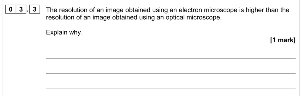
AS Paper 1 · June 2022 · Q03.3 · 1 mark
Explain why an electron microscope gives a higher resolution than an optical microscope.
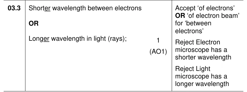
Show the mark scheme
Tap an image to open it full size.
From the examiners' report: Many focused on the microscope rather than the beam. Say electrons have a shorter wavelength than light; 'the electron microscope has a shorter wavelength' was rejected.
Question 2
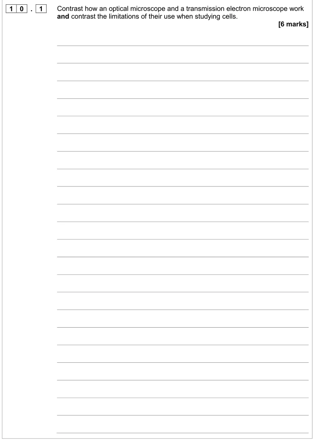
A level Paper 1 · June 2017 · Q10.1 · 6 marks
Contrast how an optical microscope and a TEM work, and their limitations.
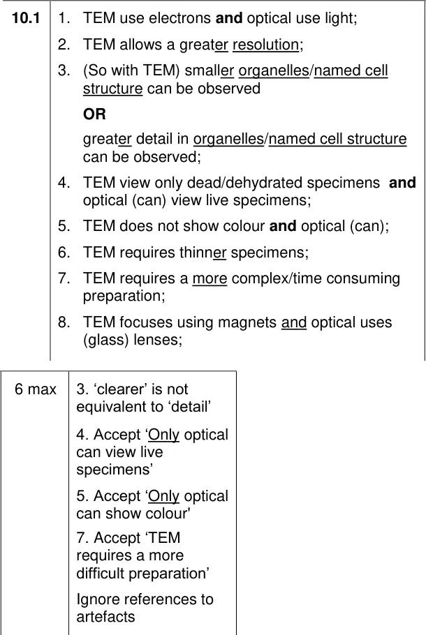
Show the mark scheme
Tap an image to open it full size.
From the examiners' report: There's no examiners' report for this paper, so this tip comes from the mark scheme. Make every point a contrast ('TEM…, whereas optical…'). 'Clearer' isn't the same as more detail, and artefacts were ignored.
Question 3
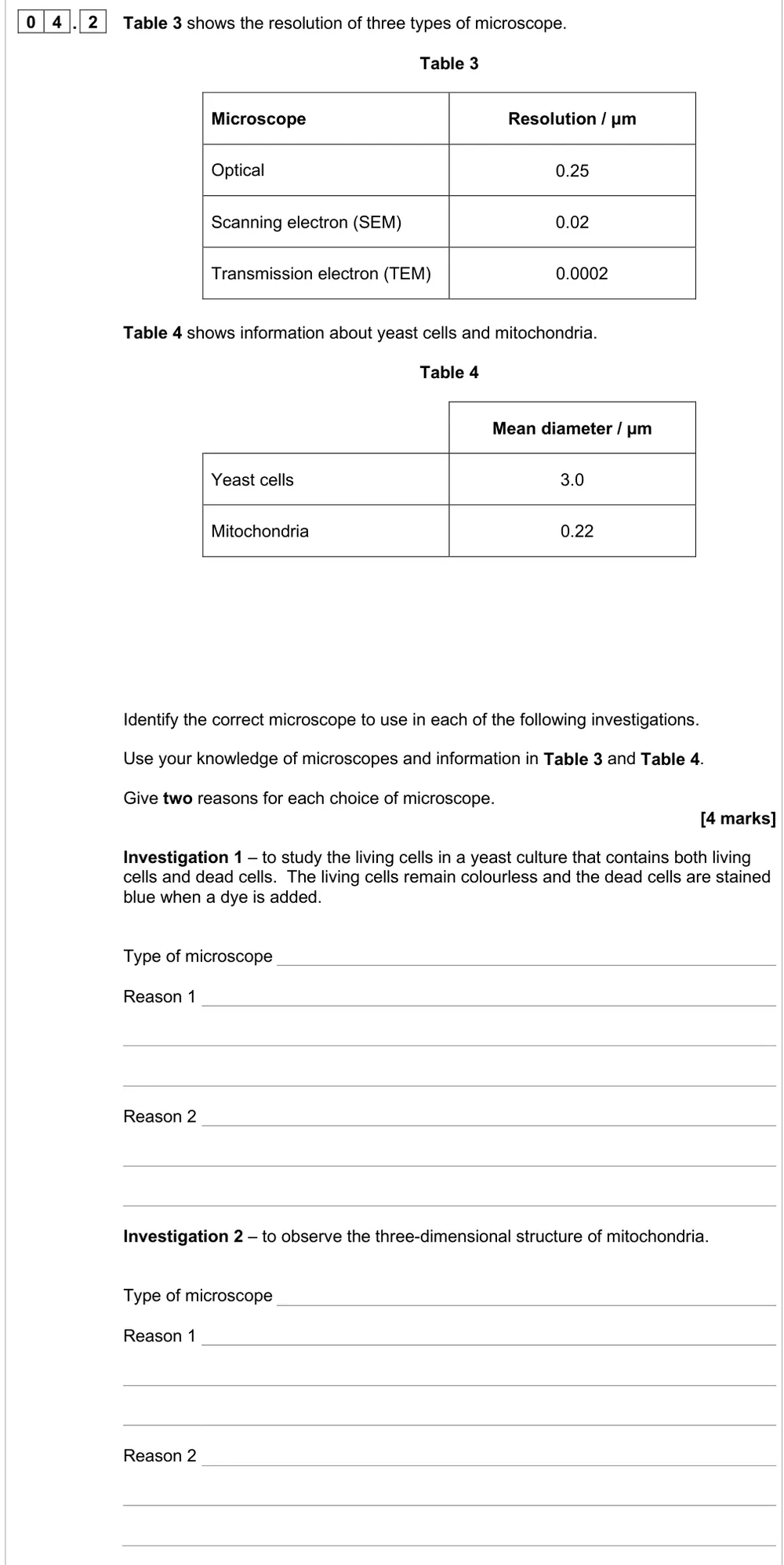
A level Paper 1 · June 2025 · Q04.2 · 4 marks · Easy
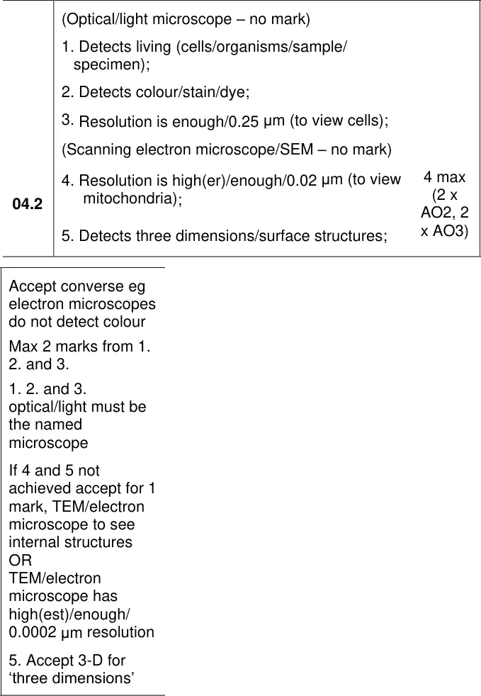
Choose a microscope for living yeast cells and for 3-D mitochondria, with reasons.
Show the mark scheme
Tap an image to open it full size.
From the examiners' report: About 55% got all four. The SEM gives 3-D images, not the TEM. A higher resolution means a smaller resolvable distance; ease of use didn't score.
Question 4
AS Paper 1 · June 2022 · Q03.2 · 2 marks · Hard
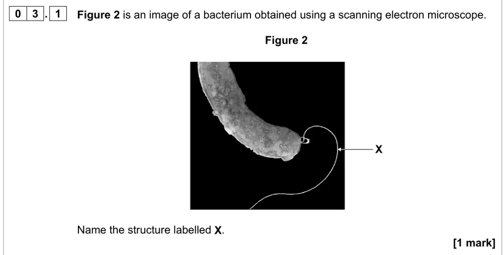
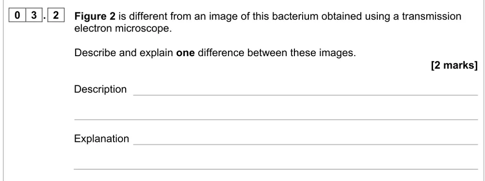
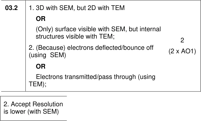
Describe and explain one difference between SEM and TEM images of a bacterium.
Information from earlier in this question:
Show the mark scheme
Tap an image to open it full size.
From the examiners' report: Only 13% got both marks. Describe both images: 'the SEM image is 3-D' needs 'the TEM image is 2-D' with it.
Question 5
AS Paper 1 · June 2023 · Q04.2 · 2 marks
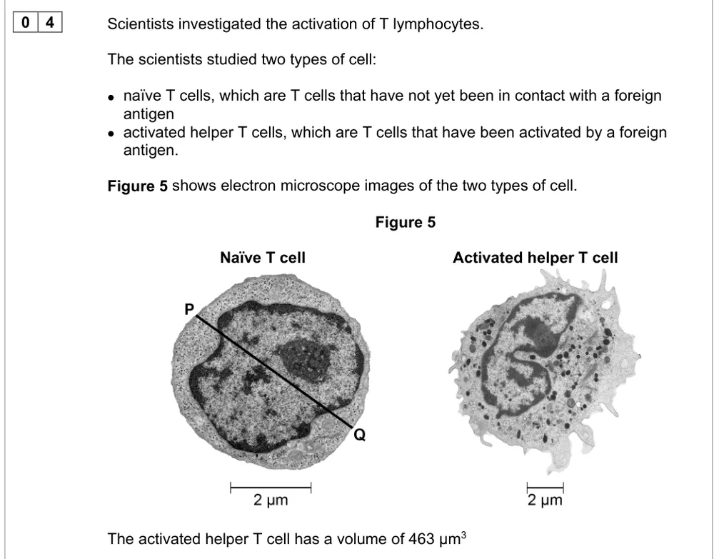
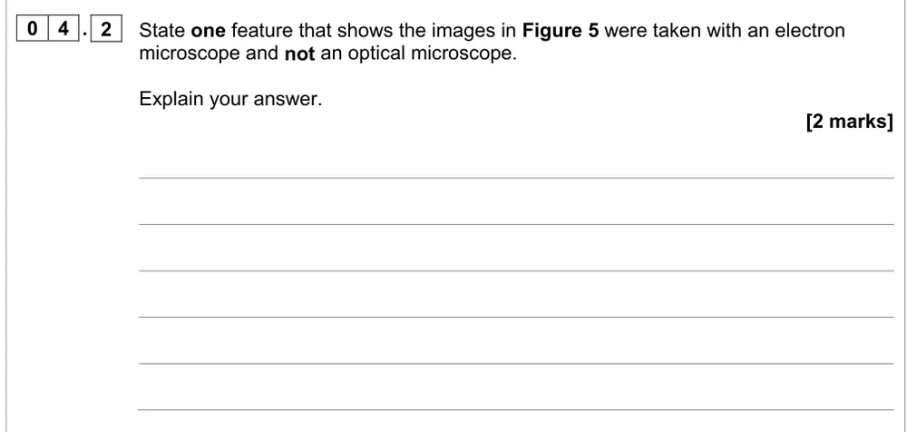
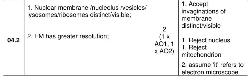
State a feature showing T cell images were taken with an electron microscope, and explain.
Information from earlier in this question:
Show the mark scheme
Tap an image to open it full size.
From the examiners' report: Pick a structure only an electron microscope can show: a nucleus or mitochondrion is visible with an optical microscope too. Black and white images and higher magnification didn't score.
Question 6
AS Paper 1 · November 2020 · Q04.2 · 1 mark · Hard
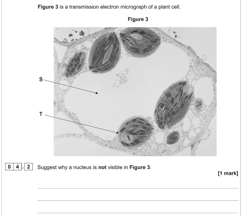
Suggest why no nucleus is visible in a TEM image of a plant cell.
Show the mark scheme

Tap an image to open it full size.
From the examiners' report: Many wrongly decided the cell must be dividing. A TEM image is a thin slice, so the nucleus can simply be in another part of the cell.
Question 7
A level Paper 1 · June 2023 · Q06.2 · 3 marks · Medium
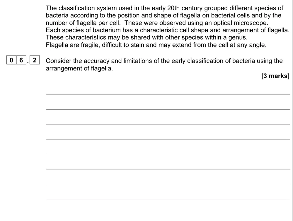
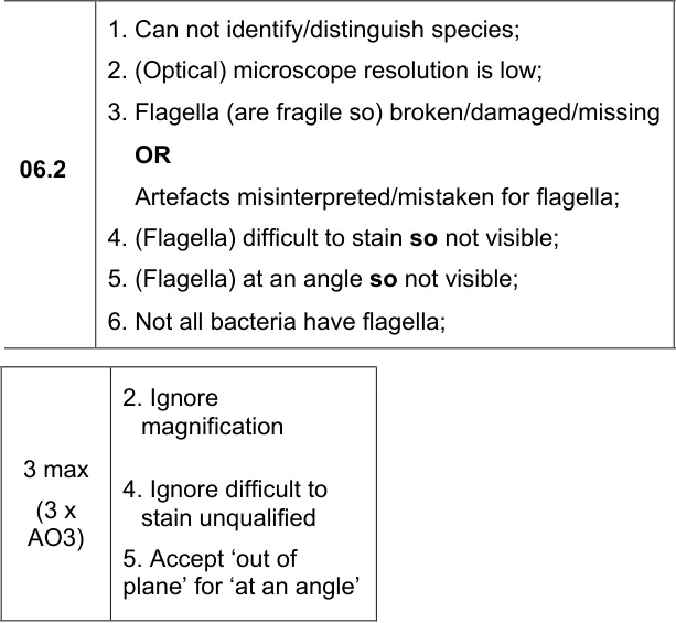
Consider the accuracy and limitations of classifying bacteria by their flagella under an optical microscope.
Show the mark scheme
Tap an image to open it full size.
From the examiners' report: A quarter got full marks. Use the information given (fragile, hard to stain, any angle) and mention low resolution; don't confuse resolution with magnification.
Cell fractionation and ultracentrifugation (3.2.1.3)
Question 8
AS Paper 2 · June 2024 · Q03.1 · 4 marks · Medium
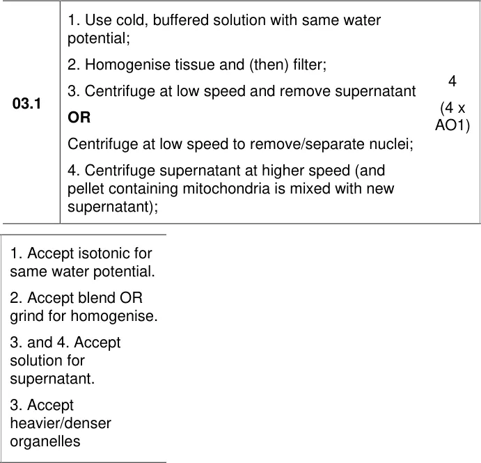
Describe how to use cell fractionation and ultracentrifugation to obtain mitochondria from muscle.
Show the mark scheme
Tap an image to open it full size.
From the examiners' report: Almost two-thirds got at least 2. Give all three solution properties, say 'filter', and say 'low speed' first. 'Centrifuge it again' is unclear: say the supernatant is spun faster.
Question 9
AS Paper 1 · June 2019 · Q05.6 · 3 marks
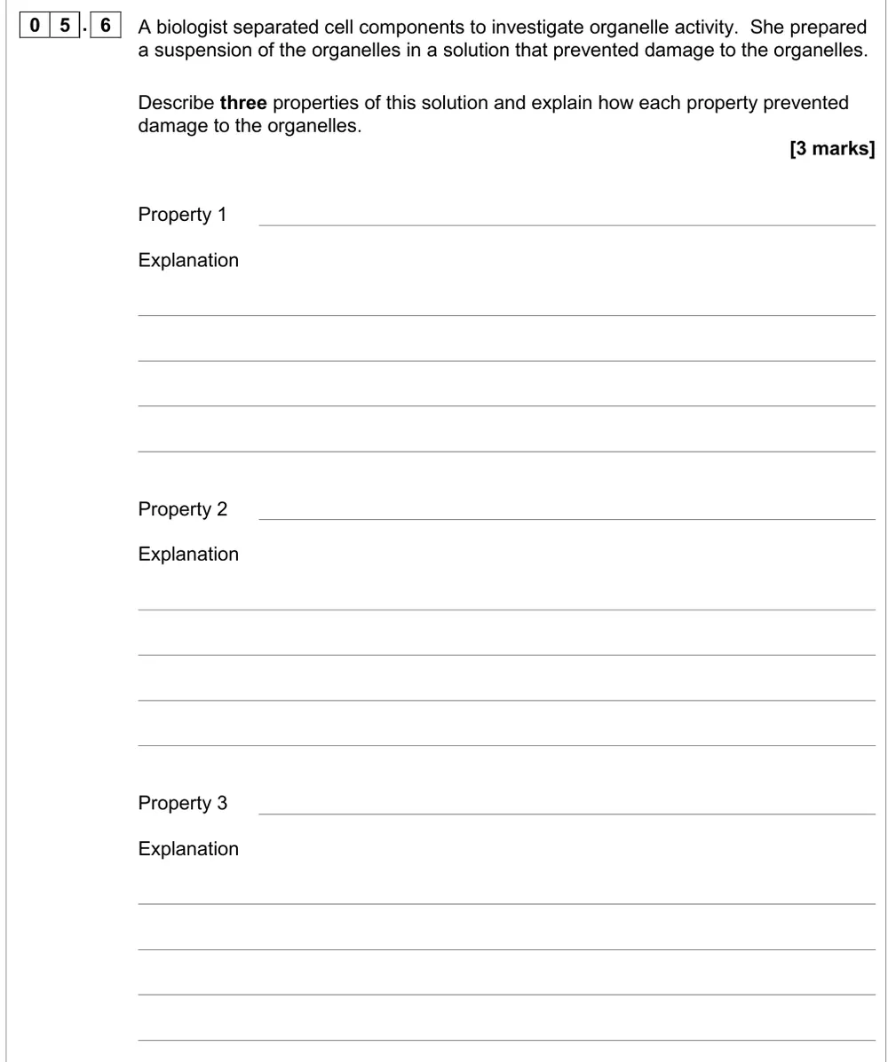
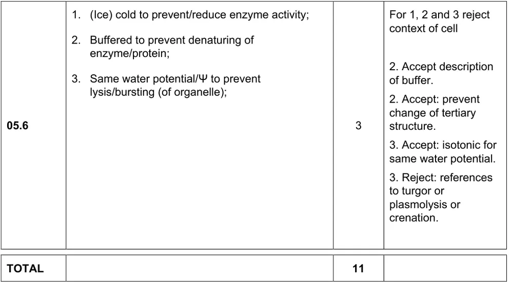
Describe three properties of the solution used to suspend organelles and explain each.
Show the mark scheme
Tap an image to open it full size.
From the examiners' report: There's no examiners' report for this paper, so this tip comes from the mark scheme. Each property needs its reason, and the reason must be about organelles, not cells.
Question 10
A level Paper 2 · June 2017 · Q04.1 · 2 marks · Easy
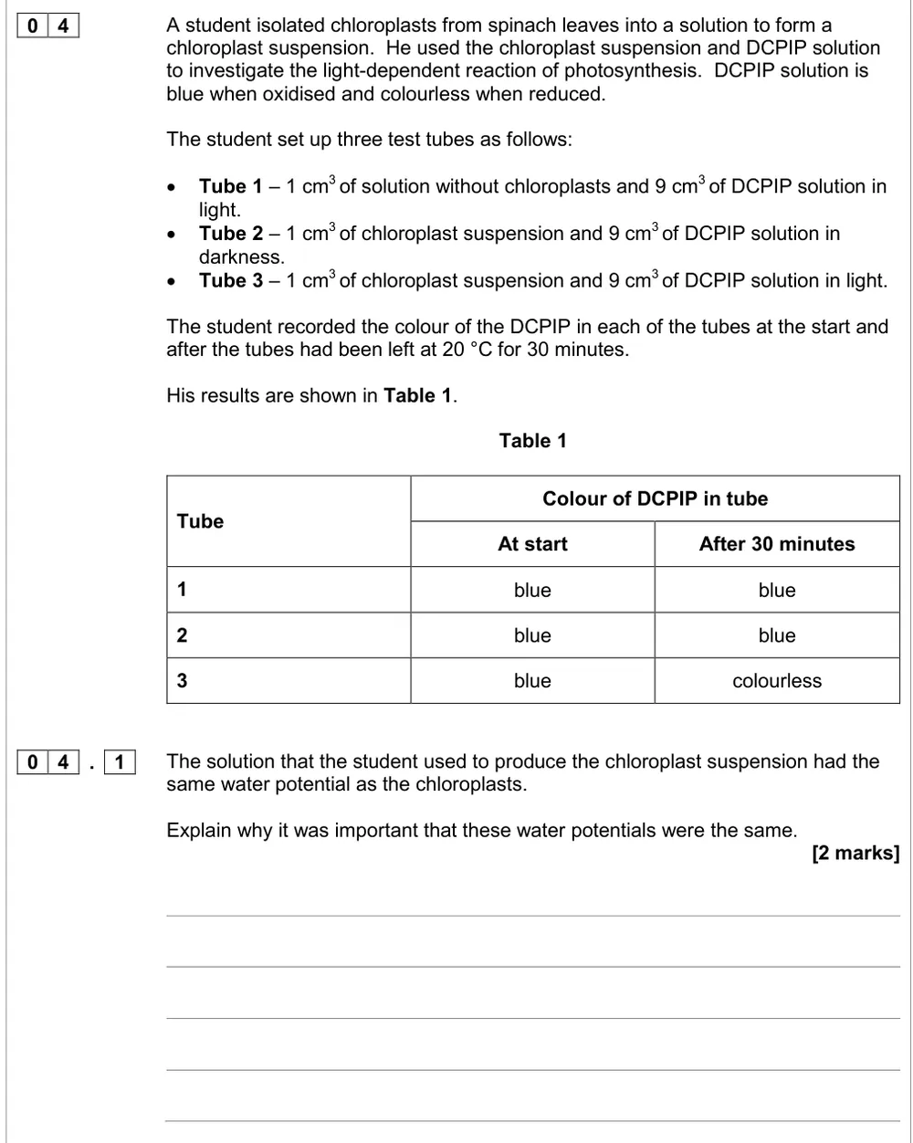
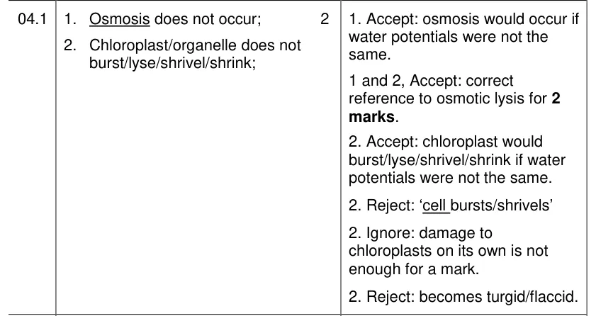
Explain why the chloroplast suspension had the same water potential as the chloroplasts.
Show the mark scheme
Tap an image to open it full size.
From the examiners' report: Over 80% got at least one mark. Write about the chloroplasts, not 'the cell', and avoid plasmolysed and turgid.
Question 11
A level Paper 1 · June 2022 · Q07.2 · 2 marks
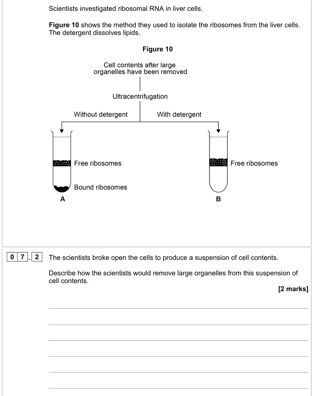
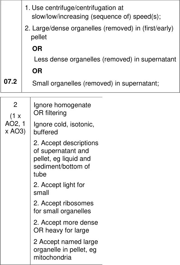
Describe how to remove large organelles from a suspension of liver cell contents.
Show the mark scheme
Tap an image to open it full size.
From the examiners' report: Say low speed first, then say where the large organelles end up: in the pellet at the bottom of the tube. Filtering alone scored nothing.
Question 12
A level Paper 1 · June 2022 · Q07.3 · 3 marks · Hard
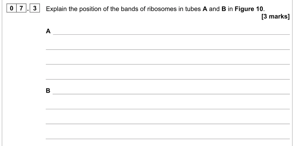
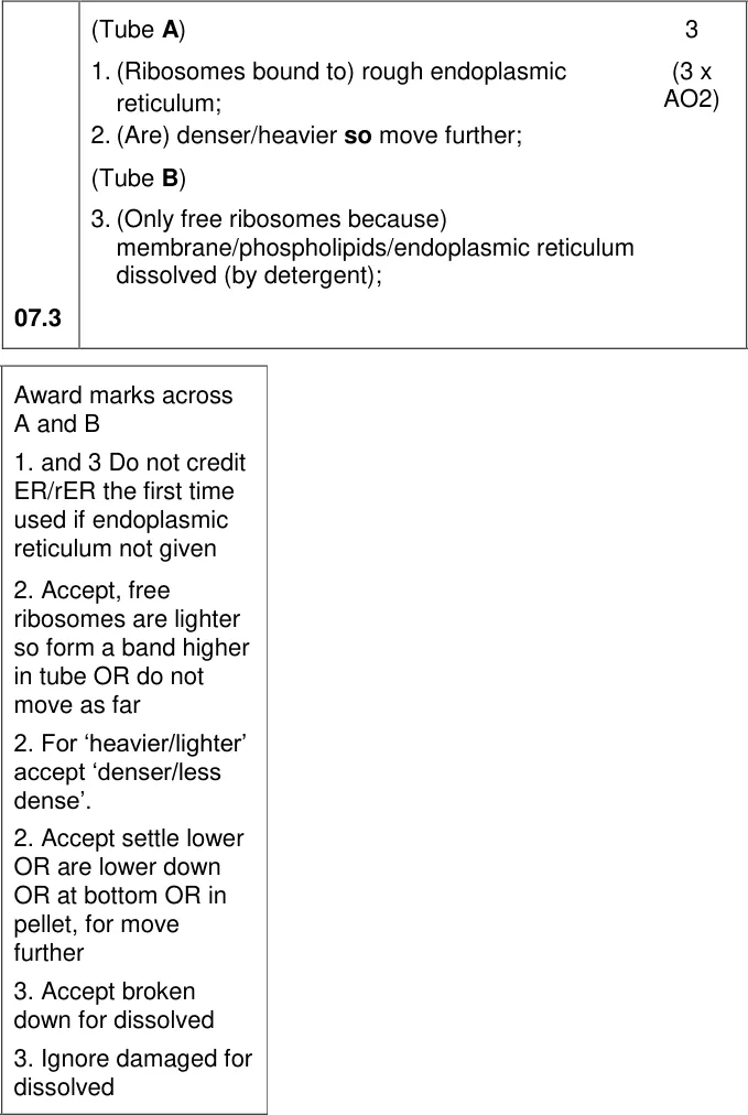
Explain the position of the bands of ribosomes in tubes A and B.
Show the mark scheme
Tap an image to open it full size.
From the examiners' report: Bound ribosomes are on rough ER, so they're denser and sink further. Say the detergent dissolves the ER membrane (phospholipids), not just 'lipids' or the cell-surface membrane.
Question 13
A level Paper 3 · June 2018 · Q05.2 · 2 marks · Hard
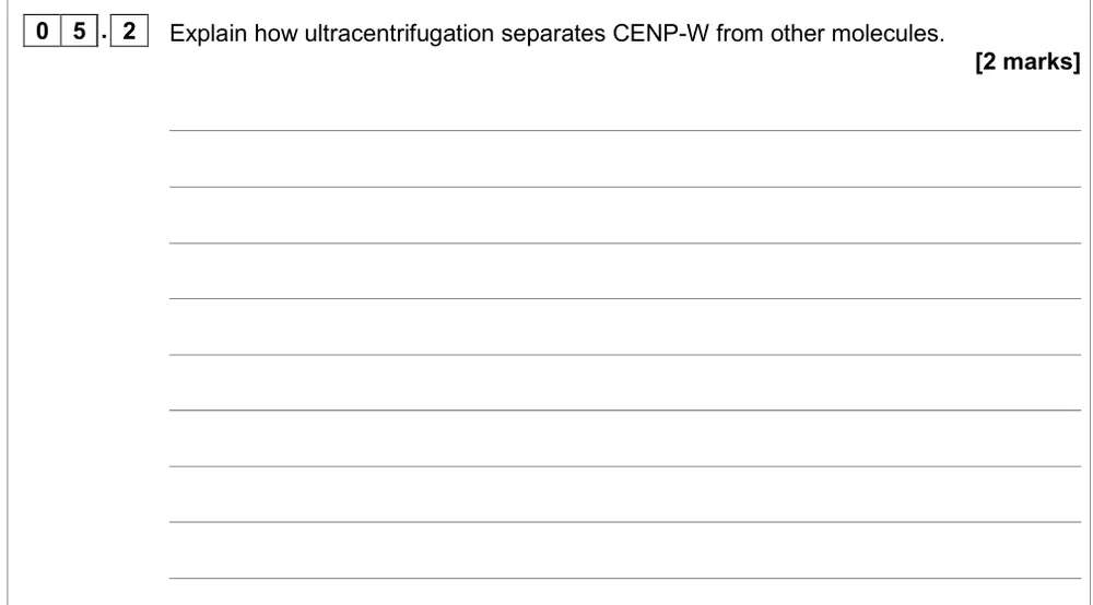
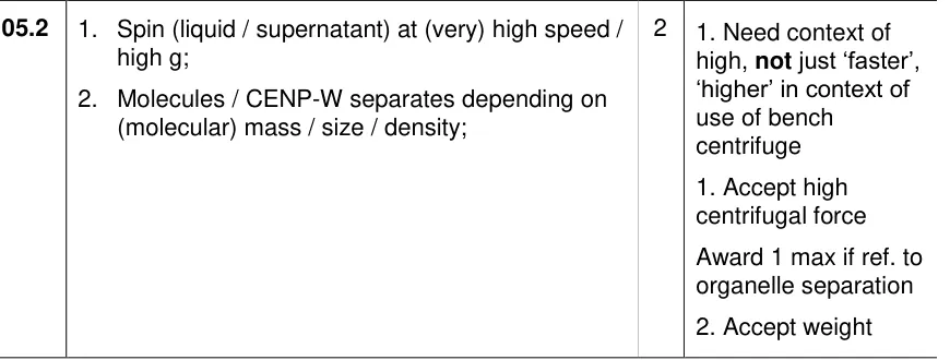
Explain how ultracentrifugation separates the protein CENP-W from other molecules.
Information from earlier in this question:

Show the mark scheme
Tap an image to open it full size.
From the examiners' report: Only 7% said very high speeds are needed to separate molecules. CENP-W is a protein, not an organelle, so don't write out the organelle method.
Preparing specimens and measuring cells (3.2.1.3)
Question 14
A level Paper 1 · June 2024 · Q03.2 · 2 marks
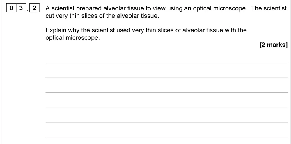
Explain why very thin slices of alveolar tissue are used with an optical microscope.
Show the mark scheme

Tap an image to open it full size.
From the examiners' report: Don't just repeat the question ('light passes through because it's thin'): say a thin slice is a single layer of cells. Thin slices don't improve resolution or magnification.
Question 15
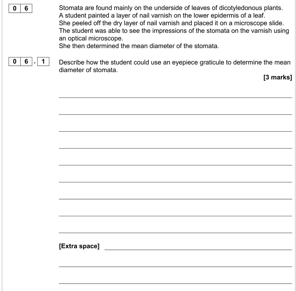
AS Paper 2 · June 2017 · Q06.1 · 3 marks
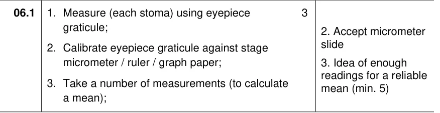
Describe how to use an eyepiece graticule to find the mean diameter of stomata.
Show the mark scheme
Tap an image to open it full size.
From the examiners' report: There's no examiners' report for this paper, so this tip comes from the mark scheme. Calibrating the graticule against a stage micrometer is the mark most often missed. Then measure several stomata to calculate the mean.
Question 16
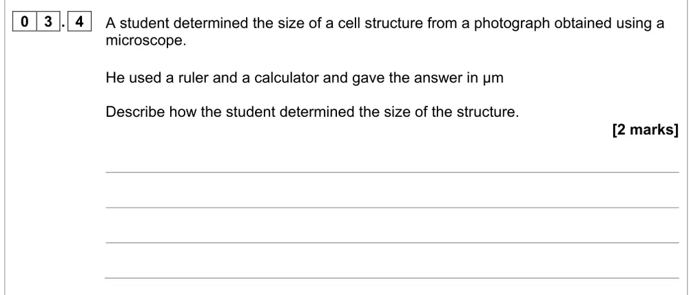
AS Paper 1 · June 2022 · Q03.4 · 2 marks · Hard
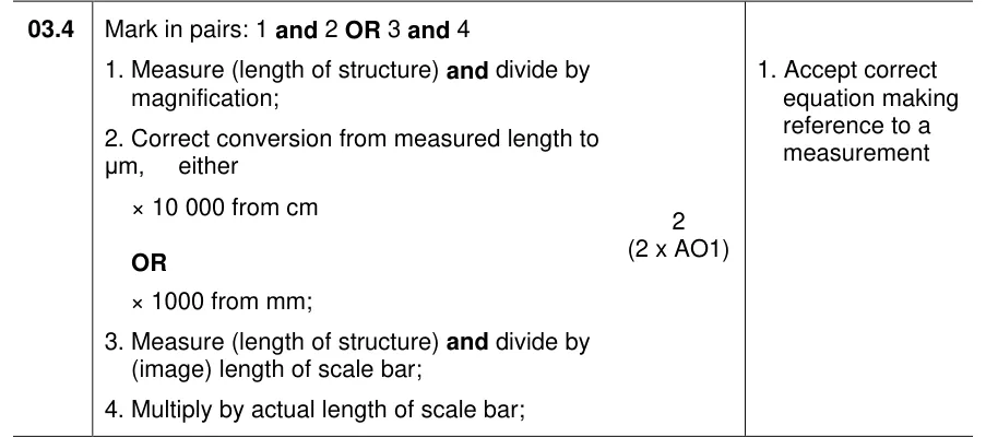
Describe how to work out the size of a structure from a photograph using a ruler.
Show the mark scheme
Tap an image to open it full size.
From the examiners' report: Only 13% got both marks. Measure, convert to µm, then divide by the magnification. If there's a scale bar, use it: divide by its measured length and multiply by its real length.
Question 17
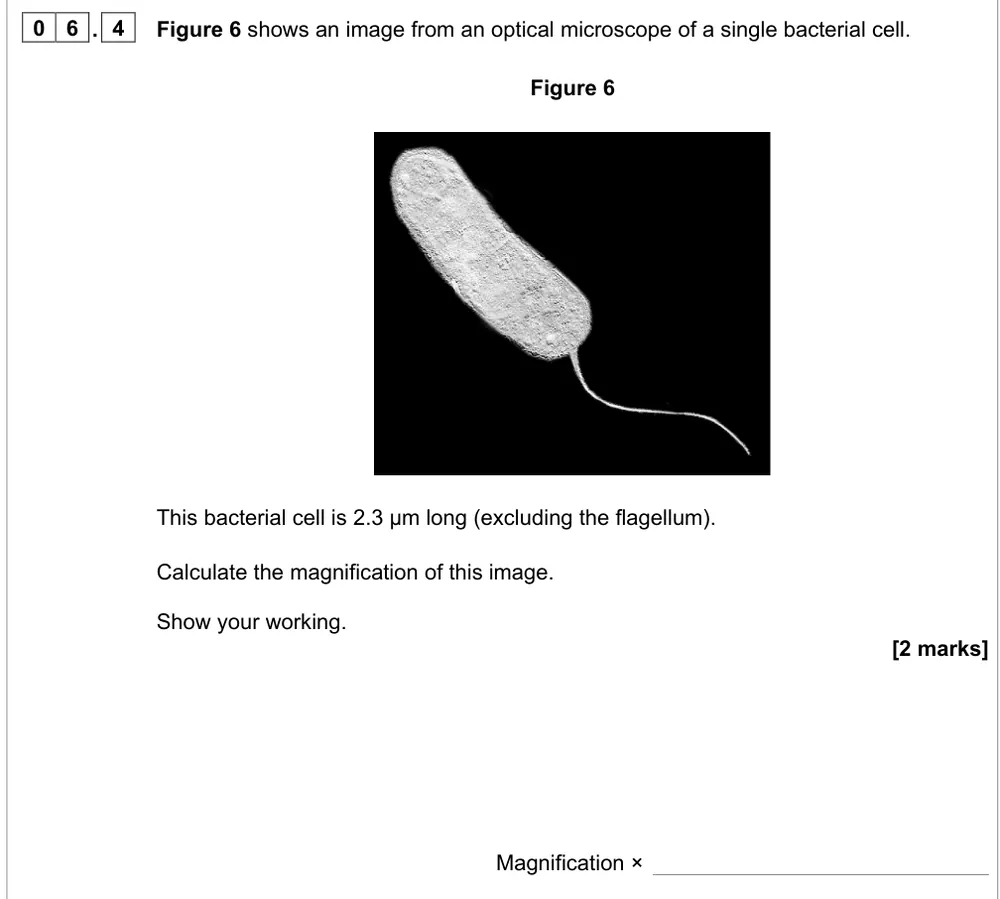
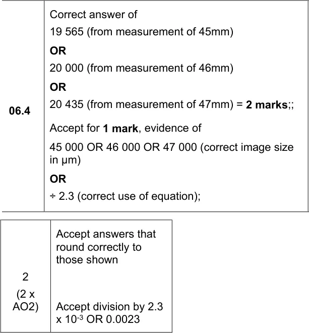
A level Paper 1 · June 2023 · Q06.4 · 2 marks · Easy
Calculate the magnification of an image of a 2.3 µm bacterium.
Show the mark scheme
Tap an image to open it full size.
From the examiners' report: About half got both marks. 46 mm is 46 000 µm, not 4600. Measure the cell on the printed paper (print the PDF at 100%), because a screen changes the size.
Question 18
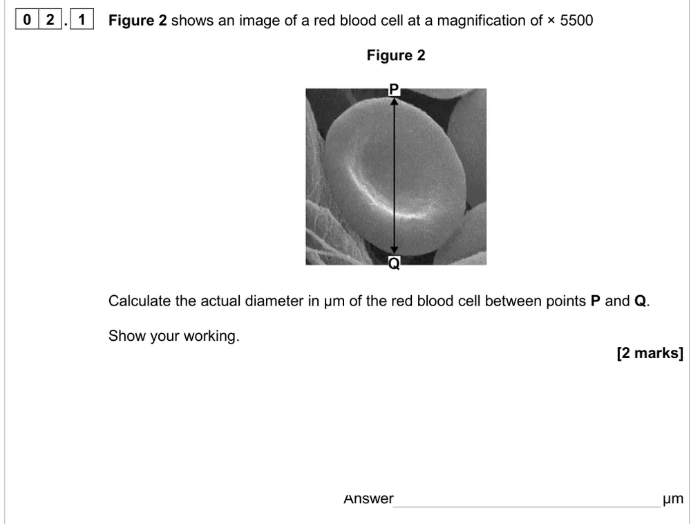
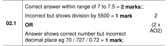
AS Paper 2 · June 2024 · Q02.1 · 2 marks · Easy
Calculate the actual diameter of a red blood cell at × 5500.
Show the mark scheme
Tap an image to open it full size.
From the examiners' report: Most got at least one mark. Measure P to Q in mm, convert to µm (× 1000) and divide by 5500; check a red blood cell is about 7 µm. Measure on the printed PDF, not the screen.
Where students lose marks (from AQA examiners' reports and mark schemes)
Each point below is followed by wording that gained marks.
Wavelength of electrons, not of the microscope: Asked why an electron microscope gives higher resolution, many wrote very brief answers about the microscope itself. The mark scheme rejects 'the electron microscope has a shorter wavelength'.
✔ Gains credit: "Shorter wavelength between electrons" or "longer wavelength in light (rays)".
Describe the SEM image and the TEM image: Only 13% got both marks for a difference between an SEM and a TEM image: many said the SEM image is 3-D but never said the TEM image is 2-D.
✔ Gains credit: "3D with SEM, but 2D with TEM; (because) electrons deflected/bounce off (SEM) or transmitted/pass through (TEM)."
Higher resolution, smaller distance: About 55% got 4 out of 4 for choosing microscopes, but some said optical microscopes have a higher resolution when they meant a larger resolvable distance, and many said a TEM gives 3-D images.
✔ Gains credit: "(SEM) resolution is high enough / 0.02 µm to view mitochondria; detects three dimensions / surface structures."
Few layers of cells, not just 'thin': Most knew light must pass through, but few said a thin slice is a single layer or a few layers of cells. Some thought thin slices improve resolution or magnification.
✔ Gains credit: "(Create a) single/few layer(s) of cells; (so) light can pass through."
Low speed first, then the supernatant faster: Many left out the low speed for the first spin, wrote 'centrifuge it again' without saying 'it' was the supernatant, or said the pellet forms at the top of the tube. Very high speeds give a pellet of mixed organelles.
✔ Gains credit: "Centrifuge at low speed to remove nuclei (in the pellet); centrifuge the supernatant at higher speed."
Cold, buffered, isotonic: give each reason: Many wrote about damage to cells rather than organelles, or used turgid and plasmolysed. 'Buffering prevents damage to organelles' was too vague.
✔ Gains credit: "Cold to prevent enzyme activity; same water potential to prevent organelles bursting/shrinking; buffered to stop enzymes/proteins denaturing."
Molecules need very high speeds: Only 7% said separating a protein needs centrifugation at very high speeds. Many wrote rote-learnt answers about separating organelles and called the protein an organelle.
✔ Gains credit: "Spin (supernatant) at (very) high speed / high g; molecules separate depending on (molecular) mass / size / density."
Millimetres × 1000 for micrometres: About half got both marks for the magnification of a bacterium. A common slip was converting 46 mm to 4600 µm; a few divided the real size by the image size.
✔ Gains credit: 46 mm = 46 000 µm; 46 000 ÷ 2.3 = × 20 000.
Download the questions
Download the PDF pack (18 questions, mark schemes at the end)
Keep practising
- Eukaryotic cell structure: AQA A Level Biology exam questions by topic
- Prokaryotic cells and viruses: AQA A Level Biology exam questions by topic
- Arithmetic and numerical skills: AQA A Level Biology exam questions by topic
- Required practical 2, mitosis: AQA A Level Biology exam questions
- AQA A Level Biology masterclasses
Questions, mark schemes and examiners' report extracts © AQA.