

Required practical 2 asks you to prepare a stained squash of cells from a plant root tip, find the stages of mitosis under an optical microscope and calculate a mitotic index. In class that usually means garlic or onion roots, warm hydrochloric acid, a stain such as toluidine blue and a firm press on the coverslip. On the AQA papers it comes up as short questions on why each step is done, plus a calculation from a drawing, photo or graph of one field of view.
The examiners' reports show where students lose marks: thinking the acid kills bacteria or speeds up mitosis, saying the press removes air bubbles instead of making a thin layer that light passes through, leaving out "field of view", not saying how many repeats make a result reliable, giving the mitotic index as a percentage, and forgetting to convert mm² to µm² before dividing.
Answer each question on paper before you open the mark scheme. If you'd like help with the required practicals, my AQA masterclasses run online on Tuesdays at 5.15pm for Year 12 and on Wednesdays or Thursdays at 6.30pm for Year 13.
What you need from GCSE and Year 12
Each question on this page assumes you're secure on these GCSE and Year 12 ideas. If any of them feels shaky, revise it before you start.
- GCSE: mitosis produces two genetically identical cells for growth and repair, and in plants it happens in the meristems at the root and shoot tips.
- GCSE: setting up a light microscope: start on low power, focus with the coarse then fine focus, then switch to high power.
- GCSE: magnification = size of image ÷ size of real object, and converting between mm and µm.
- Year 12 (AQA 3.2.1.1): the structure of plant cells, including the cellulose cell wall. See the Eukaryotic cell structure page.
- Year 12 (AQA 3.2.1.3): the principles and limitations of optical microscopes, magnification and resolution, and measuring with an eyepiece graticule.
- Year 12 (AQA 3.2.2): the cell cycle (interphase, then mitosis and cytokinesis) and the appearance of chromosomes in prophase, metaphase, anaphase and telophase.
Key terms
These words come up in the questions and mark schemes. Examiners expect you to use them exactly.
- Mitotic index: the proportion of cells undergoing mitosis: number of cells in mitosis ÷ total number of cells in the field of view, given as a decimal.
- Squash: a slide made by pressing a softened, stained root tip under a coverslip so the cells spread into a single thin layer that light can pass through.
- Stain: a dye, such as toluidine blue, that binds to DNA so the chromosomes show up clearly.
- Field of view: the circle of the specimen you can see through the microscope at one time; counts are made in several fields of view.
- Graticule: a scale in the eyepiece, calibrated against a stage micrometer at each magnification, used to measure cells.
- Independent variable: the variable you change, such as distance from the root tip or species.
- Dependent variable: the variable you measure, here usually the mitotic index.
- Control variable: a variable kept the same so the comparison is valid, such as root age, growing conditions, acid concentration and time in acid.
- Representative sample: a sample that is typical of the whole tissue, from many fields of view chosen at random.
- Resolution and uncertainty: resolution: the smallest change an instrument can show, or for a microscope the smallest distance at which two points can be told apart; uncertainty: the range either side of a reading within which the true value probably lies.
- Null hypothesis: a statement that there is no difference (or no correlation) between the samples, tested with a statistical test such as the t-test.
What these questions cover (AQA required practical 2)
The questions come from the second required practical in the AQA AS and A-level Biology specification (7401/7402), and test these statements and practical skills:
- Required practical 2: Preparation of stained squashes of cells from plant root tips; set-up and use of an optical microscope to identify the stages of mitosis in these stained squashes and calculation of a mitotic index (every question on this page; none asks you to name the stages of mitosis directly, but the anaphase question needs you to spot them)
- AT d use of light microscope at high power and low power, including use of a graticule
- PS 4.1 Know and understand how to use a wide range of experimental and practical instruments, equipment and techniques appropriate to the knowledge and understanding included in the specification
- PS 2.1 Comment on experimental design and evaluate scientific methods
- PS 3.3 Consider margins of error, accuracy and precision of data
- MS 0.3 Use ratios, fractions and percentages
- PS 3.2 Process and analyse data using appropriate mathematical skills
- MS 1.9 Select and use a statistical test
How to use this page
Write your answer on paper before you open the mark scheme. Allow about 1.3 minutes per mark: the 13 questions are worth 25 marks, so about 32 minutes in total. Then mark your answer strictly against the mark scheme and read the examiners' tip underneath it. If you'd rather print the questions, download the PDF pack.
Preparing a root tip squash
Question 1
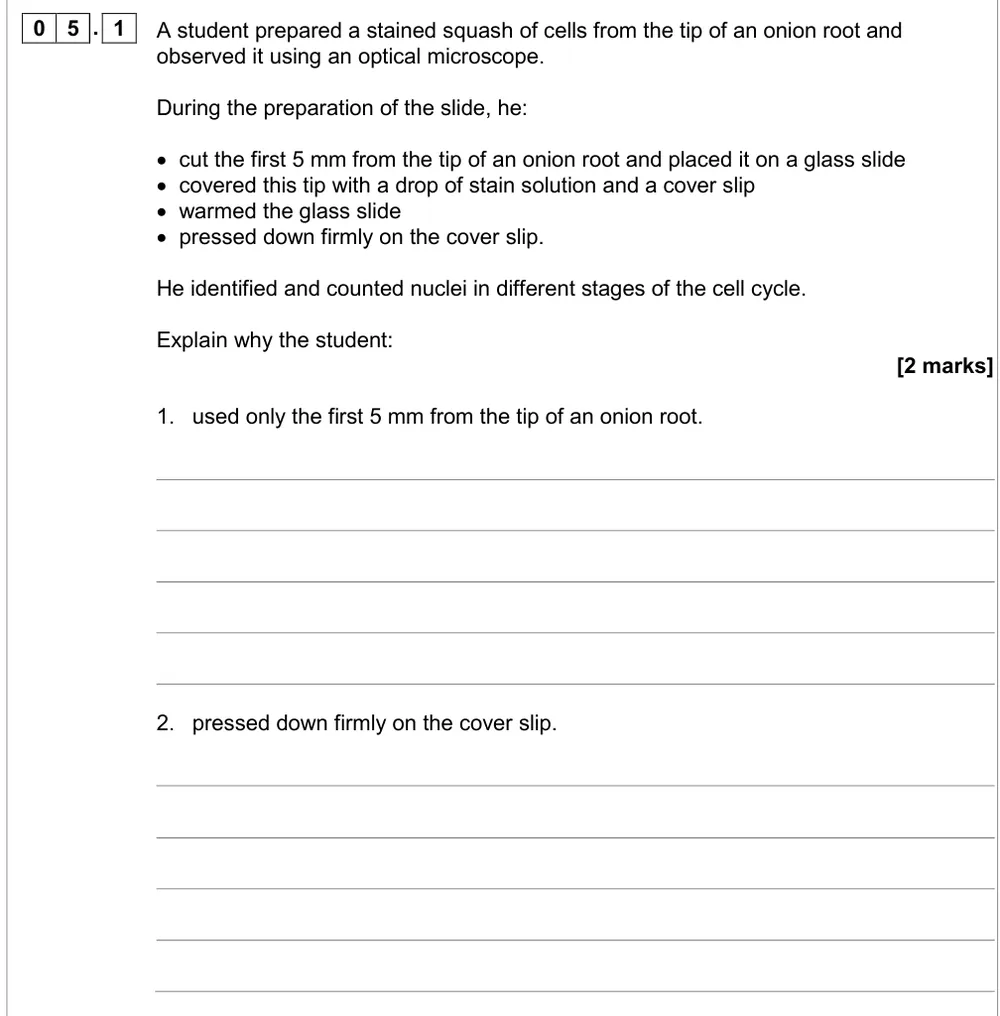
AS Paper 2 · June 2018 · Q05.1 · 2 marks
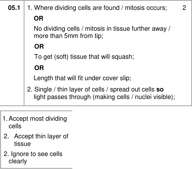
Explain why only the first 5 mm of an onion root is used and why the cover slip is pressed down firmly.
Show the mark scheme
Tap an image to open it full size.
From the examiners' report: Most got at least one mark, usually for mitosis happening at the tip. Root tips divide by mitosis, not meiosis. For pressing down, mention a thin layer of cells and light passing through; removing air bubbles was ignored.
Question 2
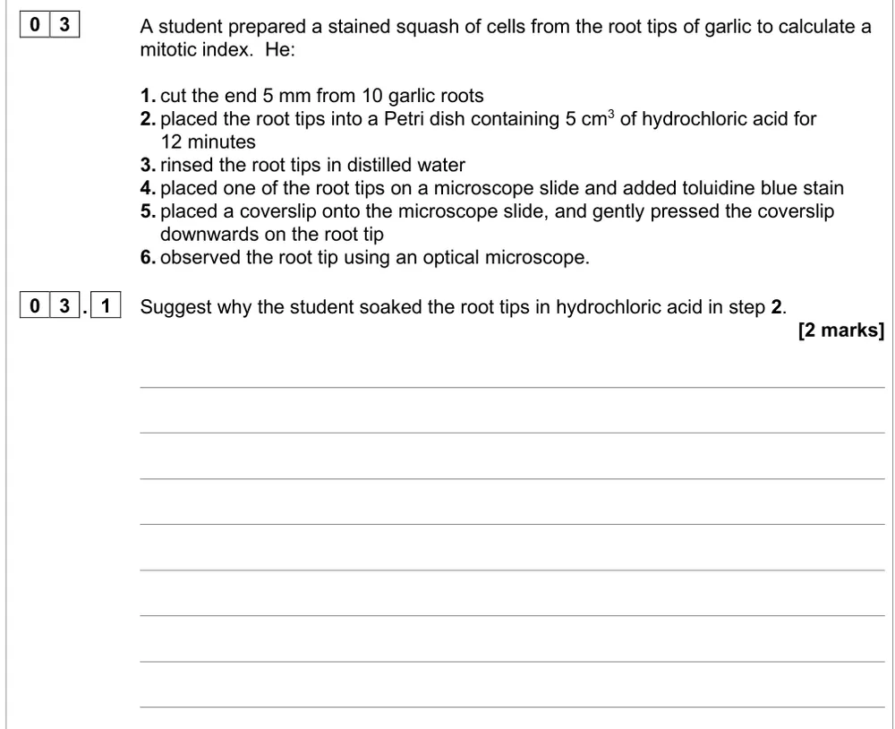
A level Paper 3 · June 2022 · Q03.1 · 2 marks · Medium
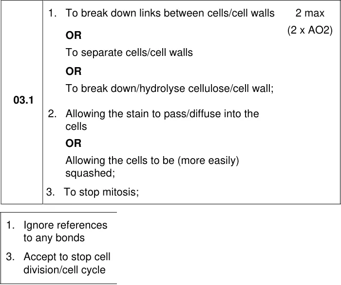
Suggest why garlic root tips are soaked in hydrochloric acid before staining.
Show the mark scheme
Tap an image to open it full size.
From the examiners' report: Just over 65% got a mark, and the best answers mentioned the middle lamella. It isn't to kill bacteria: the acid separates the cells so they can be squashed and stained.
Question 3
AS Paper 1 · June 2023 · Q08.1 · 1 mark · Hard
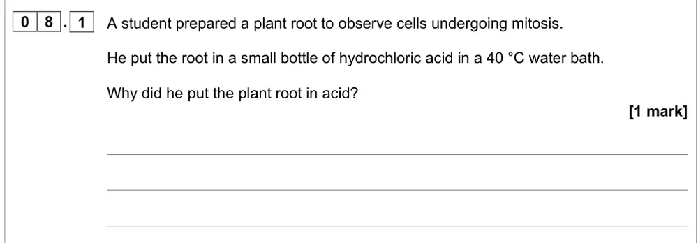
Give a reason for putting a plant root in warm hydrochloric acid before observing mitosis.
Show the mark scheme
Tap an image to open it full size.
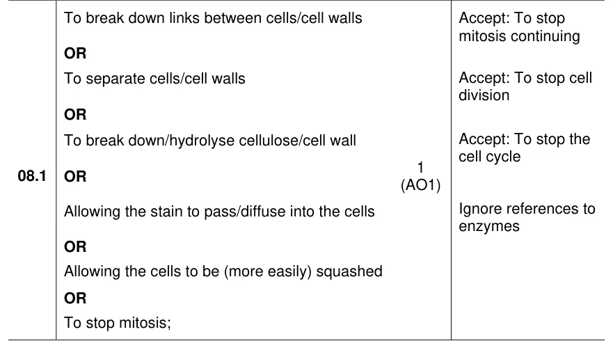
From the examiners' report: Only about a third got the mark. Wrong ideas included speeding up mitosis, neutralising the root, sterilising it, denaturing enzymes or breaking the cell membrane.
Question 4
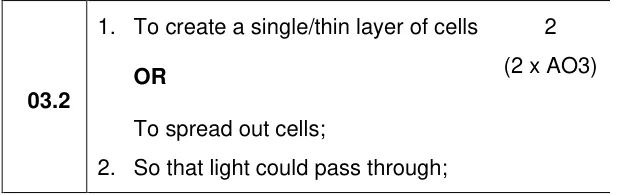
A level Paper 3 · June 2022 · Q03.2 · 2 marks · Easy
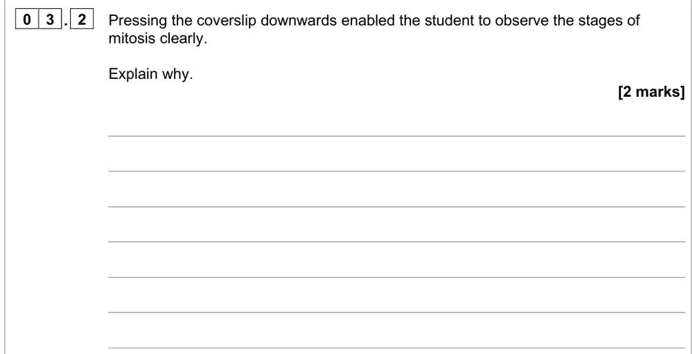
Explain why pressing down the coverslip helps the student see the stages of mitosis.
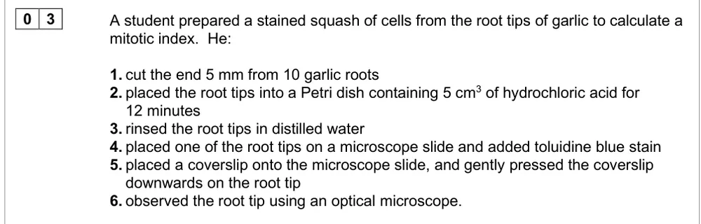
Information from earlier in this question:
Show the mark scheme
Tap an image to open it full size.
From the examiners' report: 56% got both marks. Wrong answers were about removing bubbles, making the cells themselves thinner or squashing organelles.
Question 5
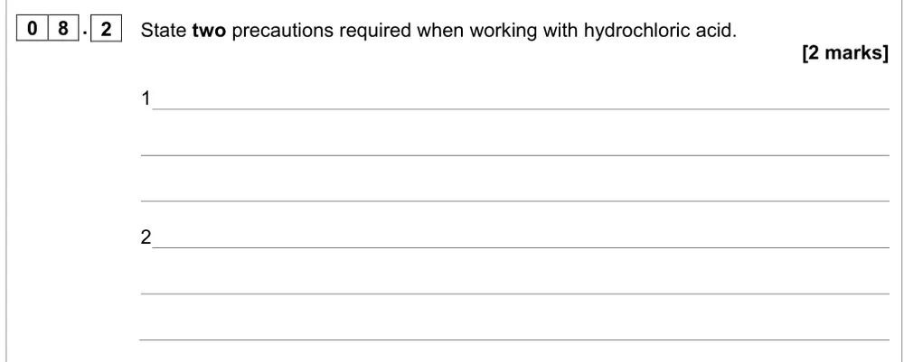
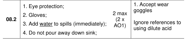
AS Paper 1 · June 2023 · Q08.2 · 2 marks
State two safety precautions when using hydrochloric acid.
Show the mark scheme
Tap an image to open it full size.
From the examiners' report: Most wrote goggles and gloves. For a spill, "clear it up" didn't score: add water to it.
Counting cells and the mitotic index
Question 6
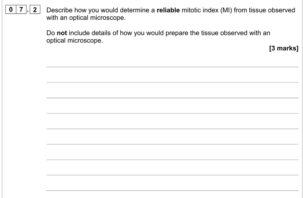
AS Paper 1 · June 2022 · Q07.2 · 3 marks
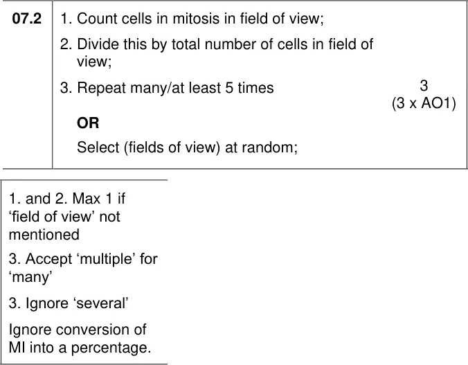
Describe how to find a reliable mitotic index from tissue under an optical microscope.
Show the mark scheme
Tap an image to open it full size.
From the examiners' report: Use the term field of view. "Reliable" means repeats: say how many (at least 5), or choose fields of view at random.
Question 7
AS Paper 2 · June 2018 · Q05.4 · 2 marks
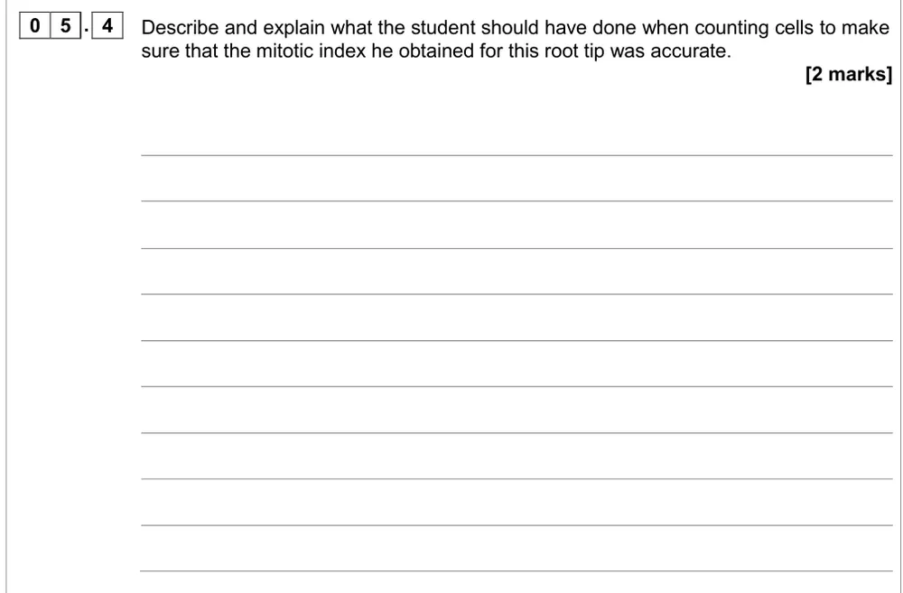
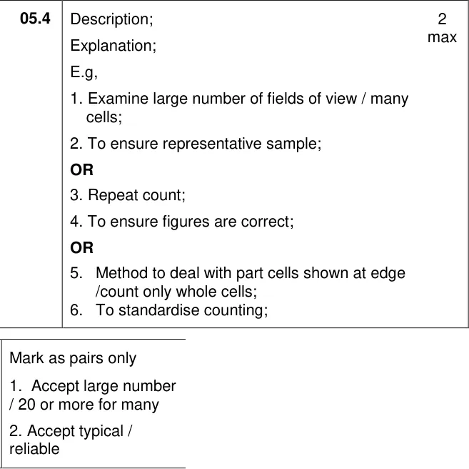
Describe and explain what to do when counting cells so the mitotic index is accurate.
Show the mark scheme
Tap an image to open it full size.
From the examiners' report: Give a step and its reason. Many wrote out how to calculate the index or about other root tips instead; the cells don't keep dividing once stained and squashed.
Question 8
A level Paper 3 · June 2022 · Q03.4 · 1 mark · Easy
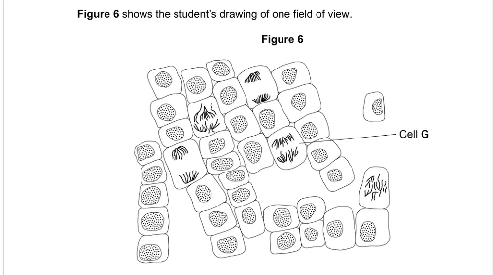
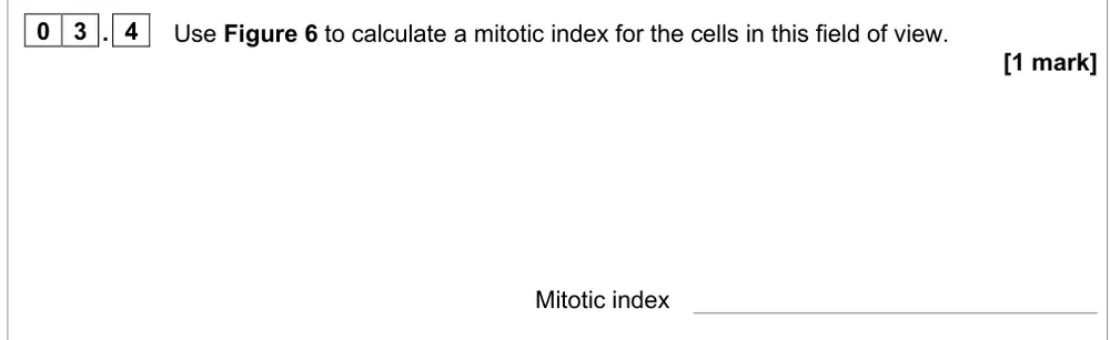
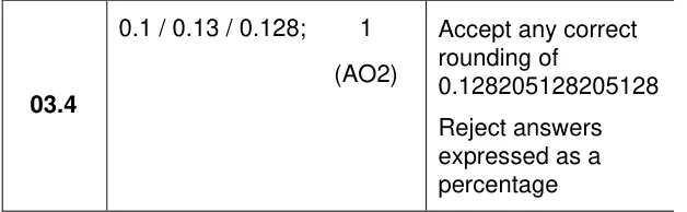
Calculate the mitotic index from a drawing of one field of view.
Information from earlier in this question:
Show the mark scheme
Tap an image to open it full size.
From the examiners' report: This was generally well answered. Mitotic index = cells in mitosis ÷ total cells in the field of view, given as a decimal, not a percentage.
Question 9
AS Paper 2 · June 2018 · Q05.2 · 2 marks · Hard
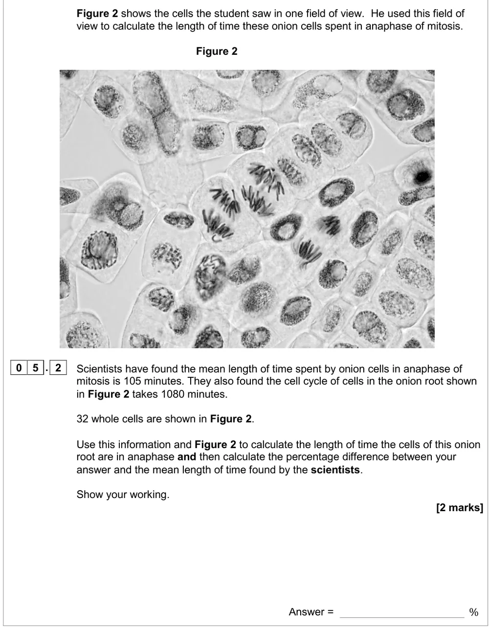
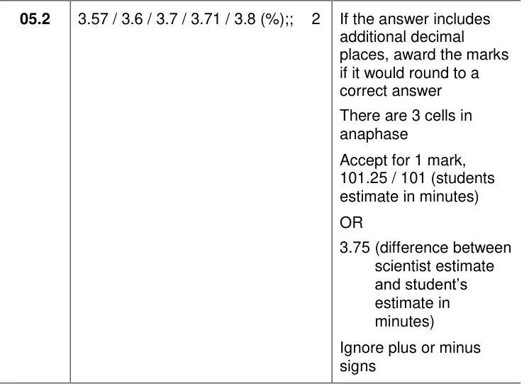
Use a photo of one field of view to estimate the time onion cells spend in anaphase.
Show the mark scheme
Tap an image to open it full size.
From the examiners' report: This discriminated well, and 8% didn't try it. Fraction of cells in anaphase (3 ÷ 32) × 1080 minutes gives the time; then find the percentage difference from 105 minutes.
Question 10
AS Paper 1 · June 2023 · Q08.4 · 2 marks · Medium
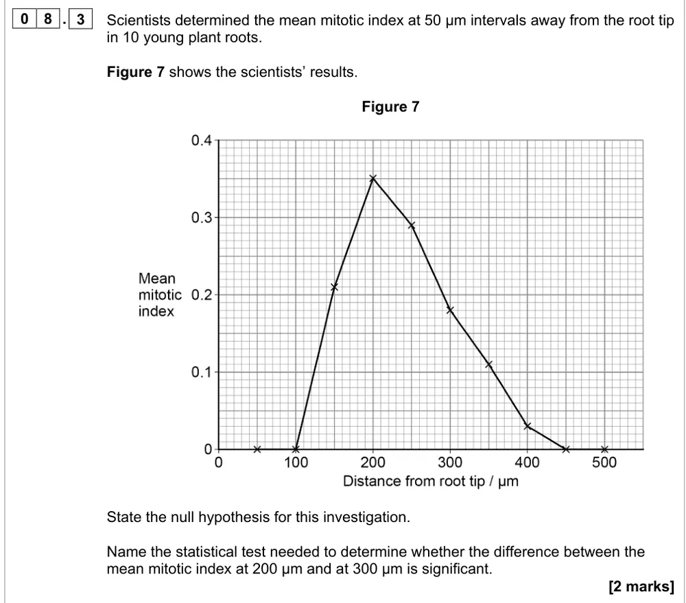
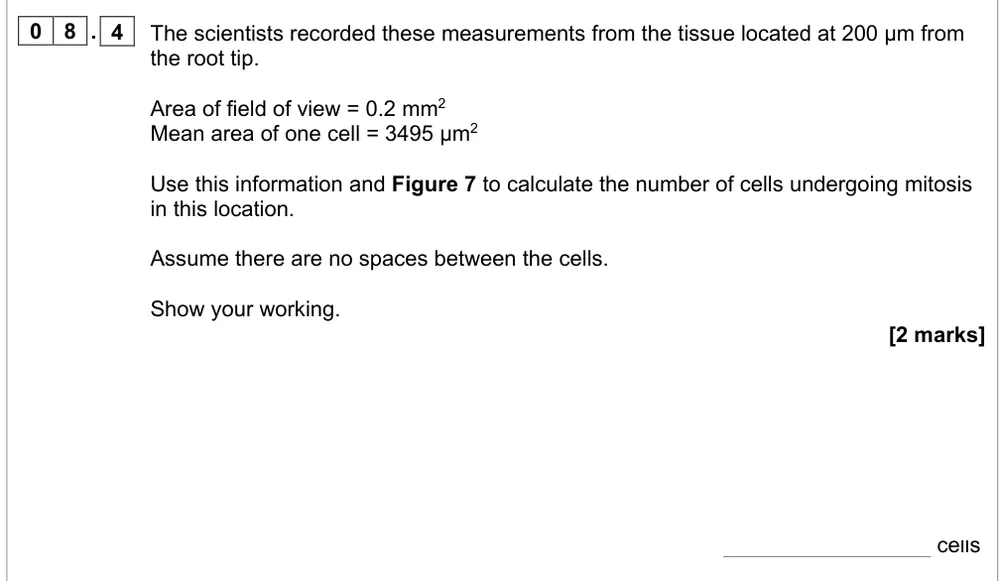
Use areas and a mitotic index to work out how many cells are in mitosis in one field of view.
Information from earlier in this question:
Show the mark scheme
Tap an image to open it full size.
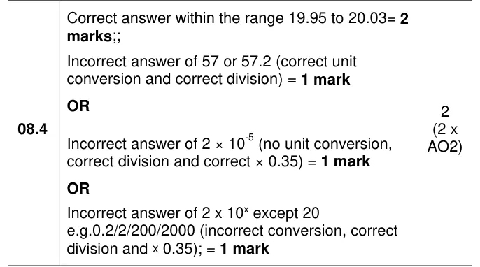
From the examiners' report: Less than a quarter got both marks. Convert 0.2 mm² to 200 000 µm² first (1 mm² = 10⁶ µm²), divide by 3495, then multiply by the index of 0.35.
Comparing and evaluating results
Question 11
A level Paper 3 · June 2022 · Q03.5 · 2 marks · Easy
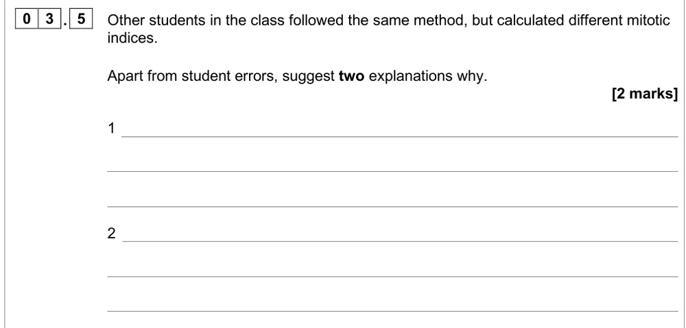
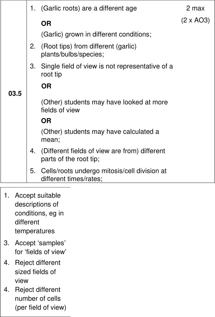
Suggest two reasons, other than student errors, why classmates got different mitotic indices.
Information from earlier in this question:
Show the mark scheme
Tap an image to open it full size.
From the examiners' report: Generally well answered. Think about the roots (age, growing conditions, plant) and the sampling (one field of view isn't representative). Different sized fields of view were rejected.
Question 12
AS Paper 1 · June 2023 · Q08.5 · 2 marks
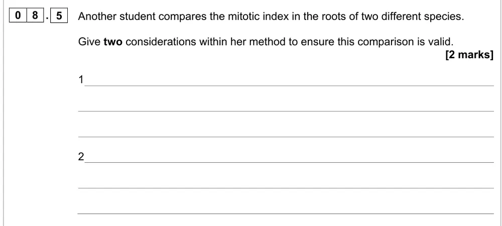
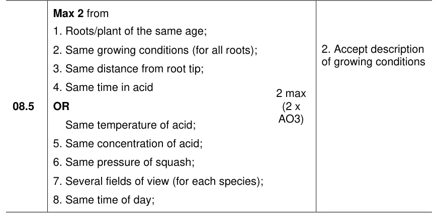
Give two things to keep the same so a comparison of mitotic index in two species is valid.
Show the mark scheme
Tap an image to open it full size.
From the examiners' report: There's no examiners' comment on this part, so this tip comes from the mark scheme. Name control variables: roots of the same age and growing conditions, and the same distance from the tip and treatment with acid.
Question 13
AS Paper 1 · June 2023 · Q08.3 · 2 marks
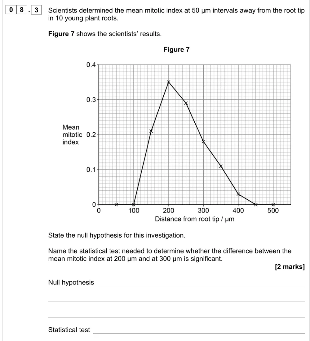
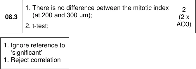
Write a null hypothesis and name the test for comparing mean mitotic index at two distances.
Show the mark scheme
Tap an image to open it full size.
From the examiners' report: Practise writing null hypotheses: "there is no difference between…". Comparing two means needs a t-test; correlation and chi-squared don't fit, and standard deviation isn't a statistical test.
Where students lose marks (from AQA examiners' reports and mark schemes)
Each point below is followed by wording that gained marks.
Acid separates the cells: Only about a third knew why a root is put in warm hydrochloric acid. Many thought it speeds up mitosis, neutralises the root, kills bacteria, sterilises the tissue or denatures enzymes.
✔ Gains credit: "To break down (hydrolyse) the cell walls and separate the cells, so the cells can be squashed and the stain can diffuse in" (or "to stop mitosis").
Press for a thin layer that light passes through: The reason for pressing down on the cover slip was not well known: answers about removing air bubbles or spreading the stain were ignored, and others never mentioned cells or light.
✔ Gains credit: "To create a single (thin) layer of cells, so that light could pass through."
Root tips divide by mitosis, not meiosis: Some students said meiosis happens in root tip cells, or described mitosis as the whole cell cycle.
✔ Gains credit: "The first 5 mm is where dividing cells are found; there is no mitosis further from the tip."
Count within a field of view: Students are expected to know the term field of view; without it, the counting and dividing steps got only 1 mark between them.
✔ Gains credit: "Count the cells in mitosis in a field of view and divide by the total number of cells in the field of view."
Say how many repeats: Many missed the bold word reliable, which should have prompted repeats, and those who did repeat often didn't say how many.
✔ Gains credit: "Repeat for at least 5 fields of view, selected at random."
Mitotic index as a decimal: The mark scheme rejects a mitotic index written as a percentage.
✔ Gains credit: "Mitotic index = 0.13" (cells in mitosis ÷ total cells).
Convert mm² to µm² before dividing: Less than a quarter got both marks for the number of cells in mitosis. The main problems were not converting between mm and µm, and rounding and dividing wrongly.
✔ Gains credit: 0.2 mm² = 200 000 µm²; 200 000 ÷ 3495 = 57.2 cells; 57.2 × 0.35 = 20 cells.
Two means need a t-test: Students need to practise writing a null hypothesis. Correlation and chi-squared were also suggested, and some named standard deviation, which isn't a statistical test.
✔ Gains credit: "There is no difference between the mitotic index at 200 and 300 µm"; "t-test".
Download the questions
Download the PDF pack (13 questions, mark schemes at the end)
Keep practising
- Arithmetic and numerical skills: AQA A Level Biology exam questions by topic
- Eukaryotic cell structure: AQA A Level Biology exam questions by topic
- Required practical 1, enzyme rates: AQA A Level Biology exam questions
- Mastering AQA A Level Biology 3.4.3: genetic diversity via mutation and meiosis
- Cell division and the cell cycle: AQA A Level Biology exam questions by topic
- AQA A Level Biology masterclasses
Questions, mark schemes and examiners' report extracts © AQA.