

OCR A PAG 6 · 18 questions · mark schemes and examiners’ tips included
PAG 6 covers two ways of separating biological molecules. In chromatography, a solvent carries substances such as amino acids, sugars or photosynthetic pigments through paper or a thin layer of silica, and you identify them from their Rf values. In gel electrophoresis, an electric current moves charged DNA fragments or proteins through agarose gel, and the fragments separate by size.
Most of the marks here are for practical details: keeping the origin line above the solvent, letting a spot dry before adding more, measuring to the middle of a spot, and giving an Rf value to a sensible number of figures. For electrophoresis, the examiners want you to explain each step, such as why you add a buffer or a dye, not just name it.
Work through the eighteen questions below in order. They start with running a chromatogram, then Rf calculations and interpreting chromatograms, and finish with electrophoresis and DNA profiling. If you'd like to go through questions like these with me each week, my OCR A masterclasses run on Wednesdays at 5.15pm for Year 12 and on Tuesdays at 6.30pm or Thursdays at 5.15pm for Year 13.
What these questions cover (OCR PAG 6: chromatography or electrophoresis)
PAG 6 is one of the twelve practical activity groups in the OCR A Level Biology A specification (H420). The questions test these parts of the specification:
- 1.2.2(g) separation of biological compounds using thin layer/paper chromatography or electrophoresis
- 2.1.2(s)(i) the principles and uses of paper and thin layer chromatography to separate biological molecules / compounds (to include calculating Rf values)
- 2.1.2(s)(ii) practical investigations to analyse biological solutions using paper or thin layer chromatography
- 5.2.1(c)(ii) practical investigations using thin layer chromatography (TLC) to separate photosynthetic pigments
- 6.1.3(c) the principles of DNA profiling and its uses (including how electrophoresis is used in a DNA profile)
- 6.1.3(e) the principles and uses of electrophoresis for separating nucleic acid fragments or proteins
How to use this page
Write your answer on paper before you open the mark scheme. Allow about 1.2 minutes per mark: the 18 questions are worth 40 marks, so about 48 minutes in total. Then mark your answer strictly against the mark scheme and read the examiners' tip underneath it. If you'd rather print the questions, download the PDF pack.
Running a chromatogram
Question 1
A level Paper 3 · June 2023 · Q2(a)(i) · 1 mark · Easy
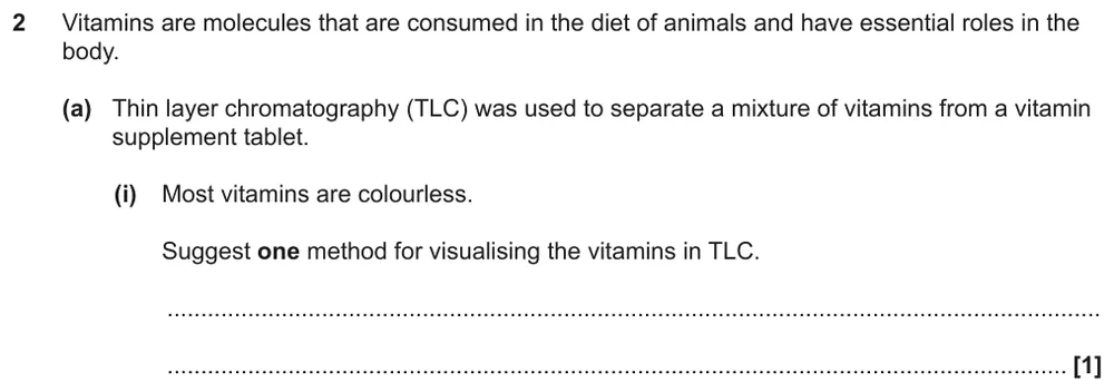
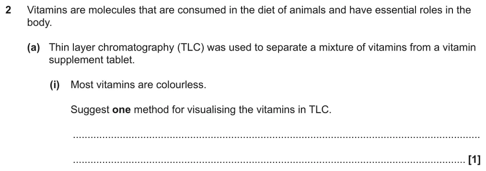
Suggest one way to see colourless vitamins on a TLC plate.
Show the mark scheme

Tap an image to open it full size.
From the examiners' report: Most students got this. Mistakes were 'a solvent', or attaching a chemical or a tagged antibody to the vitamins before running the TLC, which would change their Rf values.
Question 2
A level Paper 3 · June 2017 · Q1(b)(i) · 1 mark · Medium
State a material that can be used as the stationary phase in TLC.

Show the mark scheme

Tap an image to open it full size.
From the examiners' report: Only a small number of students could name one. Many wrongly said paper, which is used in paper chromatography, not TLC, or named a solvent.
Question 3
AS Paper 2 · June 2025 · Q1(c) · 2 marks · Medium
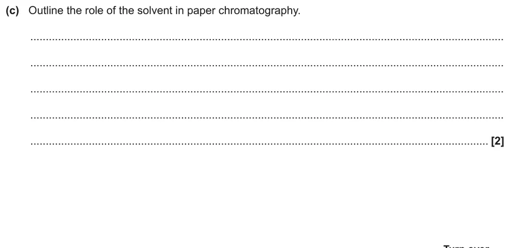
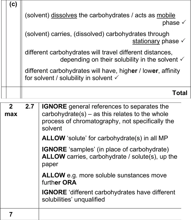
Outline the role of the solvent in paper chromatography.
Show the mark scheme
Tap an image to open it full size.
From the examiners' report: Many students knew the solvent is the mobile phase and dissolves the solute. Weaker answers talked vaguely about separating colours or dyes, or said the solvent 'pushes' or 'drags' the solute along.
Question 4
AS Paper 2 · June 2018 · Q1(c)(i) · 4 marks · Hard
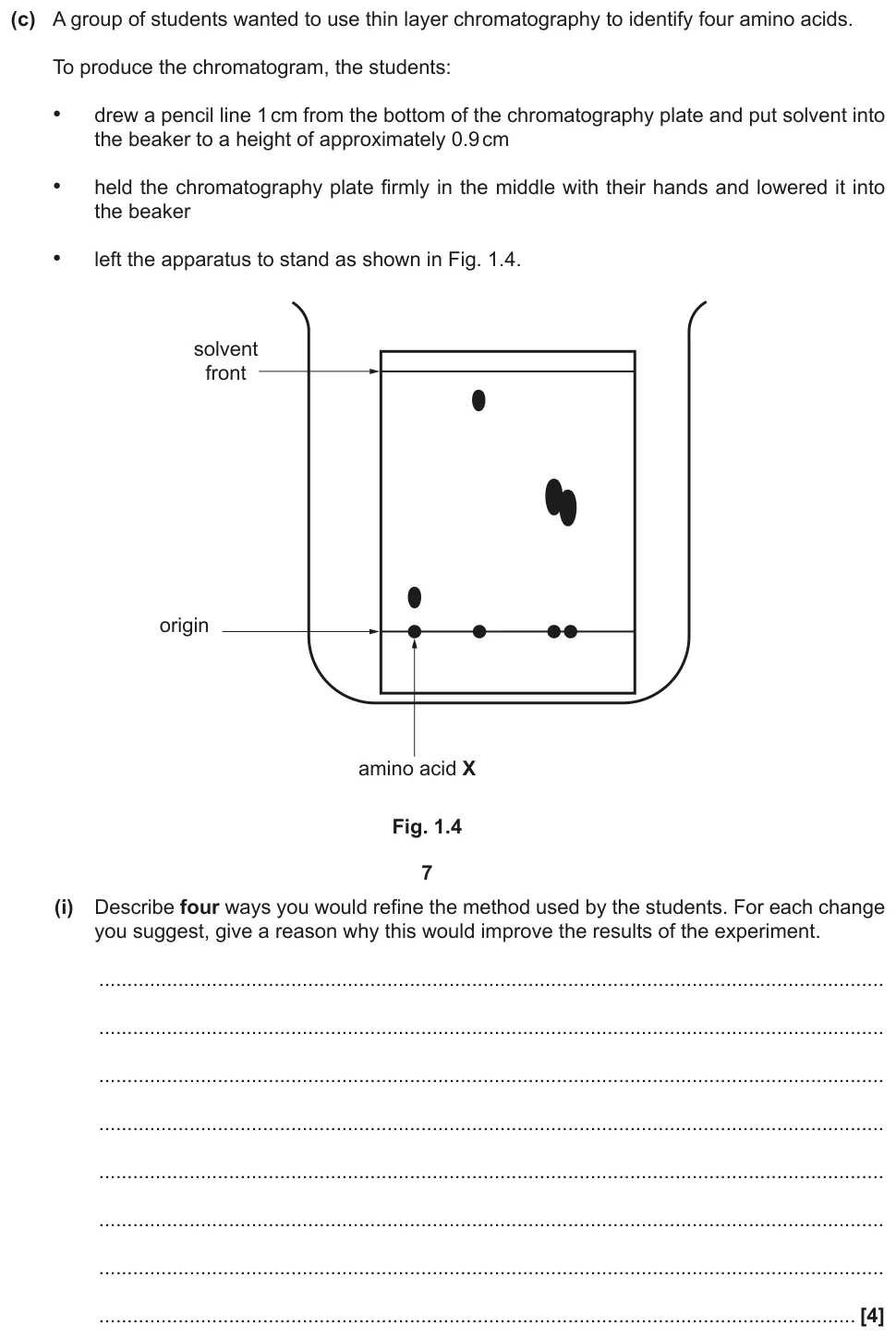
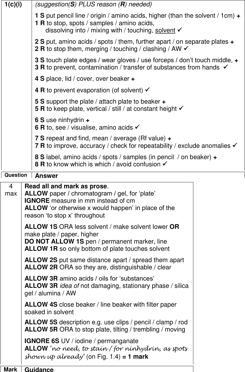
Describe four ways to refine a TLC method for amino acids, with a reason for each.
Show the mark scheme
Tap an image to open it full size.
From the examiners' report: Most students made two sensible suggestions, but very few made four. Every point needs a reason as well as a change, so write each one as 'do this, because of that'.
Calculating Rf values
Question 5
AS Paper 2 · June 2018 · Q1(c)(ii) · 2 marks · Easy
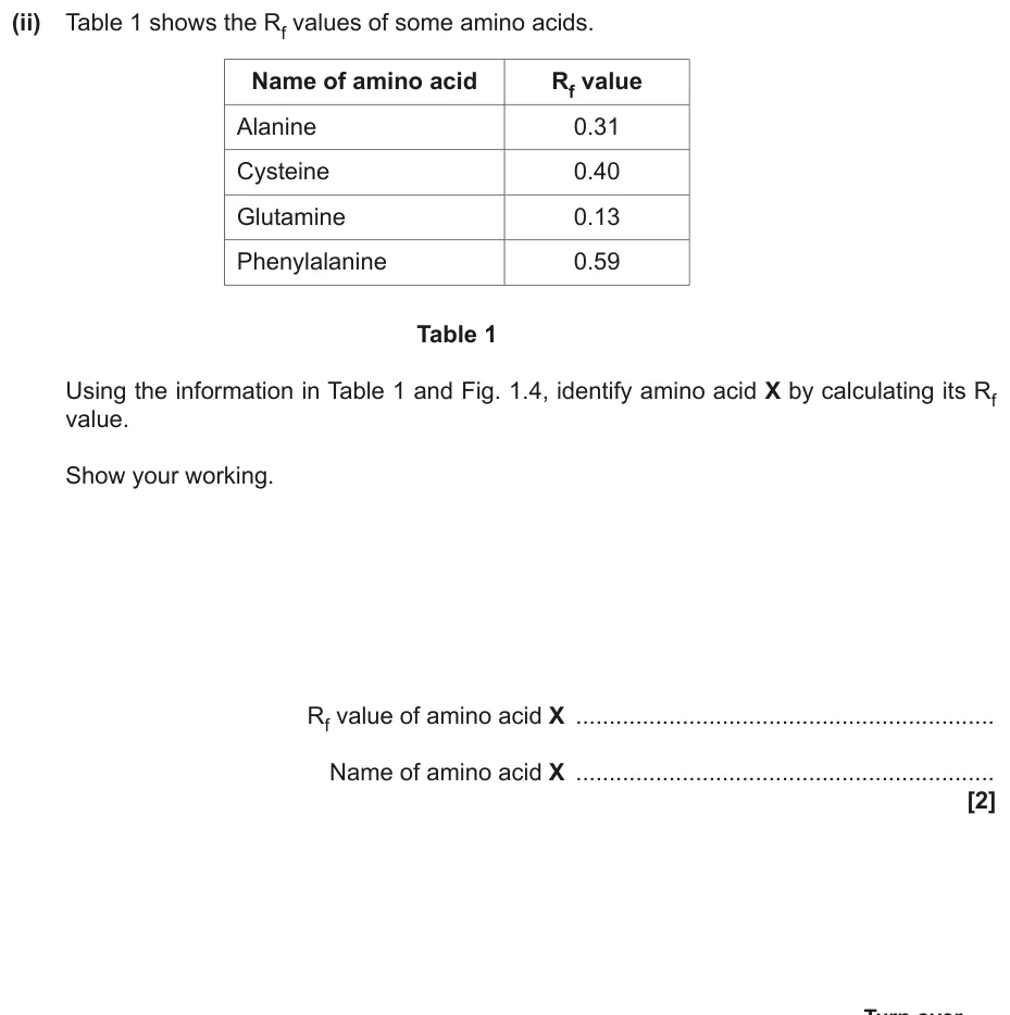
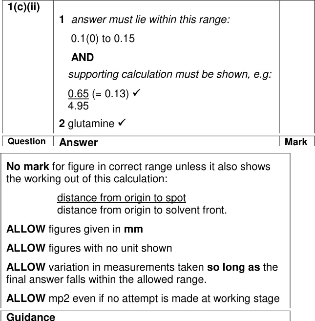
Calculate the Rf value of amino acid X and use it to identify X.
Show the mark scheme
Tap an image to open it full size.
From the examiners' report: Most students chose glutamine, but not all showed an Rf calculated from the figure to back it up. Some measured so carelessly that their value fell outside the generous range allowed.
Question 6
A level Paper 1 · June 2025 · Q16(b)(i) · 3 marks · Easy
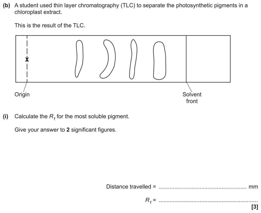
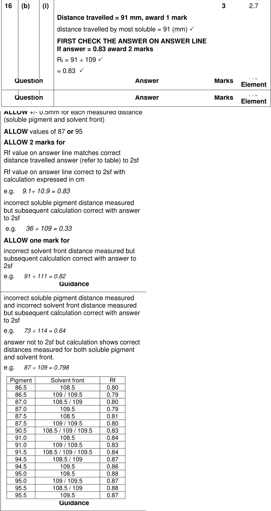
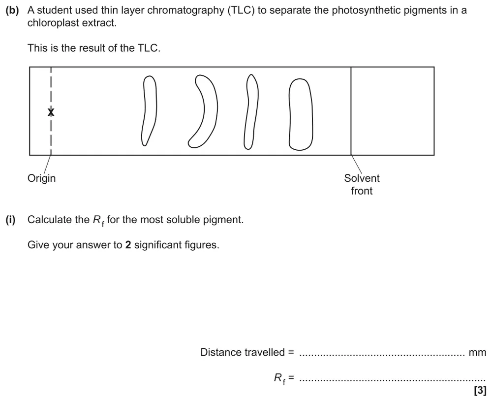
Measure a TLC plate and calculate the Rf of the most soluble pigment, to 2 significant figures.
Show the mark scheme
Tap an image to open it full size.
From the examiners' report: Most students picked the pigment nearest the solvent front. Measure to the middle of the spot, and to the solvent front line rather than the end of the plate. Many measured in cm even though the answer line said mm.
Question 7
AS Paper 1 · June 2023 · Q23(b)(i) · 2 marks · Medium
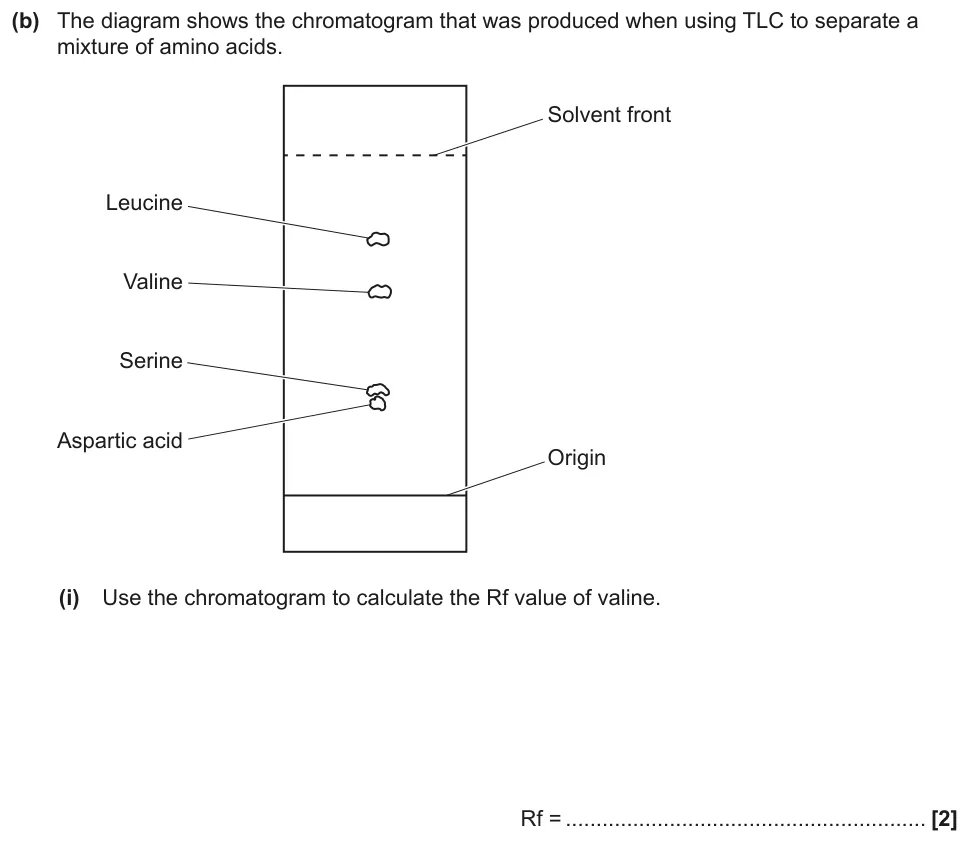
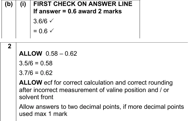
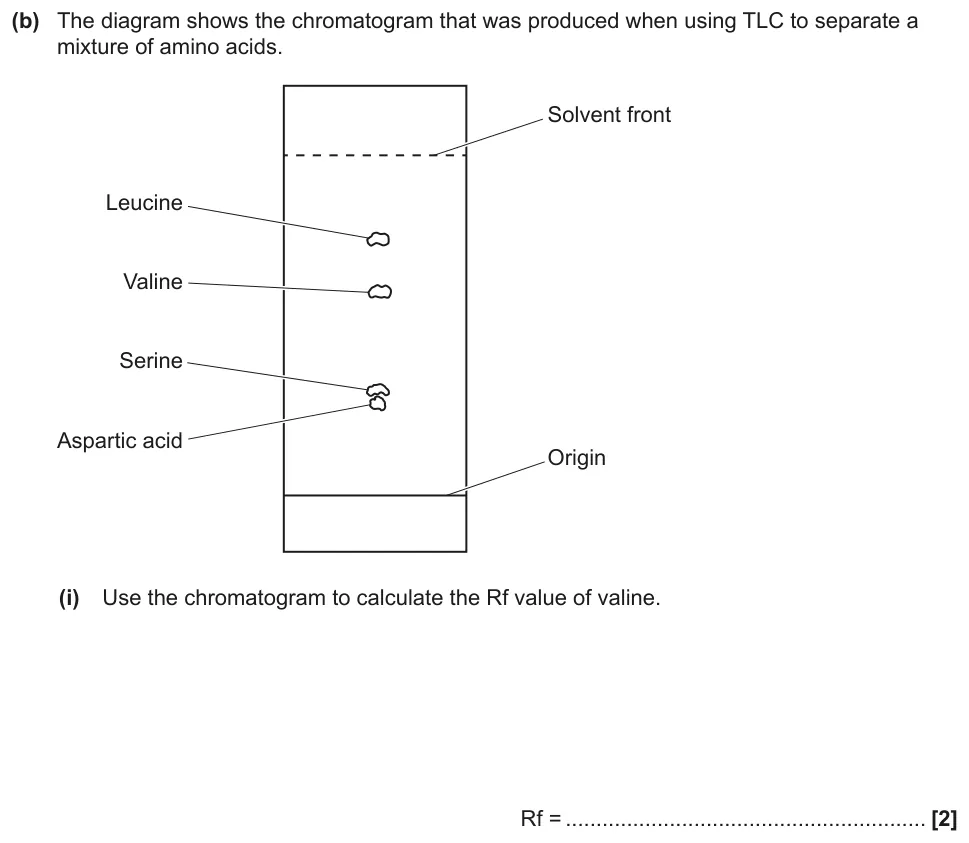
Calculate the Rf value of valine from a TLC chromatogram.
Show the mark scheme
Tap an image to open it full size.
From the examiners' report: This was very well answered. A few students lost a mark by not rounding to a sensible number of decimal places, and some worked out the Rf for leucine instead of valine.
Question 8
A level Paper 3 · June 2023 · Q2(a)(ii) · 2 marks · Hard
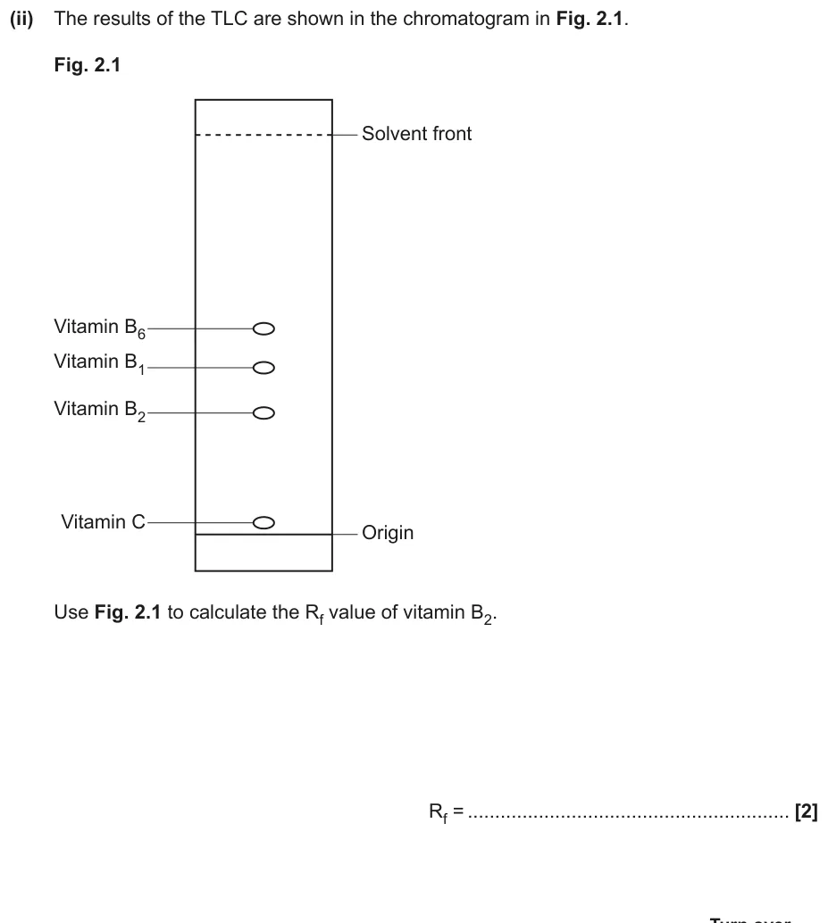
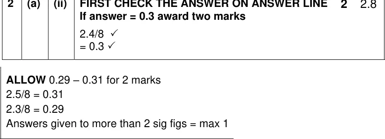
Calculate the Rf value of vitamin B2 from a chromatogram.
Information from earlier in this question:
Show the mark scheme
Tap an image to open it full size.
From the examiners' report: Most students got both marks. Those who scored nothing usually divided the wrong way round (8 ÷ 2.4) or measured the wrong vitamin. Some lost a mark by giving more than 2 significant figures.
Interpreting and improving chromatograms
Question 9
AS Paper 1 · June 2023 · Q23(b)(ii) · 1 mark · Medium
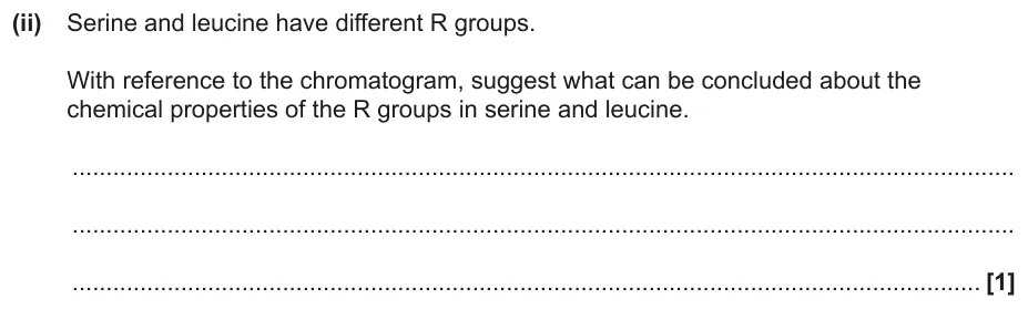
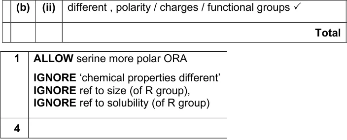
Suggest what the chromatogram shows about the R groups of serine and leucine.
Information from earlier in this question:
Show the mark scheme
Tap an image to open it full size.
From the examiners' report: Only a few students scored. Many just described differences in size or solubility. TLC of amino acids uses an organic solvent, so how far each one travels reflects the polarity of its R group.
Question 10
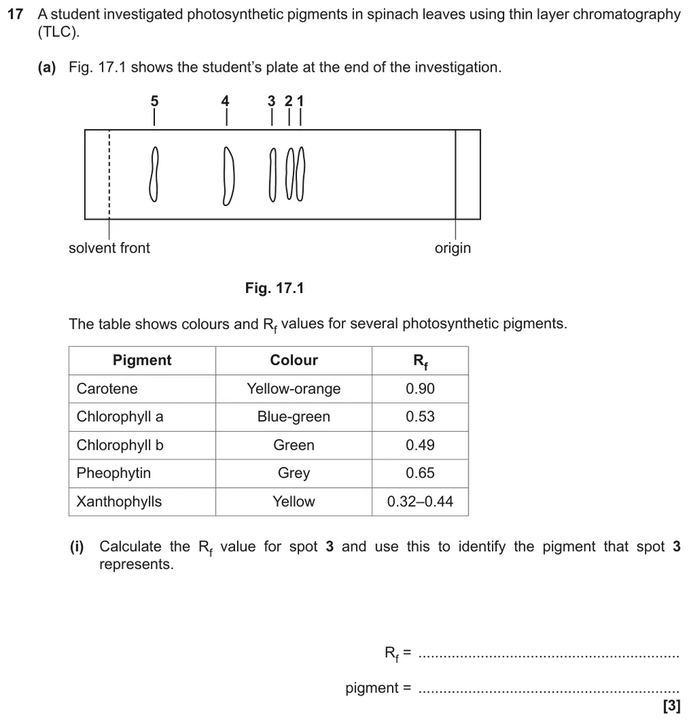
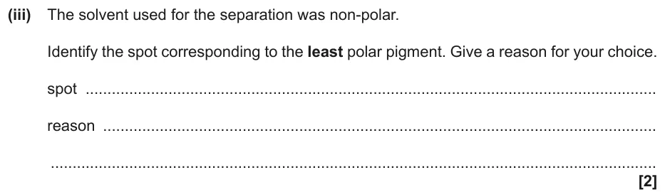
A level Paper 1 · November 2020 · Q17(a)(iii) · 2 marks · Medium
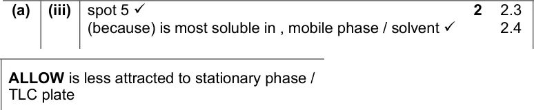
Identify the spot for the least polar pigment when the solvent is non-polar, with a reason.
Information from earlier in this question:
Show the mark scheme
Tap an image to open it full size.
From the examiners' report: Use the mark scheme wording: the least polar pigment is the most soluble in the non-polar solvent, so it travels furthest, to the spot nearest the solvent front.
Question 11
A level Paper 1 · June 2025 · Q16(b)(ii) · 2 marks · Medium
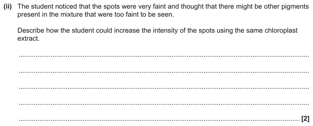
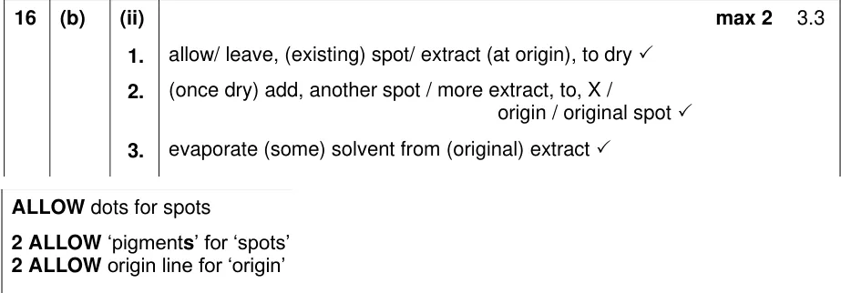
Describe how to make faint pigment spots more intense using the same extract.
Information from earlier in this question:
Show the mark scheme
Tap an image to open it full size.
From the examiners' report: Answers often didn't say where the extra extract goes. Simply adding a bigger volume spreads the spot out, so let it dry between additions. A stronger solvent or a stain doesn't make these spots more intense.
Question 12
A level Paper 1 · June 2025 · Q16(b)(iv) · 1 mark · Medium
Suggest why the pigments couldn't be identified from data-book Rf values.

Show the mark scheme

Tap an image to open it full size.
From the examiners' report: This part sorted students well. Stronger answers said that Rf values depend on the conditions, such as a different solvent, temperature or plant species.
Question 13
A level Paper 1 · June 2025 · Q16(b)(v) · 2 marks · Medium
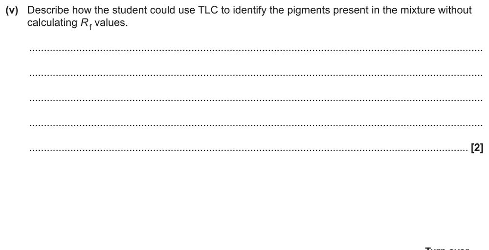
Describe how TLC could identify the pigments without calculating Rf values.
Show the mark scheme

Tap an image to open it full size.
From the examiners' report: Many students said they would compare the colours but didn't say what with. Run known pigments alongside on the same plate and compare positions or colours.
Electrophoresis and DNA profiling
Question 14
A level Paper 3 · June 2017 · Q1(c) · 3 marks · Medium
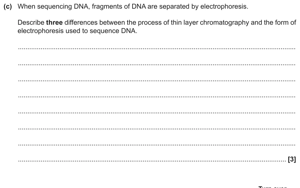
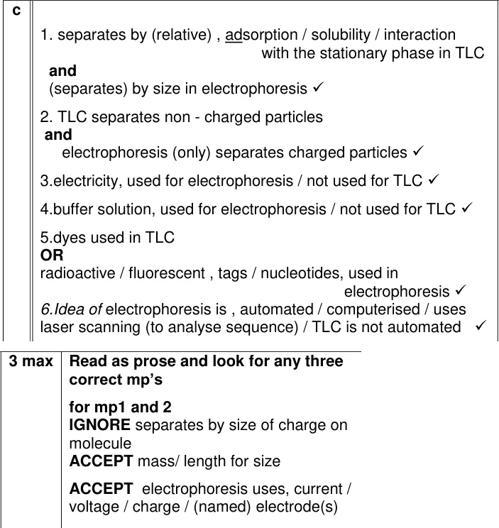
Describe three differences between TLC and the electrophoresis used to sequence DNA.
Show the mark scheme
Tap an image to open it full size.
From the examiners' report: Most students knew this well, especially that electrophoresis uses an electric current and separates DNA by size or length, while TLC separates by solubility. Tags to see the fragments and the buffer were also credited.
Question 15
A level Paper 2 · November 2021 · Q16(b)(i) · 4 marks · Medium
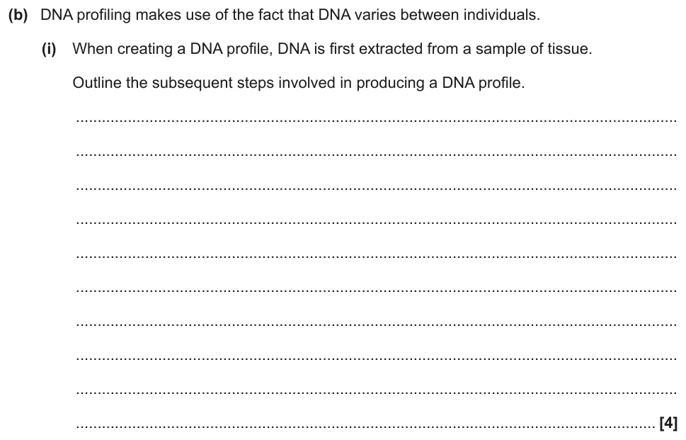
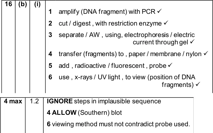
Outline the steps that follow DNA extraction in producing a DNA profile.
Show the mark scheme
Tap an image to open it full size.
From the examiners' report: Many students wrote a long description when the command word was 'outline'. Name the probe or tag: 'use radioactivity' on its own isn't enough.
Question 16
A level Paper 3 · June 2018 · Q4(c)(v) · 2 marks · Medium
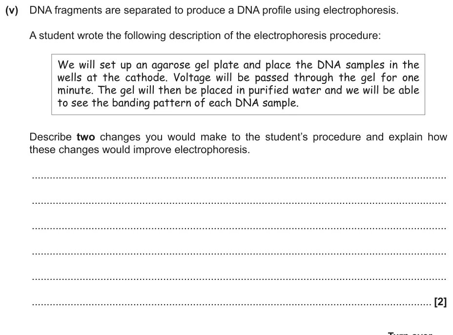
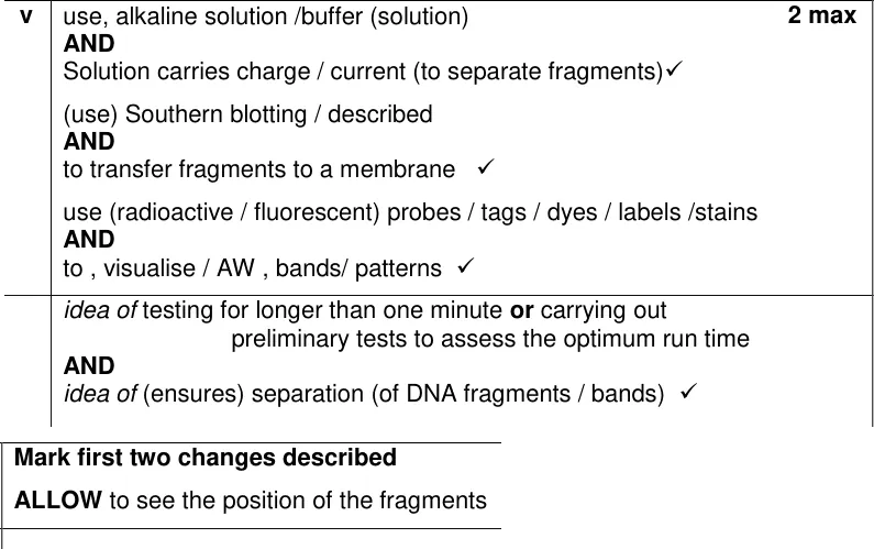
Describe and explain two changes to a student's electrophoresis procedure.
Show the mark scheme
Tap an image to open it full size.
From the examiners' report: Many students named a buffer, a longer run or a stain but didn't explain it. A longer run lets the fragments separate, not just move further, and the buffer lets the current flow. Many also mixed up the anode and the cathode.
Question 17
A level Paper 3 · June 2025 · Q4(d)(i) · 4 marks · Medium
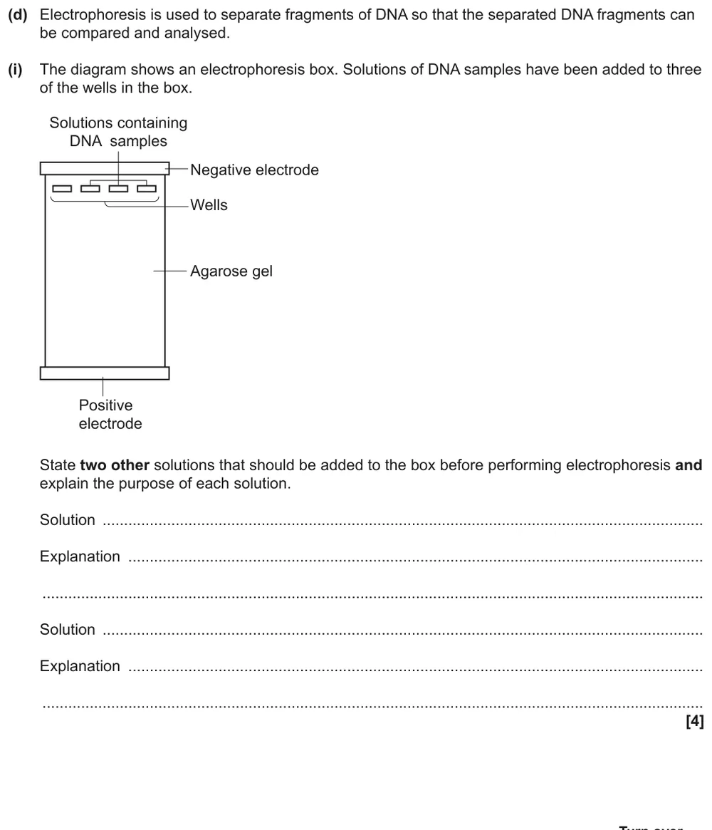
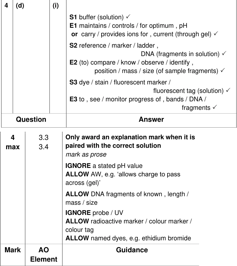
State two other solutions added to an electrophoresis box and explain the purpose of each.
Show the mark scheme
Tap an image to open it full size.
From the examiners' report: Few students got all four marks. A buffer or a dye were the most common correct answers; fewer remembered a DNA ladder of known fragment sizes to compare the samples with.
Question 18
A level Paper 3 · June 2025 · Q4(d)(ii) · 2 marks · Medium
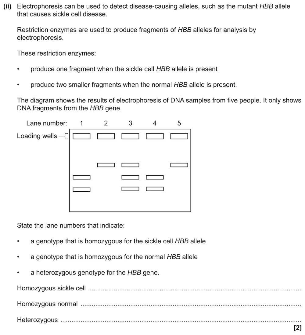
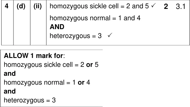
Use a gel of HBB gene fragments to identify homozygous and heterozygous genotypes.
Show the mark scheme
Tap an image to open it full size.
From the examiners' report: Students did very well on this despite the unfamiliar context. The sickle cell allele gives one larger fragment and the normal allele two smaller ones, so a heterozygote shows all three bands.
Where students lose marks (from OCR examiners' reports and mark schemes)
Each point below is followed by wording that gained marks.
Silica, not paper: Asked for the stationary phase in thin layer chromatography, many students said paper or named a solvent.
✔ Gains credit: "Silica (gel)"; aluminium oxide, cellulose or silicon dioxide are also accepted.
The solvent dissolves and carries: Vague answers about 'separating colours', or the solvent 'pushing' or 'dragging' the solute, didn't explain its role.
✔ Gains credit: "(Solvent) dissolves the carbohydrates / acts as mobile phase; carries (dissolved) carbohydrates through stationary phase".
Every change needs a reason: Most students suggested changes to a chromatography method without saying why each one would improve the results.
✔ Gains credit: "Put pencil line / origin higher (than the solvent) to stop spots dissolving into / mixing with the solvent".
Measure to the middle of the spot: Students measured to the end of the plate instead of the solvent front, and gave distances in cm when the answer line asked for mm.
✔ Gains credit: "Distance travelled by most soluble = 91 (mm); Rf = 91 ÷ 109 = 0.83".
Spot over solvent, to 2 s.f.: Some students divided the wrong way round, and answers with more than 2 significant figures lost a mark.
✔ Gains credit: "2.4/8 = 0.3" (0.29 to 0.31 allowed).
Dry between spots: To make faint spots stronger, students suggested a larger volume, a stronger solvent or a stain, none of which was credited.
✔ Gains credit: "Allow (existing) spot to dry; (once dry) add another spot / more extract to the origin".
Polarity, not size: To explain why two amino acids travelled different distances, many described differences in size or solubility of the R groups.
✔ Gains credit: "Different polarity / charges / functional groups" (serine more polar).
Say why the buffer is there: Students named a buffer, a longer run or a stain for electrophoresis but didn't explain it, and many said the buffer just keeps the pH constant.
✔ Gains credit: "Use buffer (solution) AND solution carries charge / current (to separate fragments)"; "dye / stain to see bands / DNA fragments".
Download the questions
Download the PDF pack (18 questions, mark schemes at the end)
Keep practising
- OCR A Level Biology food tests and chromatography quiz
- OCR A Level Biology cellular control and manipulating genomes quiz
- Carbohydrates: OCR A Level Biology exam questions by topic
- PAG 4 Rates of Enzyme-Controlled Reactions: OCR A Level Biology exam questions by topic
- OCR A Level Biology masterclasses
Questions, mark schemes and examiners' report extracts © OCR.