

Skeletal muscles (AQA 3.6.3) are A-level only, so this is Year 13 content. You need to know how muscles work in antagonistic pairs, the structure of a myofibril and its sarcomeres, how actin, myosin, calcium ions, tropomyosin and ATP make a myofibril contract, how phosphocreatine regenerates ATP, and the properties of slow and fast fibres.
The examiners' reports show where the marks go. Students write that muscles constrict or widen instead of contract and relax, swap actin and myosin when they explain the bands, and give actin an 'active site' instead of a binding site. They write myofibril when they mean myosin, abbreviate phosphocreatine, and describe only one type of fibre when the question wants a comparison.
Start with the structure questions, then contraction, then energy and fibre types, and finish with the data and calculation questions. Write your answer on paper before you open each mark scheme. If you'd like help with this topic, my AQA masterclasses run online on Tuesdays at 5.15pm for Year 12 and on Wednesdays or Thursdays at 6.30pm for Year 13.
What you need from GCSE and Year 12
Each question on this page assumes you're secure on these GCSE and Year 12 ideas. If any of them feels shaky, revise it before you start.
- GCSE: muscles are effectors that contract when nerve impulses arrive from motor neurones.
- GCSE: during vigorous exercise muscles respire anaerobically, producing lactic acid and much less energy per glucose than aerobic respiration.
- Year 12 (AQA 3.1.8): ATP hydrolase hydrolyses ATP to ADP and inorganic phosphate, releasing energy; ATP is resynthesised from ADP and Pi.
- Year 12 (AQA 3.1.2): glycogen is the storage polysaccharide of glucose in animals, and is hydrolysed to glucose when it's needed.
Key terms
These words come up in the questions and mark schemes. Examiners expect you to use them exactly.
- Antagonistic pair: two muscles that pull in opposite directions: when one contracts, the other relaxes.
- Sarcomere: the repeating unit of a myofibril, from one Z-line to the next; it shortens when the muscle contracts.
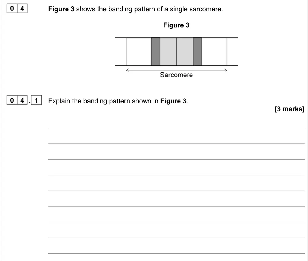
- I band and H zone: the I band (light band) contains only actin; the H zone contains only myosin; the darkest region is where actin and myosin overlap.
- Actin and myosin: actin forms the thin filaments; myosin forms the thick filaments, and its heads bind to actin and pull it.
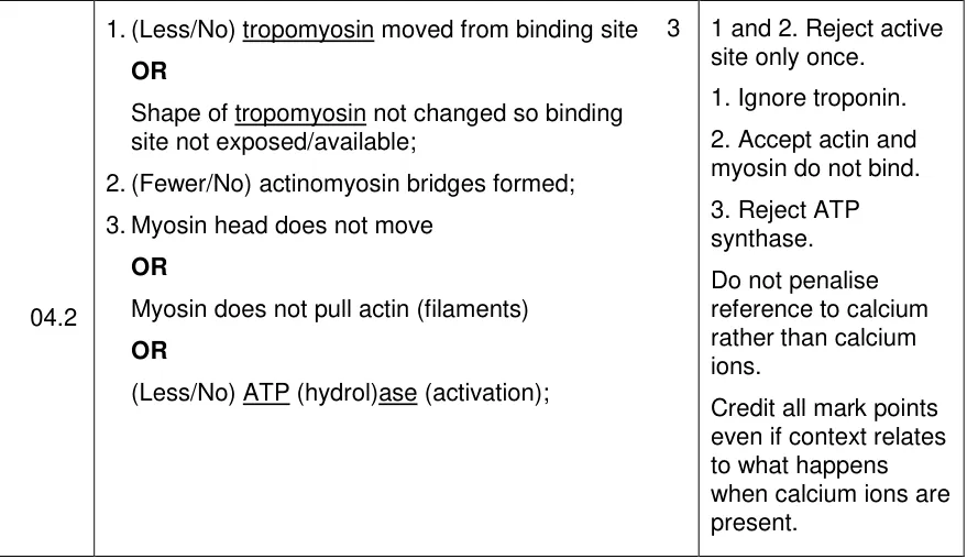
- Tropomyosin: a protein that covers the myosin binding sites on actin in resting muscle; calcium ions cause it to move, exposing the binding sites.
- Actinomyosin bridge: the cross-bridge formed when a myosin head attaches to a binding site on actin.
- Sarcoplasmic reticulum: the endoplasmic reticulum of a muscle fibre; it releases calcium ions when the fibre is depolarised and takes them back by active transport.
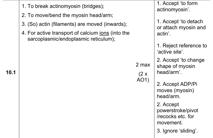
- Role of ATP: ATP is hydrolysed to move the myosin head (the power stroke), to break the actinomyosin bridge and to actively transport calcium ions back into the sarcoplasmic reticulum.
- Phosphocreatine: a store in muscle that gives phosphate to ADP to make ATP quickly, without oxygen; write it in full, as it has no accepted abbreviation.
- Slow fibres: contract slowly, used for long periods (endurance); they respire aerobically, with many mitochondria, a good blood supply and myoglobin.
- Fast fibres: contract quickly, used for short, intense exercise; they respire mainly anaerobically and store glycogen and phosphocreatine.
What these questions cover (AQA 3.6.3)
The questions test these statements from the AQA AS and A-level Biology specification (7401/7402):
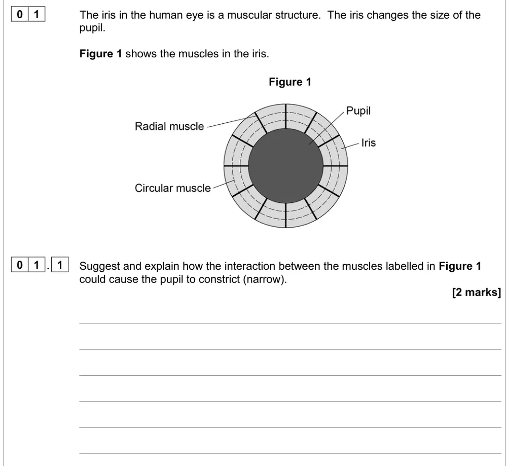
- Antagonistic pairs: Muscles act in antagonistic pairs against an incompressible skeleton. (the only question available uses the circular and radial muscles of the iris, which work as an antagonistic pair without a skeleton)
- Muscle structure: Gross and microscopic structure of skeletal muscle. The ultrastructure of a myofibril.
- Myofibril contraction: The roles of actin, myosin, calcium ions and ATP in myofibril contraction.
- Calcium ions and tropomyosin: The roles of calcium ions and tropomyosin in the cycle of actinomyosin bridge formation. (troponin isn't required, and examiners ignore it)
- ATP and phosphocreatine: The roles of ATP and phosphocreatine in muscle contraction.
- Slow and fast fibres: The structure, location and general properties of slow and fast skeletal muscle fibres.
How to use this page
Write your answer on paper before you open the mark scheme. Allow about 1.3 minutes per mark: the 12 questions are worth 31 marks, so about 40 minutes in total. Then mark your answer strictly against the mark scheme and read the examiners' tip underneath it. If you'd rather print the questions, download the PDF pack.
Muscles and myofibril structure (3.6.3)
Question 1
A level Paper 2 · November 2020 · Q01.1 · 2 marks · Medium
Explain how the circular and radial muscles of the iris constrict the pupil.
Show the mark scheme
Tap an image to open it full size.
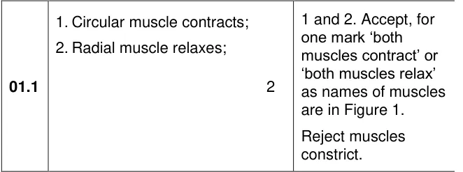
From the examiners' report: Only one in four got both marks and almost 40% scored zero. Name each muscle and say contracts or relaxes; constrict, widen and shorten don't score.
Question 2
A level Paper 2 · November 2020 · Q04.1 · 3 marks · Medium
Explain the light, dark and grey bands in a diagram of a sarcomere.
Show the mark scheme
Tap an image to open it full size.
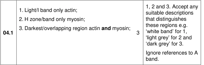
From the examiners' report: 26% got all three. For each band, say which filaments it contains: white is actin only, light grey is myosin only, and the darkest band is where they overlap. Don't swap them round.
How a myofibril contracts (3.6.3)
Question 3
A level Paper 2 · June 2023 · Q10.1 · 2 marks · Medium
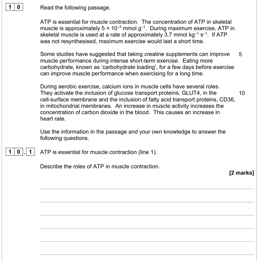
Describe the roles of ATP in muscle contraction.
Show the mark scheme
Tap an image to open it full size.
From the examiners' report: About 46% got both marks. Write myosin, not myofibril, and say 'myosin head'. Breaking the actinomyosin bridge and moving the head are the easiest two marks.
Question 4
A level Paper 2 · June 2019 · Q04.2 · 3 marks · Medium
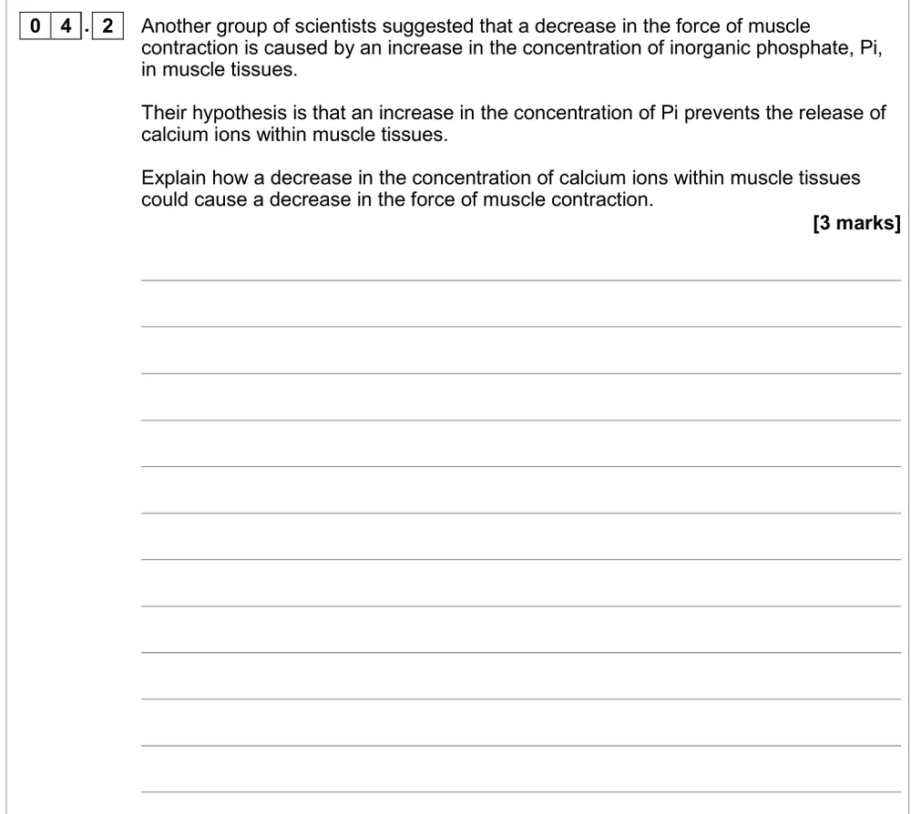
Explain how fewer calcium ions in muscle reduce the force of contraction.
Show the mark scheme
Tap an image to open it full size.
From the examiners' report: A quarter got all three. Use 'binding site', not active site, leave troponin out, and finish with the myosin head pulling the actin, not just 'filaments moving'.
Question 5
A level Paper 3 · June 2017 · Q01.2 · 2 marks · Hard
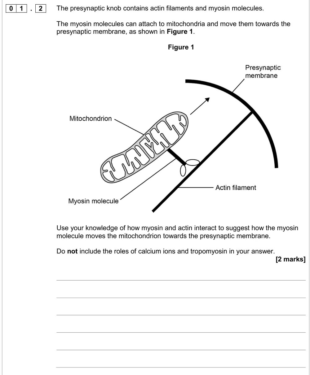
Suggest how myosin moves a mitochondrion along actin towards the presynaptic membrane.
Show the mark scheme
Tap an image to open it full size.
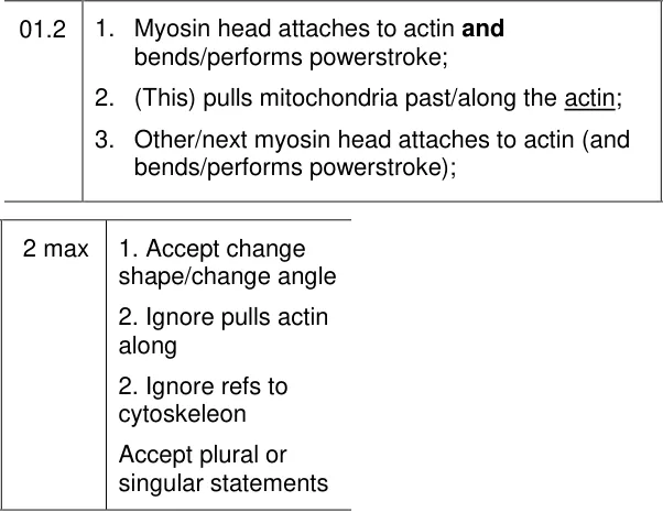
From the examiners' report: About 12% got both marks. This is application, not recall: leave the sarcomere out and say the myosin head binds actin, bends and pulls the mitochondrion along.
Question 6
A level Paper 3 · June 2017 · Q01.3 · 2 marks · Medium
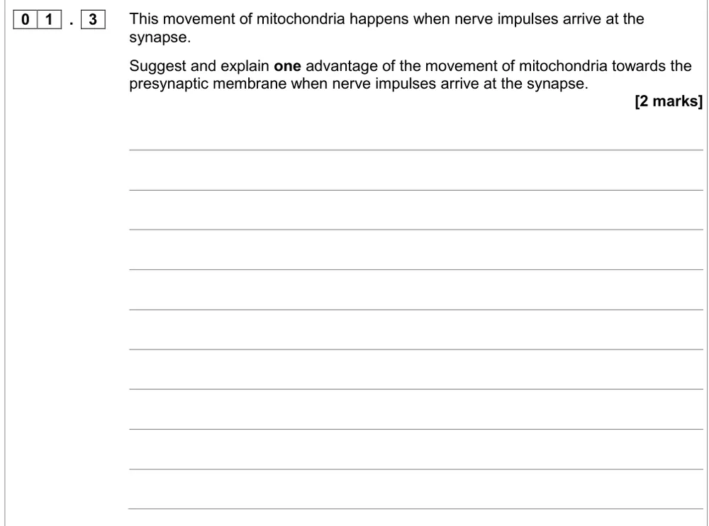
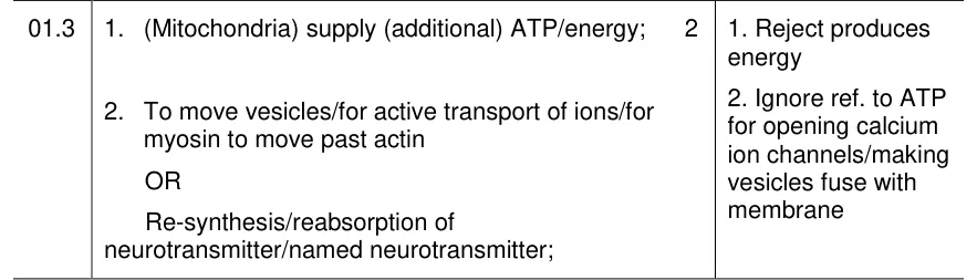
Suggest one advantage of mitochondria moving towards the presynaptic membrane.
Show the mark scheme
Tap an image to open it full size.
From the examiners' report: About half got the ATP mark and just over 40% a second mark. Say mitochondria supply ATP, not 'produce energy', then name one use such as active transport of ions or myosin moving along actin.
Energy and fibre types (3.6.3)
Question 7
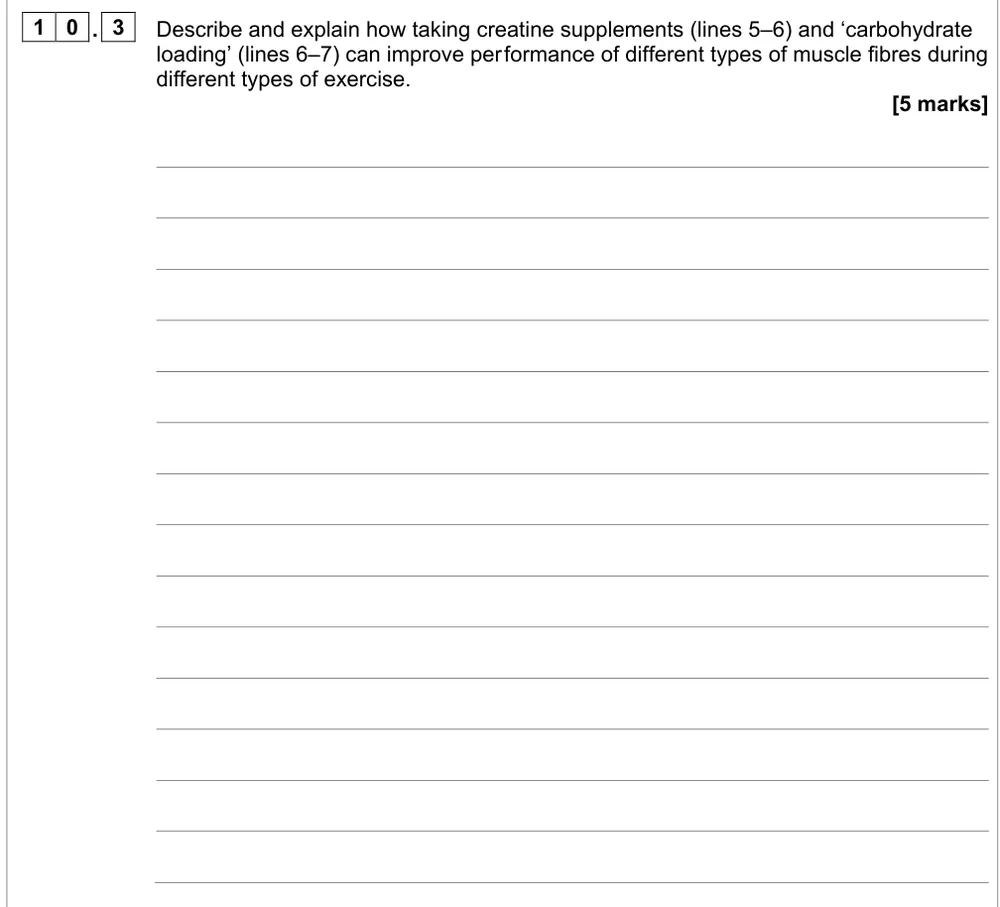
A level Paper 2 · June 2023 · Q10.3 · 5 marks · Medium
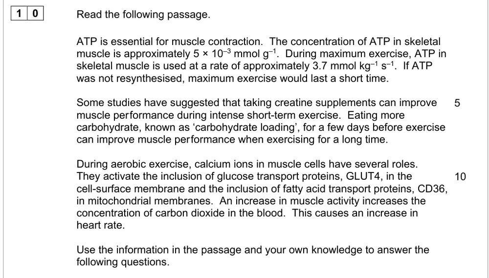
Explain how creatine supplements and carbohydrate loading help different fibres in different exercise.
Information from earlier in this question:
Show the mark scheme
Tap an image to open it full size.
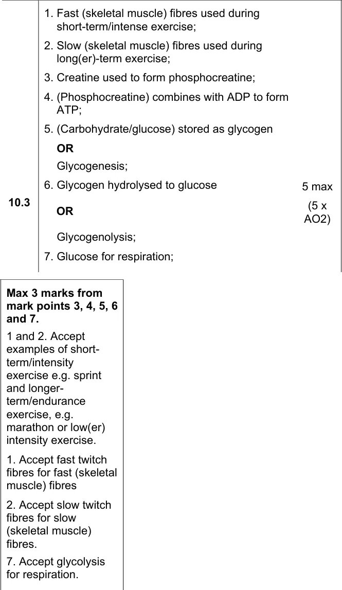
From the examiners' report: Only 11% got all five. Match fast fibres to sprints and slow fibres to endurance, write phosphocreatine in full (PCr isn't accepted), and say it gives phosphate to ADP to make ATP.
Question 8
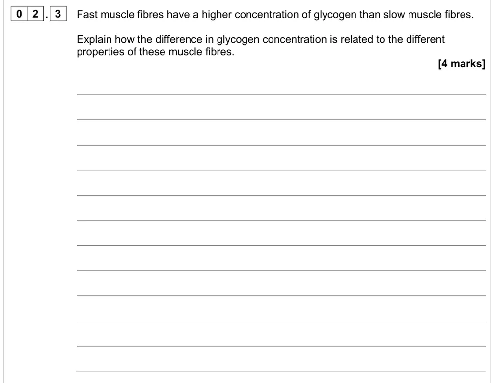
A level Paper 2 · June 2024 · Q02.3 · 4 marks · Medium
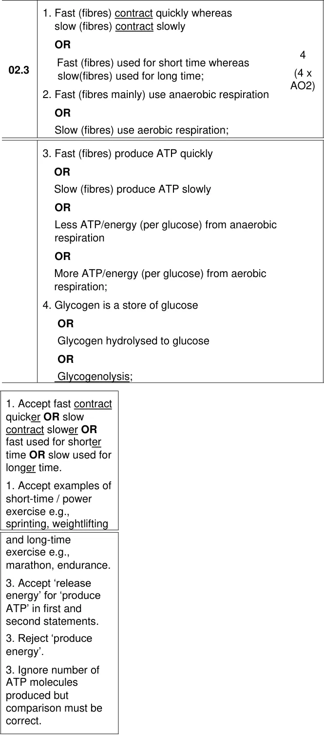
Explain why fast muscle fibres store more glycogen than slow fibres.
Show the mark scheme
Tap an image to open it full size.
From the examiners' report: Only about 5% got all four. Compare both fibre types in each point, name anaerobic respiration rather than 'working anaerobically', and say 'produce ATP', never 'produce energy'.
Question 9
A level Paper 2 · June 2017 · Q03.1 · 1 mark
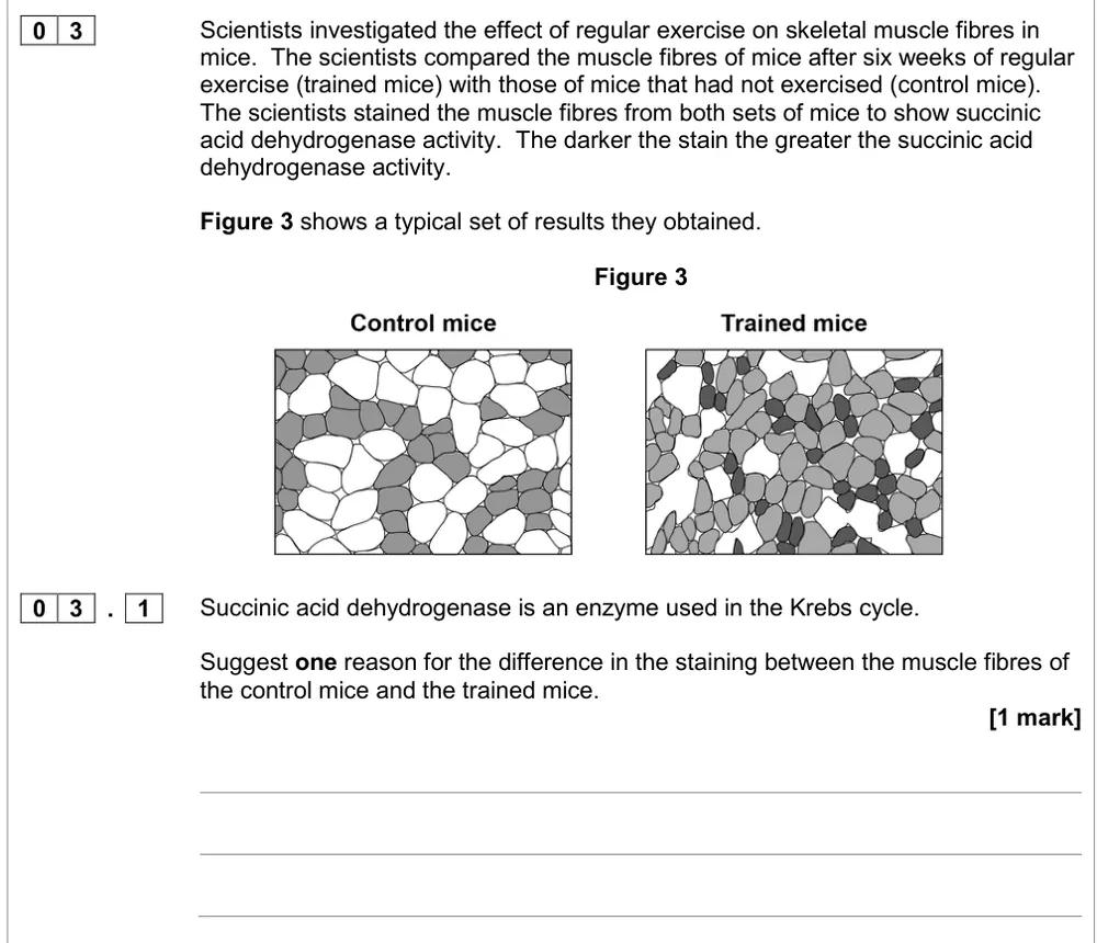
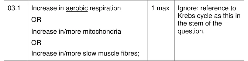
Suggest why muscle fibres from trained mice stain more darkly for a Krebs cycle enzyme.
Show the mark scheme
Tap an image to open it full size.
From the examiners' report: About two-thirds didn't get this. Don't repeat the stem with 'more Krebs cycle'; say more aerobic respiration, more mitochondria or more slow fibres.
Practical skills and maths (3.6.3)
Question 10
A level Paper 2 · June 2019 · Q04.1 · 4 marks · Medium
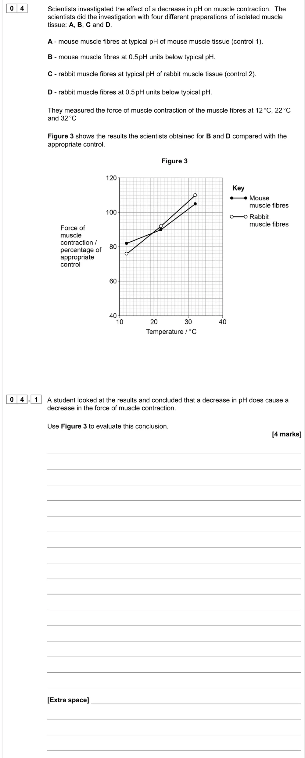
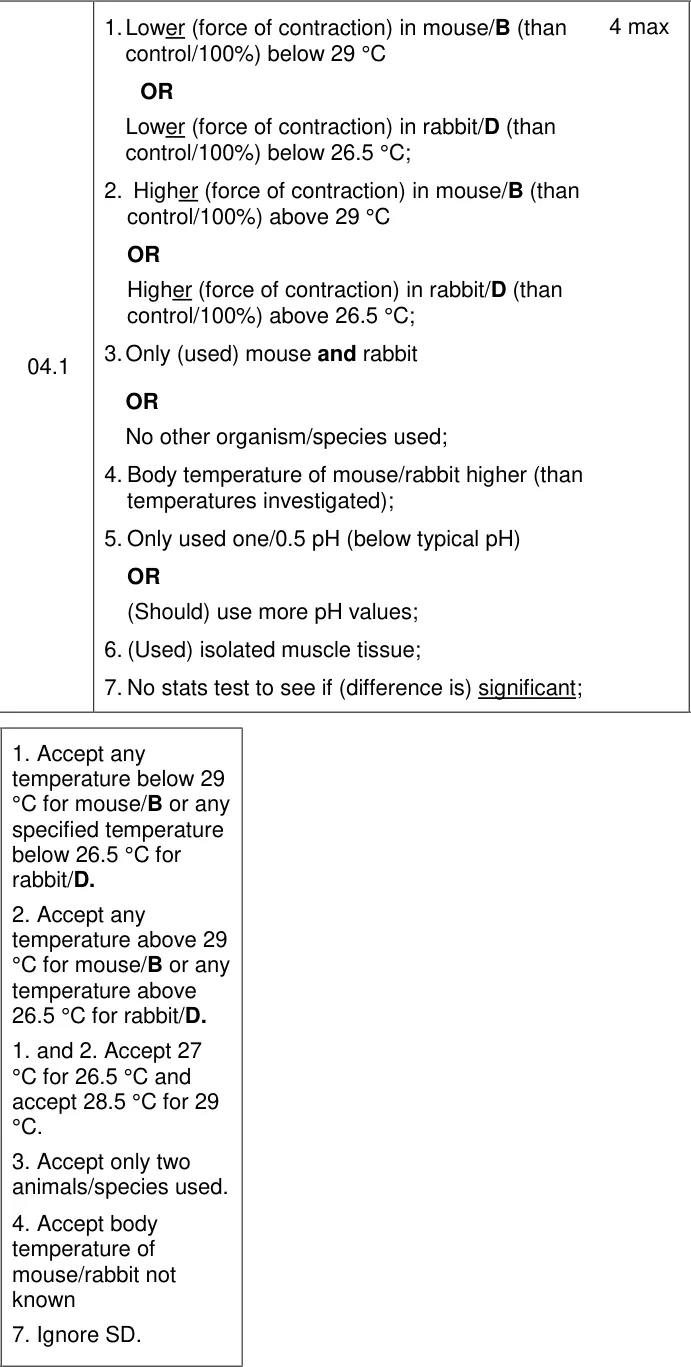
Evaluate the conclusion that a lower pH reduces the force of muscle contraction.
Show the mark scheme
Tap an image to open it full size.
From the examiners' report: Only about 6% got all four. The y-axis is already a percentage of the control, so read where each line crosses 100%, then add limits: two species, one pH, isolated tissue, no test of significance.
Question 11
A level Paper 2 · June 2024 · Q02.1 · 2 marks · Medium
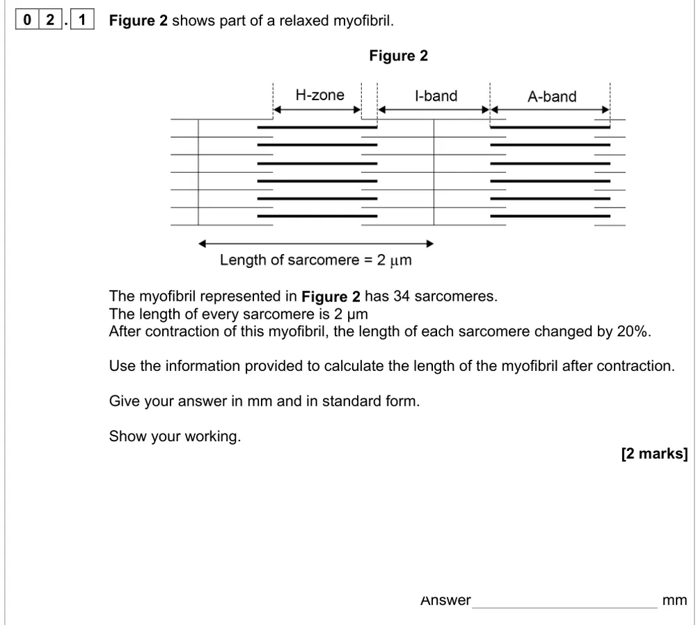
Calculate the length of a contracted myofibril in mm in standard form.
Show the mark scheme

Tap an image to open it full size.
From the examiners' report: About 45% got both marks. Each sarcomere shortens to 1.6 µm, so 34 × 1.6 = 54.4 µm; then convert to mm and write it in standard form. Don't add the 20% on.
Question 12
A level Paper 2 · June 2023 · Q10.2 · 1 mark · Hard
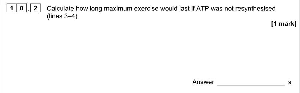
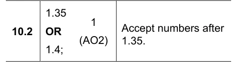
Calculate how long maximum exercise would last without resynthesising ATP.
Information from earlier in this question:
Show the mark scheme
Tap an image to open it full size.
From the examiners' report: Only about 28% got this. Convert 5 × 10⁻³ mmol g⁻¹ to 5 mmol kg⁻¹ first, then divide by 3.7 mmol kg⁻¹ s⁻¹.
Where students lose marks (from AQA examiners' reports and mark schemes)
Each point below is followed by wording that gained marks.
Muscles contract or relax; they don't constrict: Only one in four got both marks on the iris and almost 40% scored zero, often by giving the converse or not naming the muscles. 'Constrict', 'expand', 'widen', 'lengthen' and 'shorten' weren't accepted.
✔ Gains credit: "Circular muscle contracts; radial muscle relaxes."
I band is actin only; H zone is myosin only: 26% got all three on the banding pattern. Students reversed actin and myosin, or named the bands and lines correctly without saying which filaments they contain. References to the A band were ignored.
✔ Gains credit: "Light (I) band only actin; H zone only myosin; darkest (overlapping) region actin and myosin."
Tropomyosin covers a binding site, not an active site: A quarter got all three on calcium ions and force. Some wrote 'active site', many explained troponin, which isn't required, and many only said the filaments 'move' or gave a 'power stroke' with no detail.
✔ Gains credit: "Less tropomyosin moved from the binding site; fewer actinomyosin bridges formed; myosin head does not move or pull the actin filaments."
Write myosin head, not myofibril: About 46% got both marks on the roles of ATP. Quite a few wrote 'myofibril' instead of myosin, and others left out 'head', so the mark for moving the myosin head was lost.
✔ Gains credit: "To break actinomyosin bridges; to move or bend the myosin head; for active transport of calcium ions into the sarcoplasmic reticulum."
Phosphocreatine in full, then make ATP from ADP: Only 11% got all five on creatine and carbohydrate loading. 'PCr' wasn't accepted, many didn't link fibre type to the type of exercise, and weaker answers mixed up glycogen and glucagon.
✔ Gains credit: "Fast fibres used during short-term intense exercise; creatine used to form phosphocreatine; phosphocreatine combines with ADP to form ATP; glycogen hydrolysed to glucose for respiration."
Compare slow and fast fibres in one sentence: Only about 5% got all four on glycogen in fast fibres. A significant minority described only one fibre type, 'working anaerobically' wasn't accepted, and 'produce energy' was rejected.
✔ Gains credit: "Fast fibres contract quickly whereas slow fibres contract slowly; fast fibres mainly use anaerobic respiration; fast fibres produce ATP quickly; glycogen hydrolysed to glucose."
Apply actin and myosin to the new structure: Only about 12% got both marks on myosin moving a mitochondrion. Many treated it as recall and described the sarcomere shortening, instead of myosin moving along actin.
✔ Gains credit: "Myosin head attaches to actin and bends (power stroke); this pulls the mitochondrion along the actin."
Read what the y-axis compares with: Only about 6% got all four on the pH conclusion. Many didn't see that the y-axis is a percentage of the control, so they said there were no controls; few mentioned more pH values or a statistical test of significance.
✔ Gains credit: "Lower force in mouse fibres below 29 °C but higher above 29 °C; only mouse and rabbit used; no statistical test to see if the difference is significant."
Download the questions
Download the PDF pack (12 questions, mark schemes at the end)
Keep practising
- Nervous coordination: AQA A Level Biology exam questions by topic
- AQA 3.6.3 muscle contraction: self-marking quiz
- Respiration: AQA A Level Biology exam questions by topic
- ATP: AQA A Level Biology exam questions by topic
- AQA A Level Biology masterclasses
Questions, mark schemes and examiners' report extracts © AQA.