

The heart and circulation is the middle part of AQA 3.3.4.1, mass transport in animals. You need the general pattern of blood circulation in a mammal, the names of the coronary arteries and the vessels entering and leaving the heart, lungs and kidneys, the gross structure of the heart, and how pressure changes in the atria, ventricles and arteries open and close the valves during the cardiac cycle. You also need to read pressure and volume graphs and work out stroke volume and cardiac output.
The examiners' reports show where the marks go. Students write 'coronary' on its own or name coronary veins, mix up the pulmonary artery and pulmonary vein, stop a pathway at the lungs, describe when a valve opens without explaining the pressure difference that causes it, and write about oxygenated and deoxygenated blood mixing when the question is about volume and pressure. In the calculations, many can't read the graph scale or work out the heart rate from the time of one beat.
Start with the named blood vessels, then the heart and its valves, then the cardiac cycle graphs, and finish with the calculations. Answer each question on paper before you open the mark scheme. If you'd like help with this topic, my AQA masterclasses run online on Tuesdays at 5.15pm for Year 12 and on Wednesdays or Thursdays at 6.30pm for Year 13.
What you need from GCSE and Year 12
Each question on this page assumes you're secure on these GCSE and Year 12 ideas. If any of them feels shaky, revise it before you start.
- GCSE: the heart has four chambers, two atria and two ventricles; the left ventricle has a thicker muscular wall than the right.
- GCSE: mammals have a double circulation: the right side of the heart pumps blood to the lungs and the left side pumps it round the body.
- GCSE: arteries carry blood away from the heart and veins carry it back; valves in the heart stop blood flowing backwards.
- GCSE: liquids flow from a region of higher pressure to a region of lower pressure.
- Year 12 (AQA 3.3.2): blood is oxygenated in the capillaries around the alveoli of the lungs.
Key terms
These words come up in the questions and mark schemes. Examiners expect you to use them exactly.
- Double circulation: blood passes through the heart twice in each complete circuit: once to the lungs and once to the rest of the body.
- Coronary arteries: the arteries that branch from the aorta and carry blood to the heart muscle.
- Pulmonary artery and pulmonary vein: the pulmonary artery carries blood from the right ventricle to the lungs; the pulmonary vein carries blood from the lungs to the left atrium.
- Aorta and vena cava: the aorta carries blood from the left ventricle to the body; the vena cava returns blood from the body to the right atrium.
- Renal artery and renal vein: the renal artery carries blood to a kidney; the renal vein carries it away to the vena cava.
- Atrioventricular valves: the valves between each atrium and ventricle (tricuspid on the right, bicuspid on the left), held by tendons so they don't turn inside out.
- Semi-lunar valves: the valves between the ventricles and the aorta or pulmonary artery.
- Unidirectional flow: blood flowing one way only; each valve opens when the pressure behind it is higher and closes when the pressure in front of it is higher.
- Diastole: the stage of the cardiac cycle when the heart muscle is relaxed.
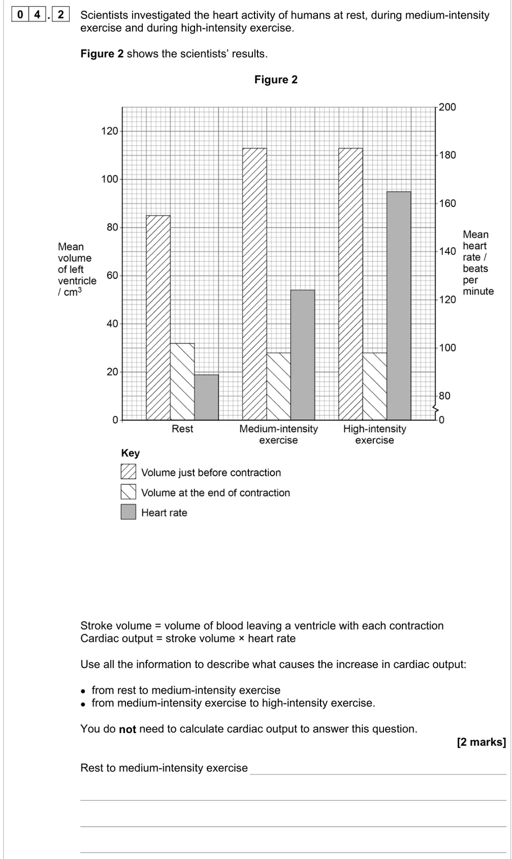
- Stroke volume: the volume of blood leaving a ventricle with each contraction; the maximum minus the minimum ventricle volume.
- Cardiac output: the volume of blood pumped by a ventricle per minute: cardiac output = stroke volume × heart rate.
What these questions cover (AQA 3.3.4.1, the heart and circulation)
The questions test these statements from the AQA AS and A-level Biology specification (7401/7402):
- Circulation: The general pattern of blood circulation in a mammal. Names are required only of the coronary arteries and of the blood vessels entering and leaving the heart, lungs and kidneys. (the fish question asks you to compare the single circulation of a fish with the double circulation of a mammal)
- The heart: The gross structure of the human heart. Pressure and volume changes and associated valve movements during the cardiac cycle that maintain a unidirectional flow of blood.
- Students should be able to analyse and interpret data relating to pressure and volume changes during the cardiac cycle (three questions ask you to calculate stroke volume or the volume pumped per minute (cardiac output) from a table or graph)
How to use this page
Write your answer on paper before you open the mark scheme. Allow about 1.3 minutes per mark: the 14 questions are worth 32 marks, so about 42 minutes in total. Then mark your answer strictly against the mark scheme and read the examiners' tip underneath it. If you'd rather print the questions, download the PDF pack.
Circulation and named blood vessels (3.3.4.1)
Question 1
AS Paper 1 · June 2018 · Q01.2 · 1 mark · Medium
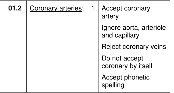
Name the blood vessels that carry blood to the heart muscle.

Show the mark scheme
Tap an image to open it full size.
From the examiners' report: Only 31.6% scored. Write coronary arteries in full: not coronary veins, and not 'coronary' on its own.
Question 2
AS Paper 2 · June 2025 · Q01.1 · 2 marks · Hard
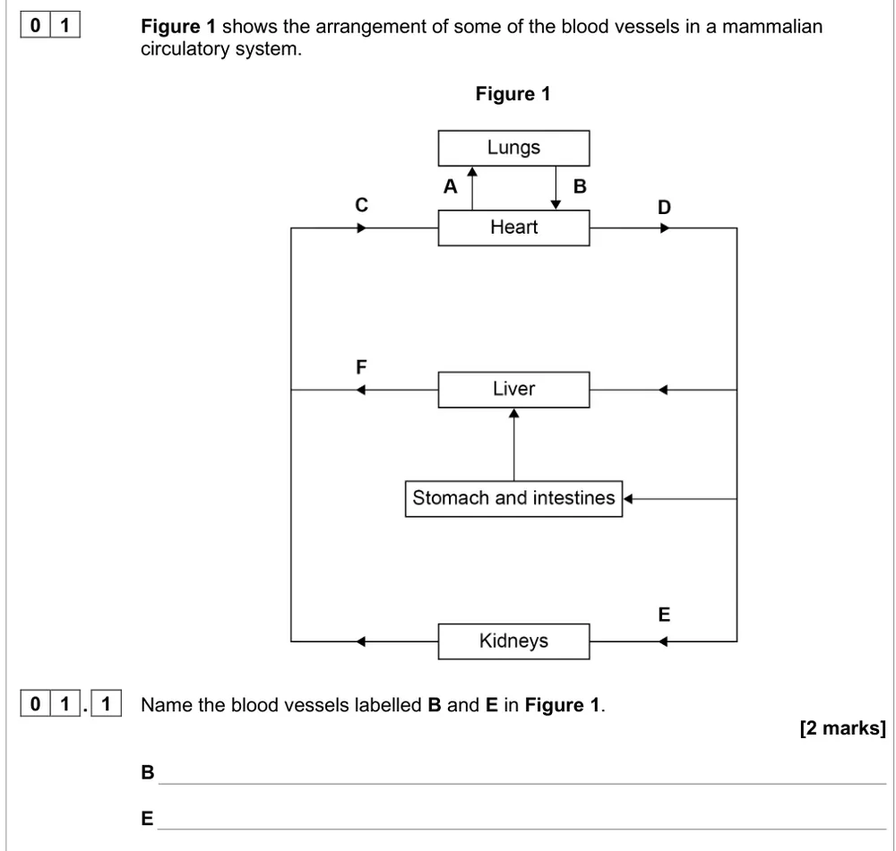
Name two blood vessels on a diagram of the mammalian circulation.
Show the mark scheme
Tap an image to open it full size.
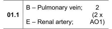
From the examiners' report: About half named both. Follow the arrows: B carries blood from the lungs back to the heart and E carries blood to a kidney, so decide artery or vein from the direction of flow.
Question 3
A level Paper 1 · June 2025 · Q10.3 · 5 marks · Medium
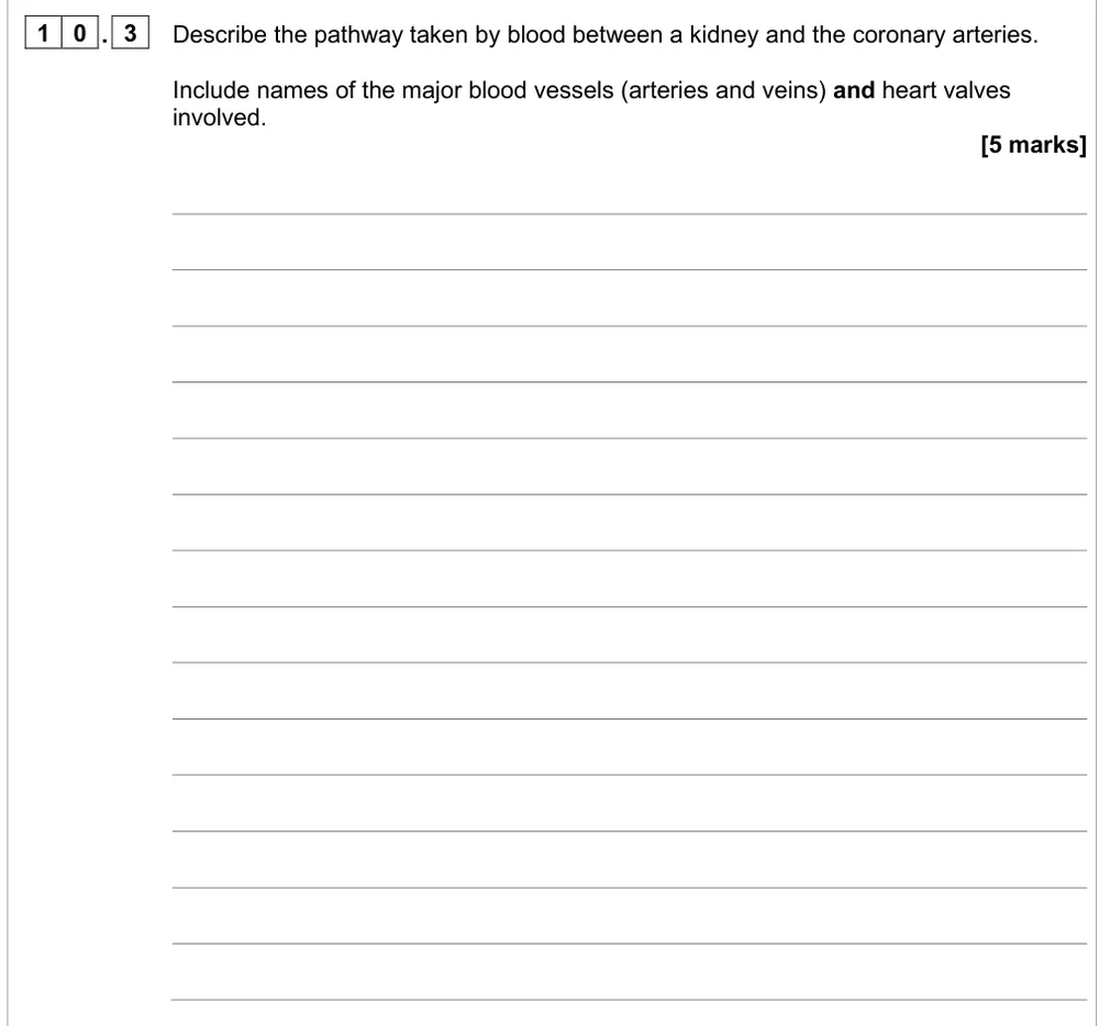
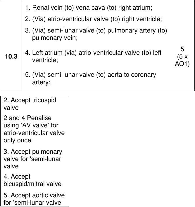
Describe the route blood takes from a kidney to the coronary arteries, naming vessels and valves.
Show the mark scheme
Tap an image to open it full size.
From the examiners' report: About 15% got all 5. Keep going after the lungs and finish at the coronary arteries. Don't swap pulmonary artery and pulmonary vein, and name both kinds of valve on each side.
Question 4
A level Paper 1 · June 2022 · Q08.4 · 2 marks
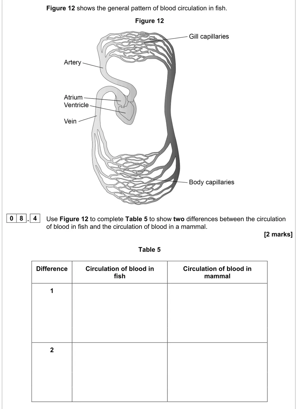
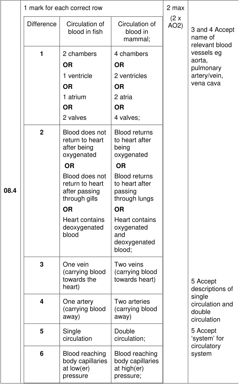
Use a diagram of fish circulation to give two differences from circulation in a mammal.
Show the mark scheme
Tap an image to open it full size.
From the examiners' report: It discriminated well. Compare the circulation, such as the chambers or single v double circulation. Counter-current flow and gills v lungs didn't score.
Heart structure and valves (3.3.4.1)
Question 5
A level Paper 3 · June 2019 · Q03.3 · 2 marks · Hard
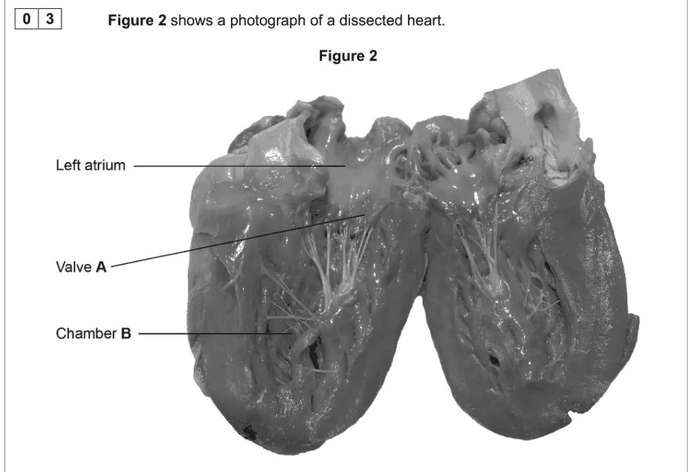
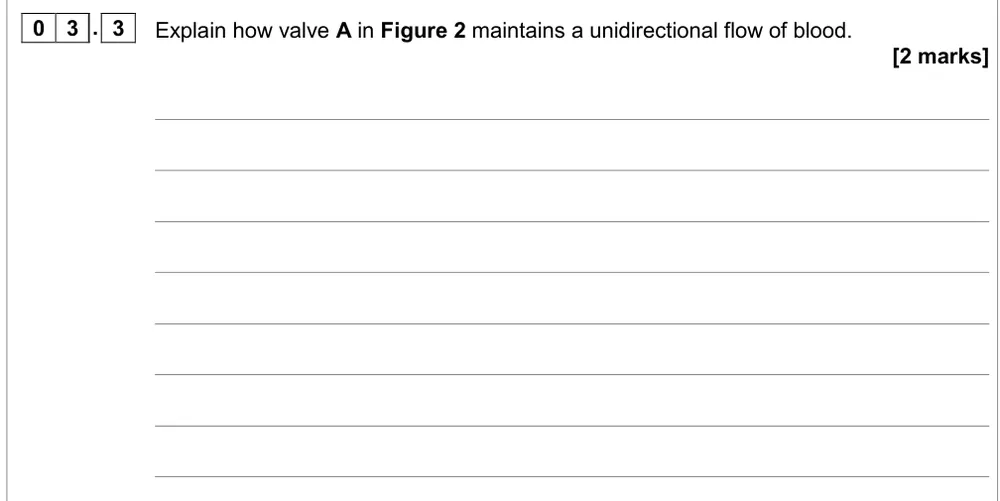
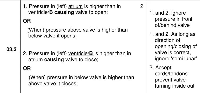
Explain how valve A in a dissected heart maintains a one-way flow of blood.
Information from earlier in this question:
Show the mark scheme
Tap an image to open it full size.
From the examiners' report: More than two-thirds scored zero. Look at the photo: valve A has tendons, so it's between the atrium and ventricle. Explain both the opening and the closing with pressures.
Question 6
AS Paper 2 · November 2020 · Q03.2 · 4 marks
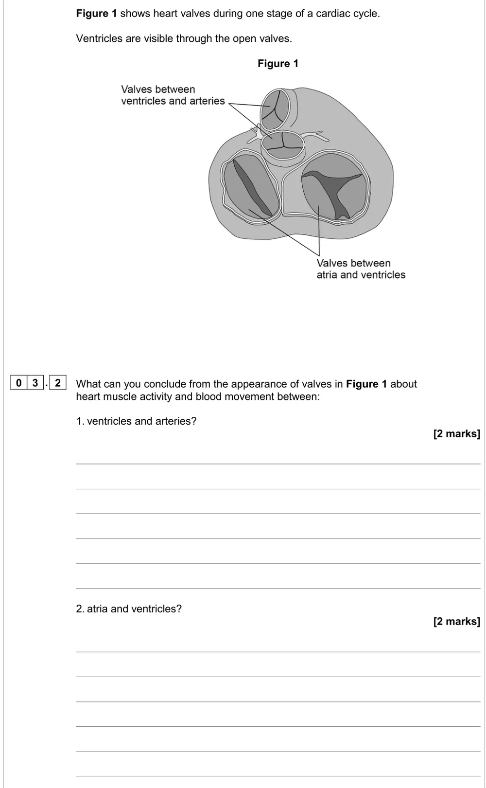
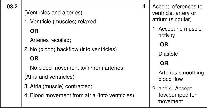
Use a drawing of open and closed heart valves to conclude what the muscle and blood are doing.
Show the mark scheme
Tap an image to open it full size.
From the examiners' report: About 20% showed a sound understanding. Base your answer on which valves are open and which are closed, not on their size.
Question 7
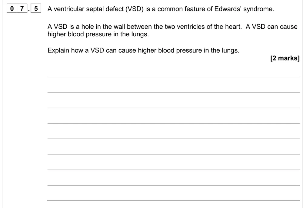
AS Paper 2 · June 2024 · Q07.5 · 2 marks · Hard
Explain how a hole between the ventricles can raise blood pressure in the lungs.
Show the mark scheme
Tap an image to open it full size.
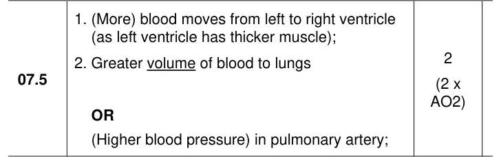
From the examiners' report: Mixing of oxygenated and deoxygenated blood is irrelevant here. Think about volume: the thicker left ventricle pushes extra blood into the right ventricle and on into the pulmonary artery.
Question 8
AS Paper 2 · June 2017 · Q08.4 · 3 marks
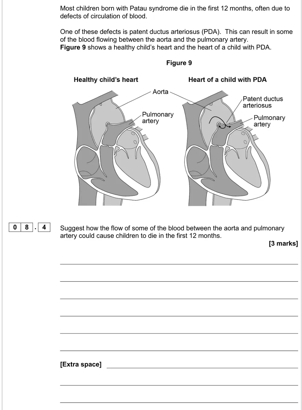
Suggest how blood flowing between the aorta and pulmonary artery could cause death in infancy.
Show the mark scheme
Tap an image to open it full size.
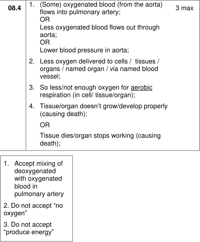
From the examiners' report: No examiners' report for this paper. Follow the chain: less oxygenated blood leaves through the aorta, so less oxygen reaches the tissues for aerobic respiration. The mark scheme doesn't accept 'no oxygen' or 'produce energy'.
Cardiac cycle data (3.3.4.1)
Question 9
A level Paper 1 · November 2020 · Q03.1 · 2 marks · Hard
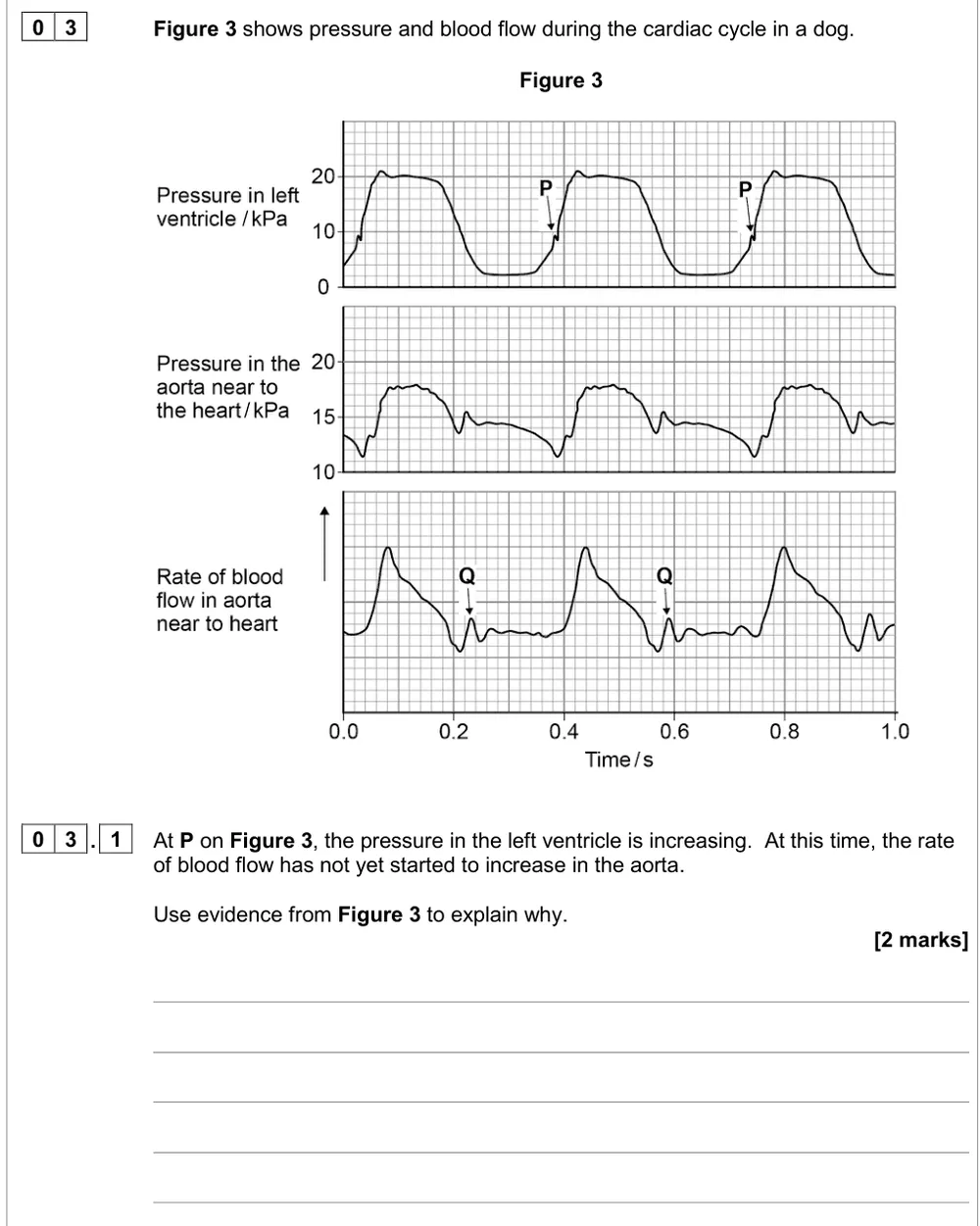
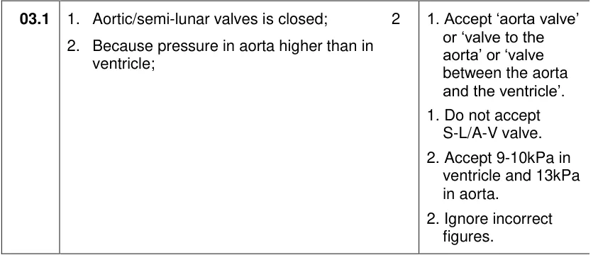
Use pressure and flow traces to explain why aortic blood flow hasn't increased while ventricle pressure rises.
Show the mark scheme
Tap an image to open it full size.
From the examiners' report: Only 16% got both marks. Name the aortic (semi-lunar) valve, not the atria, and use the figure: aortic pressure is still higher than ventricle pressure at P.
Question 10
A level Paper 1 · November 2020 · Q03.3 · 2 marks
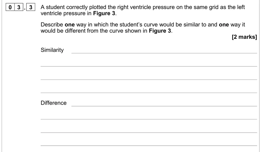
Predict one similarity and one difference between right and left ventricle pressure curves.
Show the mark scheme

Tap an image to open it full size.
From the examiners' report: Only look at the left ventricle trace. Both ventricles contract together, so the pattern is the same but the right ventricle pressure is lower; don't put its peak where the left is lowest.
Question 11
AS Paper 2 · June 2023 · Q04.2 · 2 marks
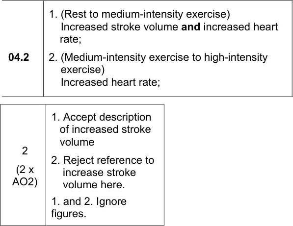
Use ventricle volume and heart rate data to explain what raises cardiac output during exercise.
Show the mark scheme
Tap an image to open it full size.
From the examiners' report: Use cardiac output = stroke volume × heart rate and look at both factors for each change. Respiration and oxygen demand are irrelevant here.
Stroke volume and cardiac output calculations (3.3.4.1)
Question 12
AS Paper 2 · June 2016 · Q09.2 · 1 mark · Medium
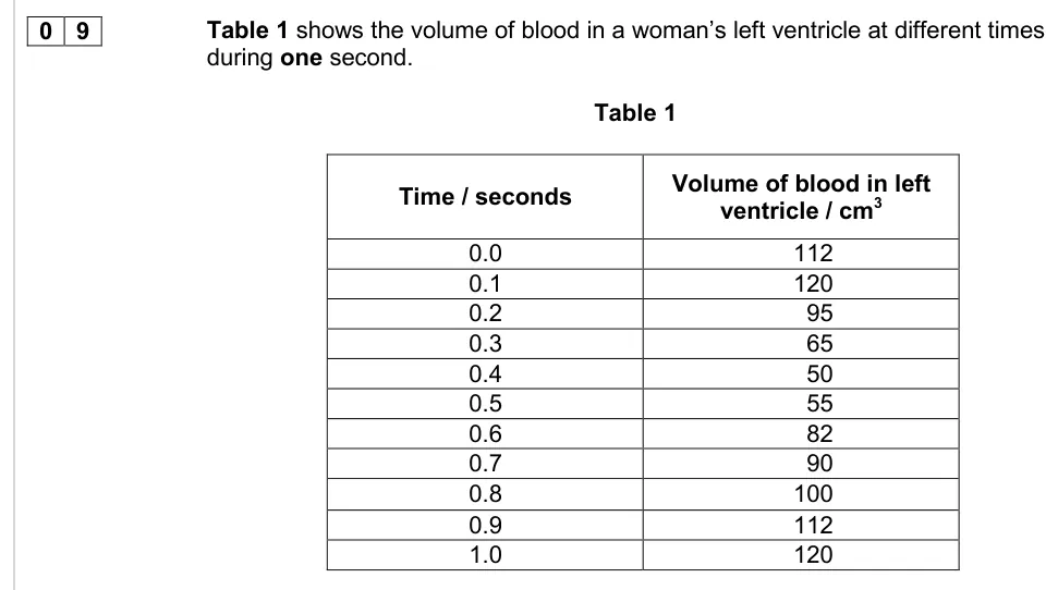
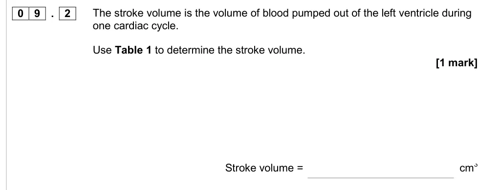
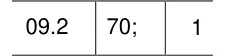
Use a table of left ventricle volumes to work out the stroke volume.
Information from earlier in this question:
Show the mark scheme
Tap an image to open it full size.
From the examiners' report: 40% got it right. Stroke volume is the volume pumped out in one beat: the largest volume in the ventricle minus the smallest.
Question 13
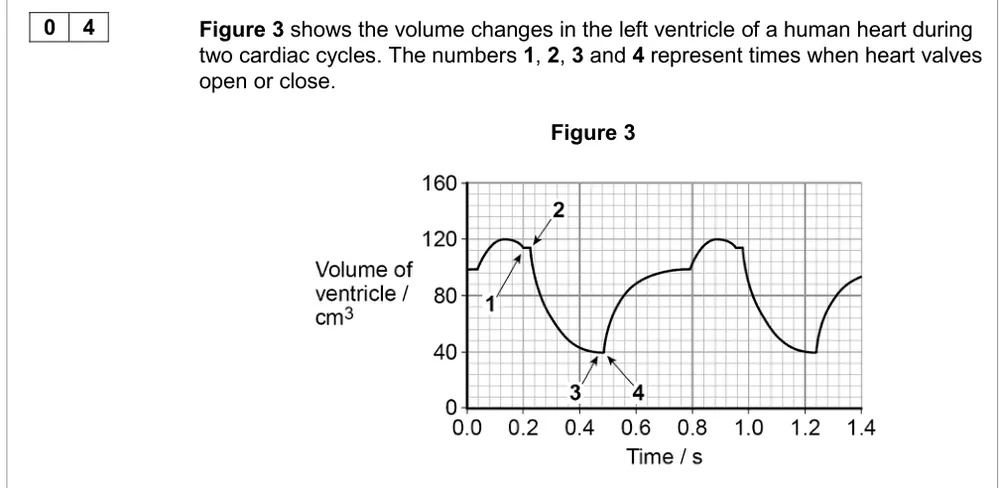
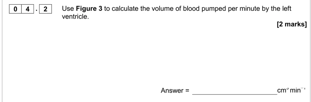
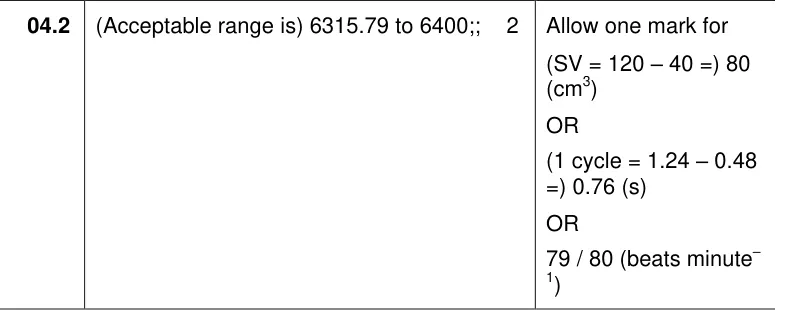
AS Paper 1 · June 2017 · Q04.2 · 2 marks · Hard
Use a ventricle volume graph to calculate the volume of blood pumped per minute.
Information from earlier in this question:
Show the mark scheme
Tap an image to open it full size.
From the examiners' report: 56% scored zero. Work it out in steps: stroke volume from the graph, the time of one cycle, beats per minute (60 ÷ time), then multiply.
Question 14
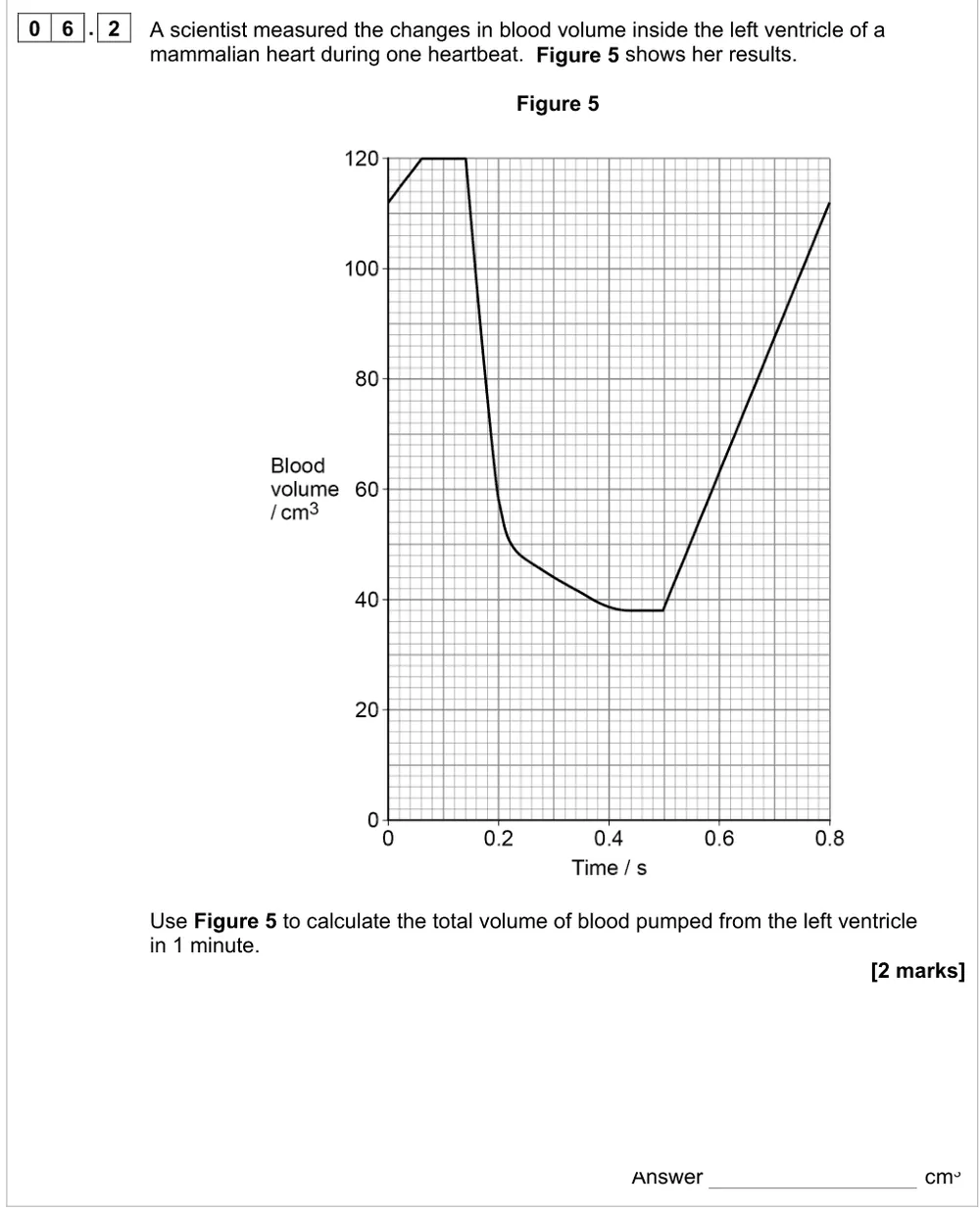
AS Paper 2 · June 2022 · Q06.2 · 2 marks · Hard
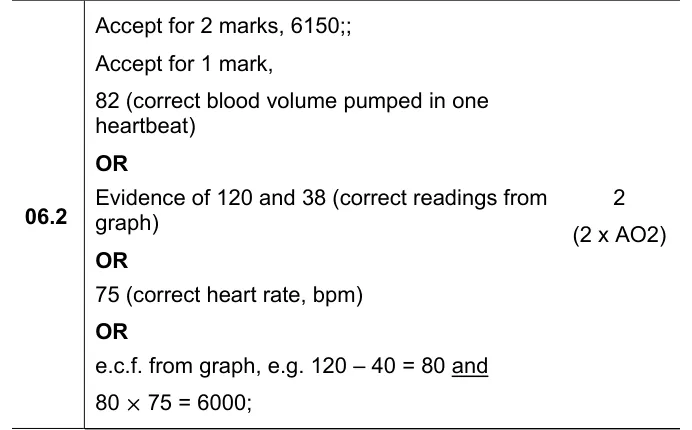
Use a graph of blood volume during one heartbeat to calculate the volume pumped in a minute.
Show the mark scheme
Tap an image to open it full size.
From the examiners' report: About 20% got both marks. Read the minimum carefully from the scale (38, not 40), and get the heart rate from the time of one beat: 60 ÷ 0.8 = 75.
Where students lose marks (from AQA examiners' reports and mark schemes)
Each point below is followed by wording that gained marks.
Coronary arteries, in full: Only 31.6% could name the vessels that carry blood to the heart muscle. The mark scheme rejects coronary veins and doesn't accept 'coronary' by itself; aorta and capillary are ignored.
✔ Gains credit: "Coronary arteries."
Follow the blood all the way to the coronary arteries: Only about 15% got all 5 marks for the route from a kidney to the coronary arteries. Students confused pulmonary arteries with pulmonary veins, stopped at the lungs, or left out or muddled the valves.
✔ Gains credit: "Renal vein to vena cava to right atrium; atrioventricular valve to right ventricle; semi-lunar valve to pulmonary artery ... pulmonary vein to left atrium; atrioventricular valve to left ventricle; semi-lunar valve to aorta to coronary artery."
Explain the pressure that moves the valve: More than two-thirds scored zero for how valve A keeps blood flowing one way. Many answered about semi-lunar valves, though the tendons show it's an atrioventricular valve, and many described when it opens without the cause.
✔ Gains credit: "Pressure in the (left) atrium is higher than in the ventricle, causing the valve to open; pressure in the ventricle is higher than in the atrium, causing the valve to close."
Read the aorta pressure off the graph: Only 16% got both marks for why blood flow in the aorta hadn't increased at P. Many confused the atria with the aorta, and the second mark needed the pressures from the figure.
✔ Gains credit: "The aortic / semi-lunar valve is closed because pressure in the aorta is higher than in the ventricle (13 kPa in the aorta, 9–10 kPa in the ventricle)."
Open or closed matters, not size: About 20% showed a sound understanding of the valves photograph. Answers about the different sizes of the valves, rather than whether they were open or closed, scored no marks or only one.
✔ Gains credit: "Ventricle muscle relaxed; no backflow into the ventricles; atria contracted; blood moves from the atria into the ventricles."
A hole between the ventricles is about volume, not mixing: On the ventricular septal defect, many were fixated on oxygenated and deoxygenated blood mixing, which was irrelevant, and some still mixed up the left and right sides and the pulmonary artery and aorta.
✔ Gains credit: "More blood moves from the left to the right ventricle (thicker muscle); a greater volume of blood to the lungs / higher pressure in the pulmonary artery."
Compare the circulations, not the gas exchange organs: On fish v mammal circulation, writing 'counter-current flow' or 'gills v lungs' scored nothing. The marks were for the heart and the route of the blood.
✔ Gains credit: "Fish: 2 chambers, single circulation; mammal: 4 chambers, double circulation"; "blood returns to the heart after passing through the lungs".
Heart rate comes from the time of one beat: 56% scored zero for the volume pumped per minute in 2017, and only about 20% got full marks in 2022. Many couldn't read the graph scale or work out the heart rate from the time of one heartbeat.
✔ Gains credit: "82 (cm³ pumped in one heartbeat)" and "75 (correct heart rate, bpm)", giving "6150" cm³.
Download the questions
Download the PDF pack (14 questions, mark schemes at the end)
Keep practising
- Haemoglobin and oxygen transport: AQA A Level Biology exam questions by topic
- Blood vessels and tissue fluid: AQA A Level Biology exam questions by topic
- Gas exchange: AQA A Level Biology exam questions by topic
- Algebra (MS 2): AQA A Level Biology exam questions
- Calculations and mathematical content in A level Biology, from AQA papers
- AQA A Level Biology masterclasses
Questions, mark schemes and examiners' report extracts © AQA.