

Cell structure is where OCR A Level Biology starts, and it underpins almost everything that follows. Spec point 2.1.1 asks you to know the organelles of eukaryotic cells and what each one does, how the rough ER, Golgi apparatus and vesicles work together to make and secrete proteins, why the cytoskeleton matters, and how prokaryotic cells differ from eukaryotic ones. It also covers the microscopy skills: choosing between light, TEM and SEM images, preparing and staining slides, drawing what you see and using the magnification formula.
Most lost marks here come from precision rather than missing knowledge. In the examiners' reports, students call the nucleolus the nucleus, say ribosomes are membrane-bound, use magnification as evidence that rules out an electron microscope, or convert centimetres to micrometres by multiplying by 1000 instead of 10,000.
Work through the seventeen questions below in order. They start with organelle recall, move on to applying it to new cells and stains, and finish with microscope images and calculations. If you'd like to practise questions like these with me each week, my OCR A masterclasses run on Wednesdays at 5.15pm for Year 12 and on Tuesdays at 6.30pm or Thursdays at 5.15pm for Year 13.
What you need from GCSE
Each question on this page assumes you're secure on these GCSE ideas. If any of them feels shaky, look it up before you start.
- Animal and plant cells have a nucleus, cytoplasm, cell membrane, mitochondria and ribosomes; plant cells also have a cell wall, chloroplasts and a permanent vacuole.
- Bacterial cells are prokaryotic: they are much smaller, have no nucleus, and their DNA is a single loop, often with plasmids.
- Electron microscopes have a much higher magnification and resolution than light microscopes, so they show sub-cellular structures in more detail.
- magnification = size of image ÷ size of real object, and 1 mm = 1000 µm.
Key terms
These words come up in the questions and mark schemes. Examiners expect you to use them exactly.
- Magnification: how many times bigger the image is than the real object.
- Resolution: the smallest distance between two points that can still be seen as separate; higher resolution shows more detail.
- Ultrastructure: the detailed internal structure of a cell, seen only with an electron microscope.
- Eyepiece graticule: a scale in the eyepiece with no units; it is calibrated against a stage micrometer for each objective lens.
- Stage micrometer: a slide with an accurate scale, used to work out what one eyepiece graticule division measures.
- Differential staining: using stains that colour different cell types or cellular components differently, to increase contrast.
- Nucleolus: the dense region inside the nucleus where ribosomes are made.
- Cytoskeleton: a network of protein fibres (microtubules and microfilaments) that supports the cell, moves organelles and vesicles, and enables cell movement.
- Exocytosis: secretion: a vesicle fuses with the cell surface membrane and releases its contents outside the cell.
- 70S ribosome: the smaller ribosome found in prokaryotes (and in mitochondria and chloroplasts); eukaryotic cytoplasm has 80S ribosomes.
What these questions cover (OCR 2.1.1)
The questions test these learning outcomes from the OCR A Level Biology A specification (H420):
- (a) the use of microscopy to observe and investigate different types of cell and cell structure in a range of eukaryotic organisms (light, transmission electron and scanning electron microscopes)
- (b) the preparation and examination of microscope slides for use in light microscopy (including the eyepiece graticule and stage micrometer)
- (c) the use of staining in light microscopy (including differential staining)
- (d) the representation of cell structure as seen under the light microscope using drawings and annotated diagrams of whole cells or cells in sections of tissue
- (e) the use and manipulation of the magnification formula
- (f) the difference between magnification and resolution
- (g) the ultrastructure of eukaryotic cells and the functions of the different cellular components
- (h) photomicrographs of cellular components in a range of eukaryotic cells (interpreting electron micrographs)
- (i) the interrelationship between the organelles involved in the production and secretion of proteins
- (j) the importance of the cytoskeleton
- (k) the similarities and differences in the structure and ultrastructure of prokaryotic and eukaryotic cells
How to use this page
Write your answer on paper before you open the mark scheme. Allow about 1.2 minutes per mark: the 17 questions are worth 39 marks, so about 47 minutes in total. Then mark your answer strictly against the mark scheme and read the examiners' tip underneath it. If you'd rather print the questions, download the PDF pack.
Recall
Question 1
A level Paper 3 · June 2018 · Q1(a) · 2 marks · Easy
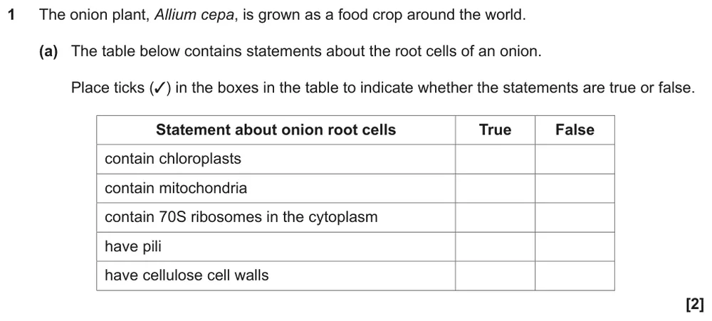
Tick true or false for five statements about onion root cells.
Show the mark scheme
Tap an image to open it full size.
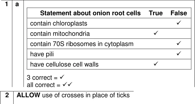
From the examiners' report: Most students got only one of the two marks, usually because they said onion root cells have 70S ribosomes or pili. Those belong to prokaryotes; root cells are eukaryotic and also have no chloroplasts.
Question 2
AS Paper 1 · June 2023 · Q28(a) · 4 marks · Medium
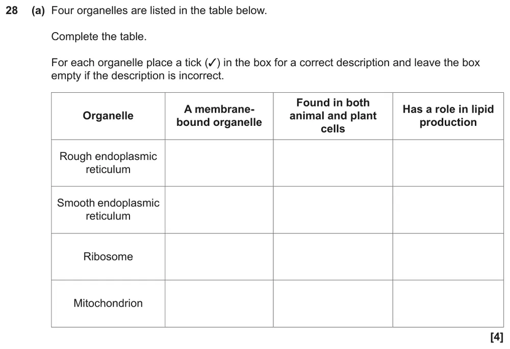
Tick which descriptions fit the rough ER, smooth ER, ribosome and mitochondrion.
Show the mark scheme
Tap an image to open it full size.
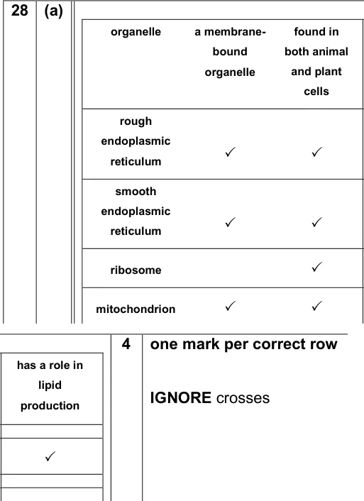
From the examiners' report: Most students got 3 or 4 marks. The usual slip was ticking the ribosome as membrane-bound: ribosomes are made of RNA and protein with no membrane around them.
Question 3
A level Paper 3 · June 2019 · Q1(a)(i) · 2 marks · Medium
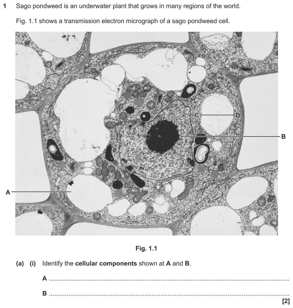
Identify the vacuole and nucleolus on an electron micrograph of a pondweed cell.
Show the mark scheme

Tap an image to open it full size.
From the examiners' report: Most students recognised the vacuole, but many called B the nucleus. Look at what the label line touches: the dark, dense patch inside the nucleus is the nucleolus.
Question 4
A level Paper 1 · June 2024 · Q21(a) · 3 marks · Medium
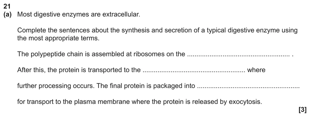
Complete sentences about where a digestive enzyme is made, processed and packaged.
Show the mark scheme

Tap an image to open it full size.
From the examiners' report: This was answered very well. Learn the route as a sequence (ribosomes on the RER, Golgi apparatus, vesicles, exocytosis) so you can drop the right term into each gap.
Question 5
A level Paper 1 · June 2023 · Q18(b)(i) · 3 marks · Easy
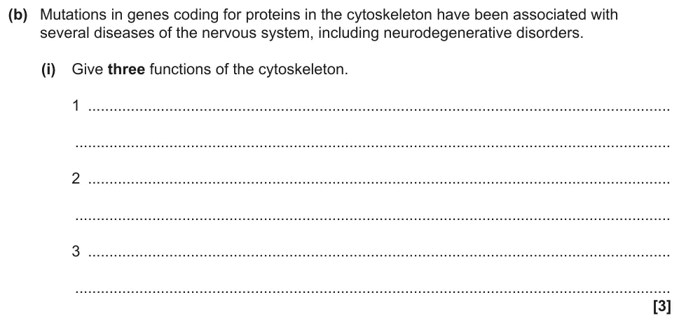
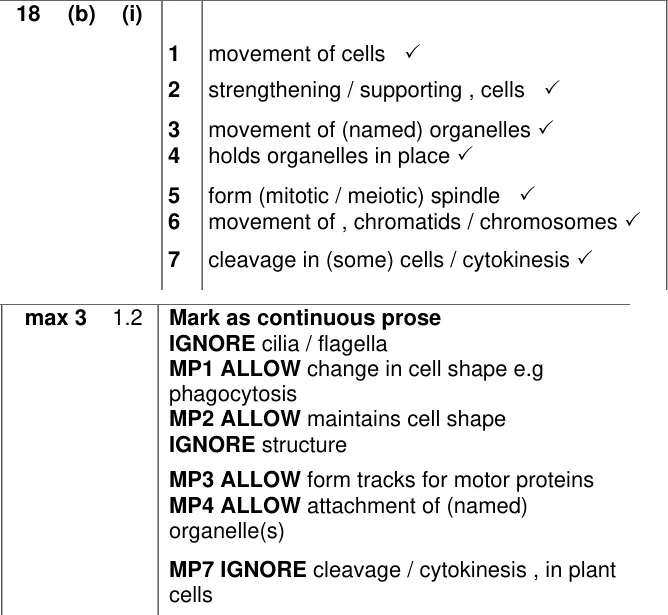
Give three functions of the cytoskeleton.
Show the mark scheme
Tap an image to open it full size.
From the examiners' report: Most students scored at least one mark, but some lost marks by giving the same point twice. Vesicles count as organelles, so "moves vesicles" and "moves organelles" earn only one mark between them.
Question 6
AS Paper 1 · June 2019 · Q25(b)(i) · 1 mark · Medium
Name the molecule found in bacterial cell walls.

Show the mark scheme

Tap an image to open it full size.
From the examiners' report: Many students knew it but spelled it badly. Learn to spell peptidoglycan; chitin (fungi) and cellulose (plants) are wrong.
Question 7
A level Paper 3 · June 2024 · Q1(a)(i) · 1 mark · Medium
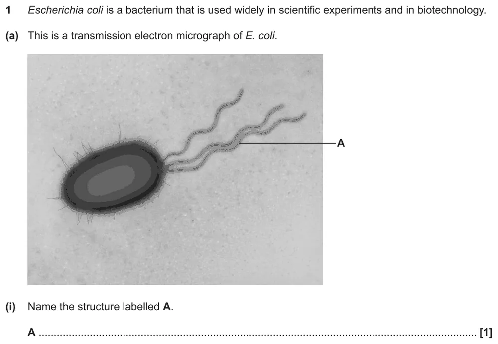
Name the structure labelled on an electron micrograph of E. coli.
Show the mark scheme

Tap an image to open it full size.
From the examiners' report: Most students got this. Wrong answers were tail, cilia or undulipodium. A bacterium has a flagellum; undulipodia are only found in eukaryotic cells.
Applying what you know
Question 8
A level Paper 3 · June 2024 · Q1(a)(ii) · 1 mark · Hard
Estimate the diameter of an E. coli cell from your own knowledge.

Show the mark scheme

Tap an image to open it full size.
From the examiners' report: Most students got this mark. There is no magnification or scale bar, so don't try to calculate: use what you know about prokaryote size and give the answer in µm, not nm.
Question 9
A level Paper 1 · June 2023 · Q18(b)(ii) · 2 marks · Hard
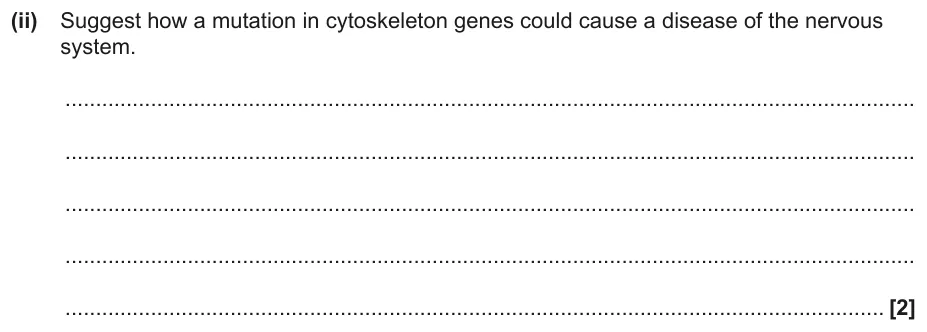
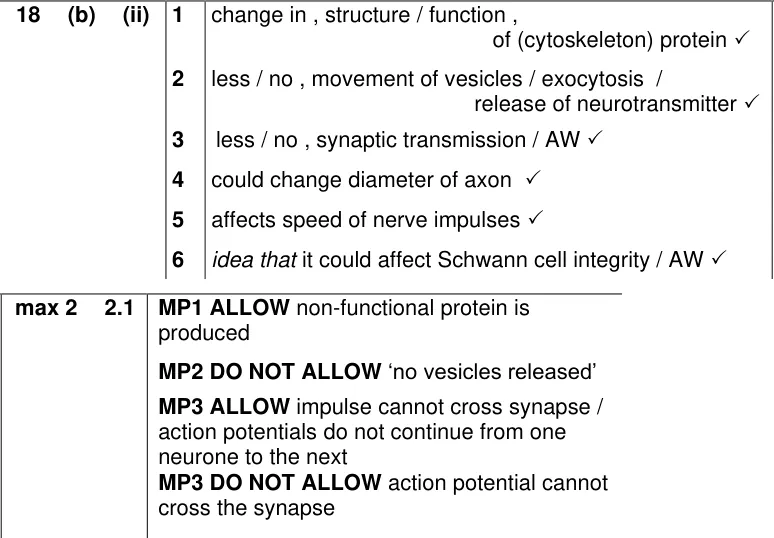
Suggest how a mutation in cytoskeleton genes could cause a nervous system disease.
Show the mark scheme
Tap an image to open it full size.
From the examiners' report: Link each step: mutation, changed cytoskeleton protein, fewer vesicles moved to the membrane, less neurotransmitter released, less transmission at the synapse. Answers about channel proteins didn't score because they aren't part of the cytoskeleton.
Question 10
AS Paper 2 · June 2019 · Q2(c)(i) · 3 marks · Hard
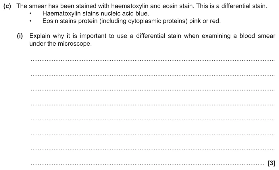
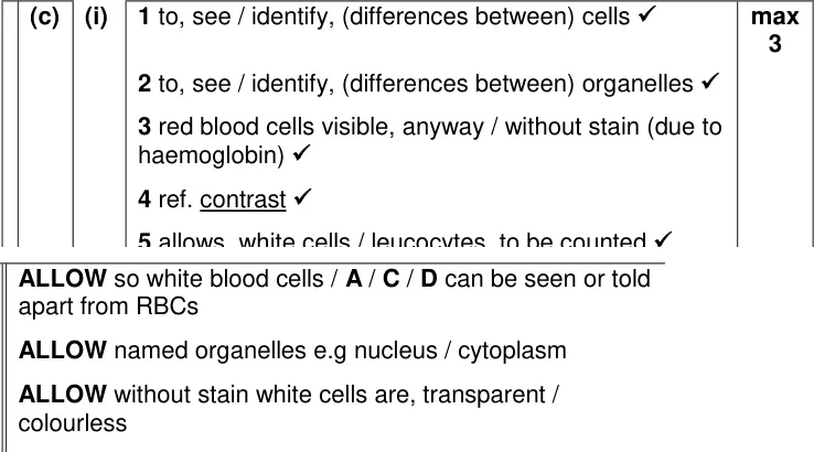
Explain why a differential stain is important when examining a blood smear.
Show the mark scheme
Tap an image to open it full size.
From the examiners' report: Strong answers said the stain lets you see both different cells and different organelles, and used the word contrast. Say "organelles" or name one, such as the nucleus, rather than "components" or "structures".
Microscopes and images
Question 11
AS Paper 2 · November 2020 · Q1(b) · 4 marks · Medium
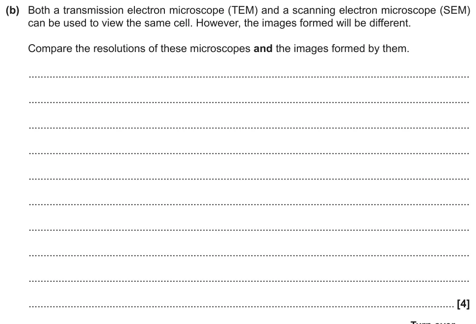
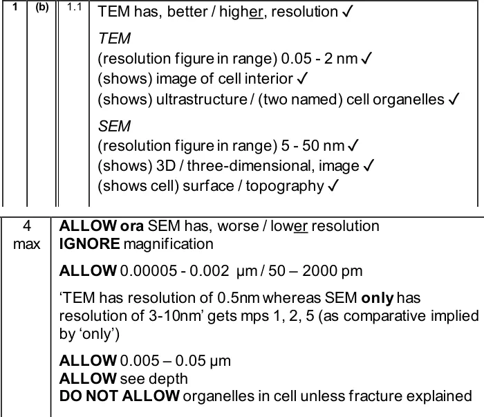
Compare the resolution and images of a transmission and a scanning electron microscope.
Show the mark scheme
Tap an image to open it full size.
Question 12
AS Paper 2 · June 2024 · Q5(b) · 2 marks · Easy
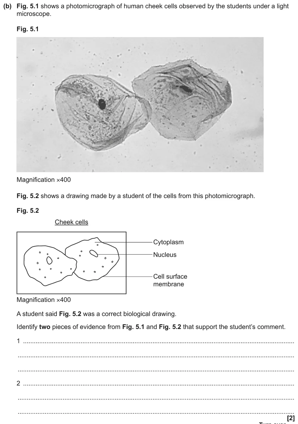
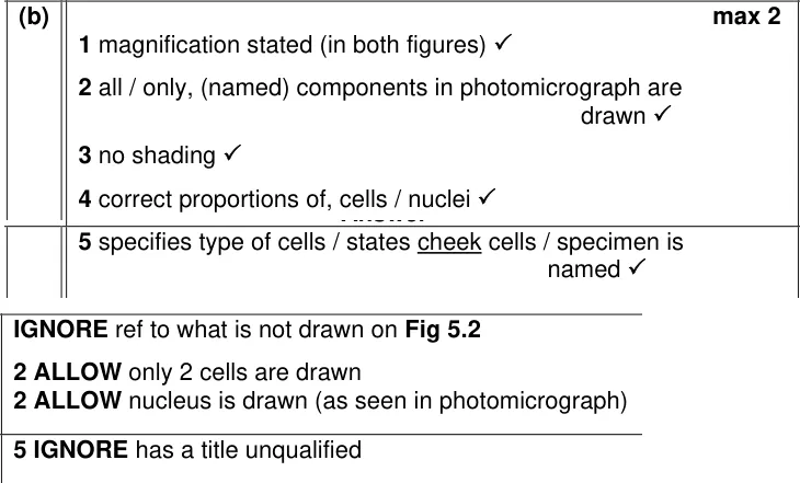
Use a photomicrograph and a student's drawing of cheek cells to support the claim it is a correct biological drawing.
Show the mark scheme
Tap an image to open it full size.
From the examiners' report: Most students mentioned no shading and the magnification. The question asks for evidence from both figures, so compare the drawing with the photo rather than listing drawing rules such as label lines.
Practical and maths
Question 13
A level Paper 2 · June 2024 · Q17(b)(iii) · 3 marks · Medium
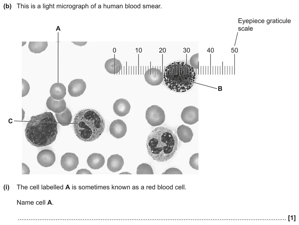
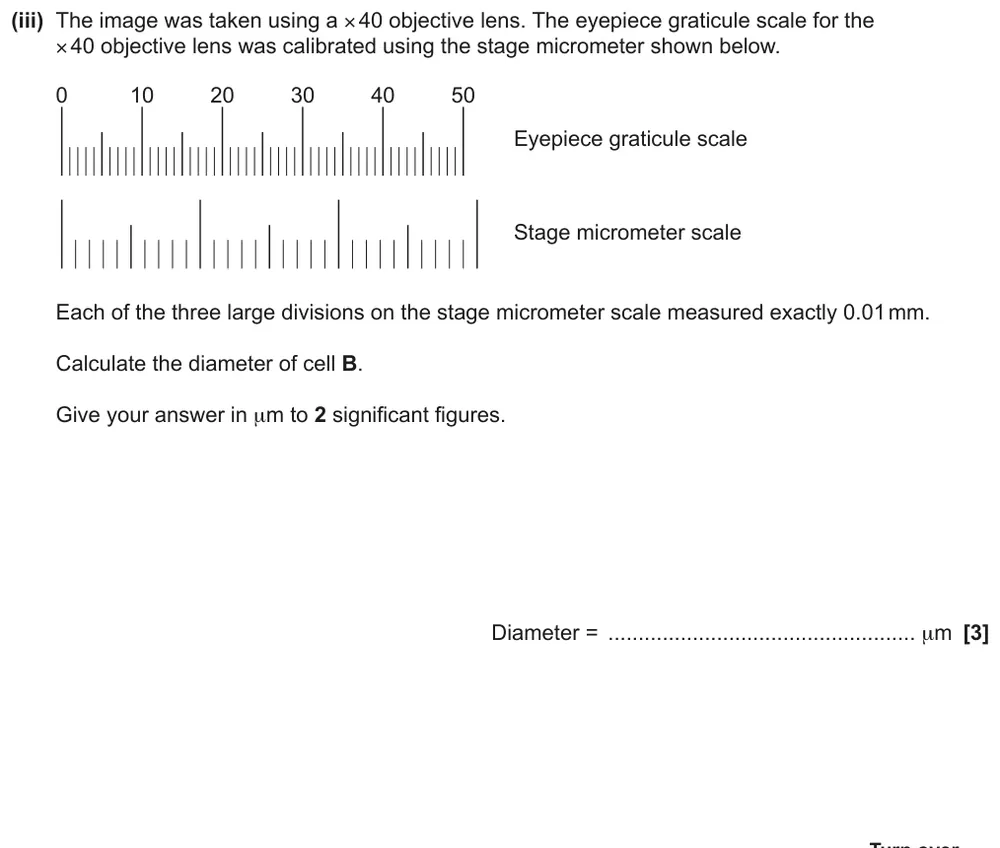
Use a calibrated eyepiece graticule to calculate a cell's diameter in µm.
Information from earlier in this question:
Show the mark scheme
Tap an image to open it full size.
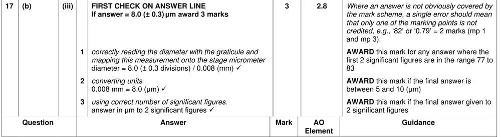
From the examiners' report: Only about half of students used the scales correctly, and only 1 in 10 got all three marks. Once you have converted the stage micrometer reading to µm, stop: don't divide by the objective magnification as well.
Question 14
A level Paper 3 · June 2022 · Q2(a)(iii) · 2 marks · Medium
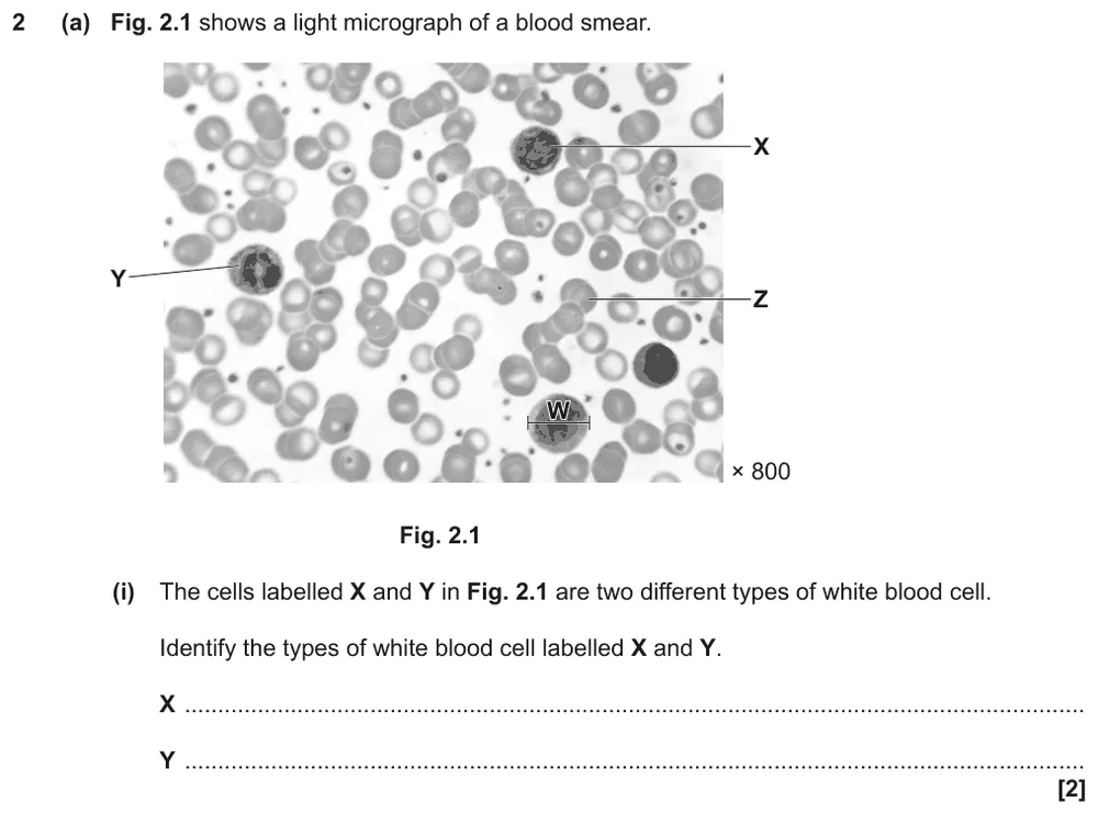
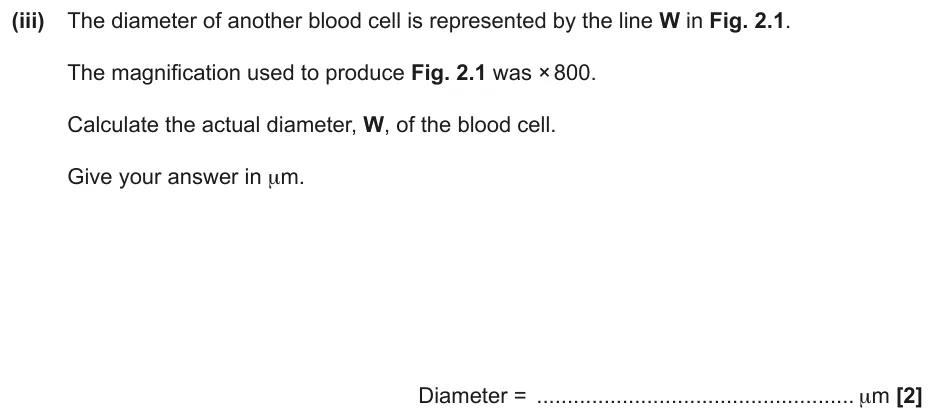
Calculate the actual diameter of a blood cell from an image at ×800.
Information from earlier in this question:
Show the mark scheme
Tap an image to open it full size.
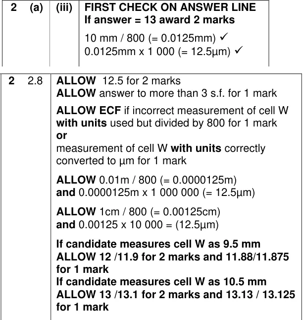
From the examiners' report: About half of students got both marks. Most errors came from measuring in cm and multiplying by 1000 to get µm; there are 10,000 µm in a cm. Measure in mm and show the measurement with its unit.
Question 15
A level Paper 3 · June 2022 · Q2(c) · 2 marks · Medium
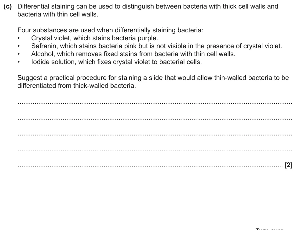
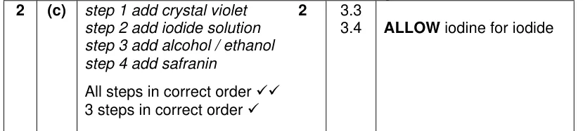
Suggest the order for using four substances to differentially stain thin- and thick-walled bacteria.
Show the mark scheme
Tap an image to open it full size.
From the examiners' report: Most students got one mark. Put the substances on one at a time, not crystal violet and iodine together, and finish with safranin: thin-walled bacteria are invisible once the alcohol has removed the crystal violet.
Question 16
A level Paper 2 · June 2025 · Q16(a)(i) · 2 marks · Hard
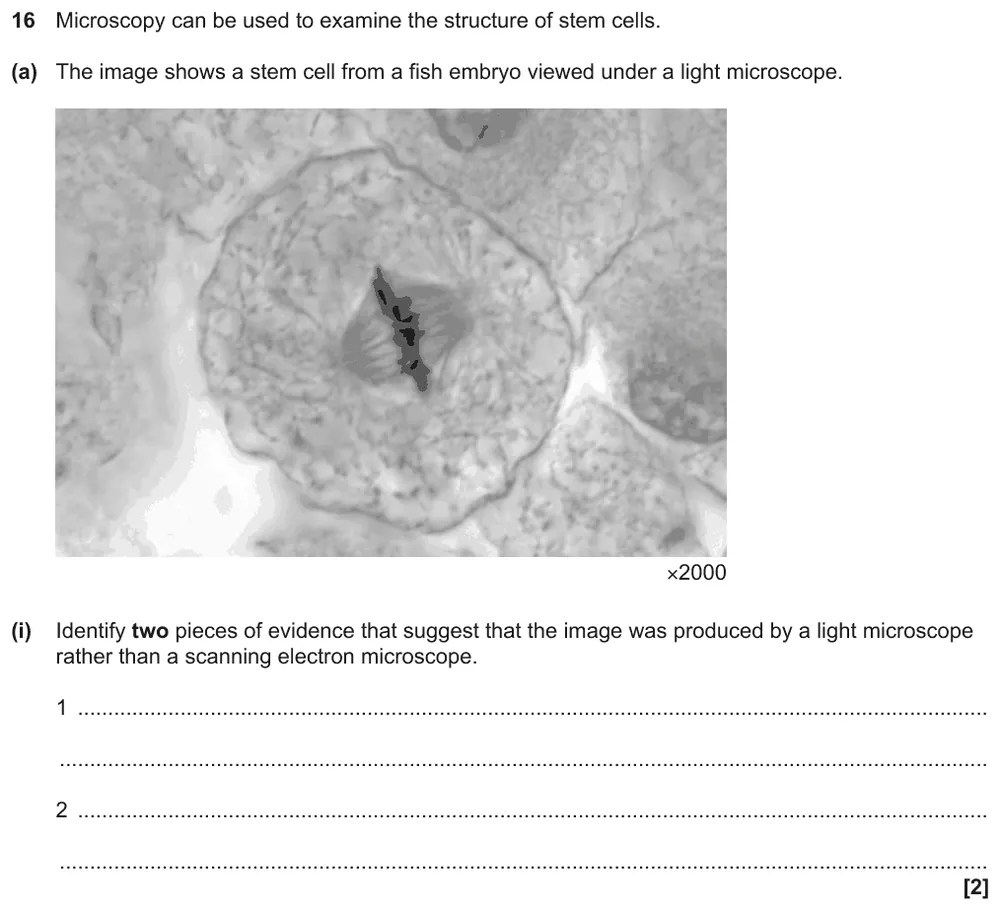
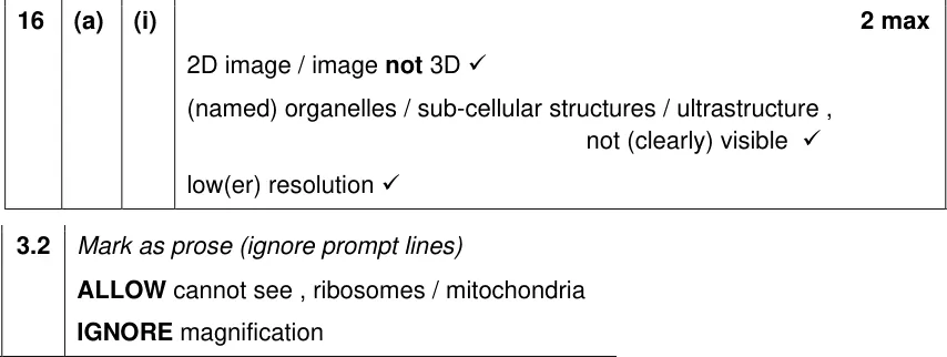
Give two pieces of evidence that an image of a stem cell came from a light microscope, not an SEM.
Show the mark scheme
Tap an image to open it full size.
From the examiners' report: Many students used the ×2000 magnification as evidence, but an electron microscope can work at ×2000 too. Use features of the image: it is 2D, and organelles aren't clearly visible because the resolution is lower.
Question 17
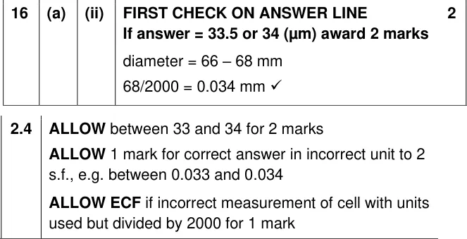
A level Paper 2 · June 2025 · Q16(a)(ii) · 2 marks · Hard
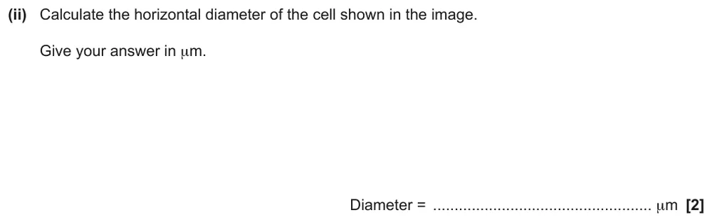
Calculate the horizontal diameter of the stem cell in µm.
Show the mark scheme
Tap an image to open it full size.
From the examiners' report: Many students didn't divide their ruler measurement by the magnification, and others slipped converting mm to µm. Measure in mm, divide by 2000, then multiply by 1000.
Where students lose marks (from OCR examiners' reports and mark schemes)
Each point below is followed by wording that gained marks.
Ribosomes have no membrane: In the organelle table, the most common error was ticking the ribosome as a membrane-bound organelle.
✔ Gains credit: Rough ER, smooth ER and mitochondria are membrane-bound; the ribosome is not. All four are found in both animal and plant cells, and only smooth ER has a role in lipid production.
Nucleolus, the dark spot inside: Many students labelled the dense region inside the nucleus as "nucleus", which the mark scheme does not allow.
✔ Gains credit: "B = nucleolus" and "A = (permanent) vacuole".
Root cells are eukaryotic, so no pili: The commonest errors about onion root cells were saying they contain 70S ribosomes in the cytoplasm and have pili.
✔ Gains credit: True: contain mitochondria; have cellulose cell walls. False: contain chloroplasts; contain 70S ribosomes in the cytoplasm; have pili.
Vesicles are organelles too: Students listed "moves vesicles" and "moves organelles" as two functions of the cytoskeleton, but these are the same marking point.
✔ Gains credit: Three different functions, such as "strengthens and supports the cell", "moves (named) organelles" and "forms the spindle to move chromatids".
×2000 doesn't rule out an SEM: Many students said the magnification proved a light microscope was used, but an electron microscope can also magnify ×2000, so the mark scheme ignores magnification.
✔ Gains credit: "The image is 2D, not 3D", "organelles are not visible" and "lower resolution".
10,000 µm in every centimetre: Many answers were 10 times too small because students measured in cm and multiplied by 1000 to convert to µm.
✔ Gains credit: "10 mm ÷ 800 = 0.0125 mm", then "0.0125 mm × 1000 = 12.5 µm".
Calibrated means finished: Students who had correctly converted the stage micrometer reading to µm then divided it by the ×40 objective magnification, spoiling a correct answer.
✔ Gains credit: "Diameter = 8.0 divisions = 0.008 mm = 8.0 µm", given to 2 significant figures.
Crystal violet, iodide, alcohol, then safranin: A common error was adding crystal violet and iodine at the same time, and many left out safranin, as if thin-walled bacteria would still be visible without a stain.
✔ Gains credit: "Add crystal violet, then iodide solution, then alcohol, then safranin", one after the other.
Download the questions
Download the PDF pack (17 questions, mark schemes at the end)
Keep practising
- Cell Structure quiz
- Microscopy and Magnification quiz
- Cell Ultrastructure, Prokaryotes and Cytoskeleton quiz
- Cell Structure and Organelles: OCR A exam questions with worked answers
- Microscopy and Staining: OCR A exam questions with worked answers
- PAG 1 Microscopy: OCR A Level Biology exam questions by topic
- Nucleotides and Nucleic Acids (2.1.3): OCR A Level Biology exam questions by topic
- Cell structure and microscopy: interactive written questions (20)
- OCR A Level Biology masterclasses
Questions, mark schemes and examiners' report extracts © OCR.