

OCR A PAG 1 · 17 questions · mark schemes and examiners’ tips included
PAG 1 is microscopy, and it is usually the first practical you do in Year 12. You make a slide, stain it, find the cells on low power, switch to high power, measure them with a calibrated eyepiece graticule and then draw what you see. Every one of those steps turns up in OCR exam questions, on the AS papers and on all three A level papers.
The examiners' reports are clear that students who have actually handled a microscope do much better. Marks go on practical detail: lowering the cover slip at an angle, using blotting paper to draw the stain through, starting on the lowest power objective and focusing with the coarse then the fine focus, and saying that a stain increases contrast. Drawings lose marks for shading, arrowheads on label lines and structures you can't actually see.
Work through the seventeen questions below in order. They start with preparing slides, then using the microscope and graticule, staining, light versus electron microscopes, and finish with scientific drawings. The magnification calculations are on my arithmetic (M0) page. If you'd like to go through questions like these with me each week, my OCR A masterclasses run on Wednesdays at 5.15pm for Year 12 and on Tuesdays at 6.30pm or Thursdays at 5.15pm for Year 13.
Want help with this topic? It comes up in my weekly OCR A masterclasses. See the timetable →
What these questions cover (OCR PAG 1: microscopy)
PAG 1 is one of the twelve practical activity groups in the OCR A Level Biology A specification (H420). The questions test these parts of the specification:
- 1.2.2(d) use of a light microscope at high power and low power, including use of a graticule
- 1.2.2(e) production of scientific drawings from observations with annotations
- 2.1.1(b) the preparation and examination of microscope slides for use in light microscopy (including the use of an eyepiece graticule and stage micrometer)
- 2.1.1(c) the use of staining in light microscopy (to include differential staining)
- 2.1.1(d) the representation of cell structure as seen under the light microscope using drawings and annotated diagrams of whole cells or cells in sections of tissue
- 2.1.1(f) the difference between magnification and resolution
How to use this page
Write your answer on paper before you open the mark scheme. Allow about 1.2 minutes per mark: the 17 questions are worth 43 marks, so about 52 minutes in total. Then mark your answer strictly against the mark scheme and read the examiners' tip underneath it. If you'd rather print the questions, download the PDF pack.
Preparing microscope slides
Question 1
A level Paper 2 · June 2024 · Q17(a) · 3 marks · Easy
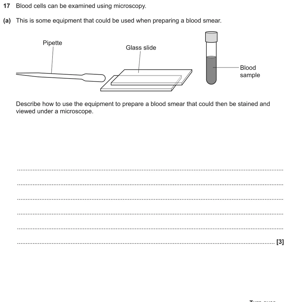
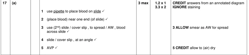
Describe how to use a pipette and glass slides to prepare a blood smear.
Show the mark scheme
Tap an image to open it full size.
From the examiners' report: Most students got at least one mark, and many got all three. Stick to making the smear: describing staining or focusing gained nothing because it wasn't asked for.
Question 2
A level Paper 3 · June 2019 · Q1(a)(iv) · 2 marks · Medium
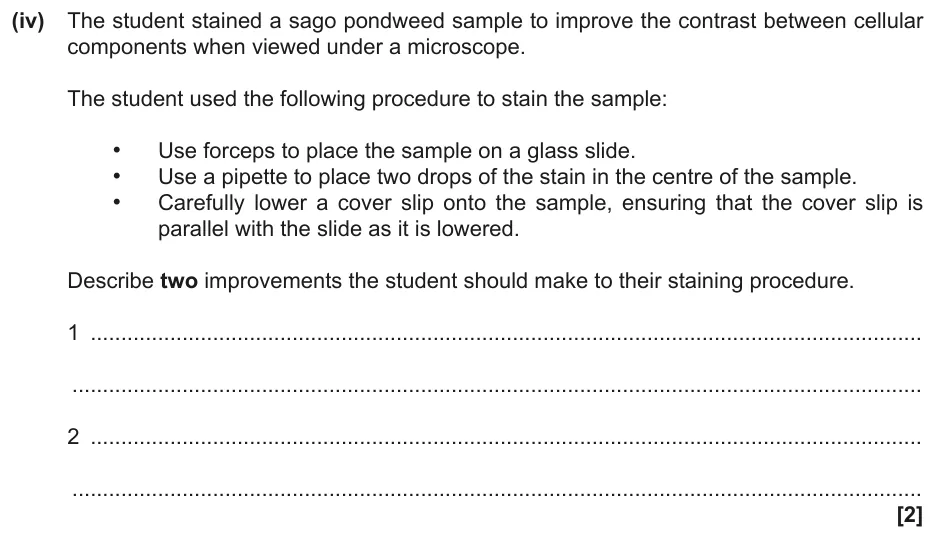
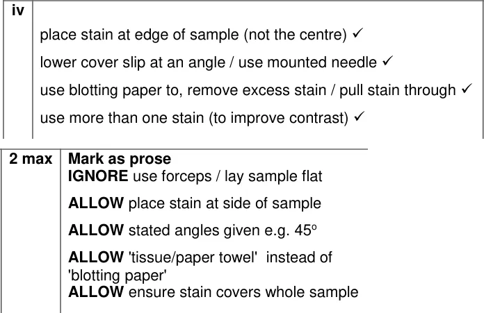
Suggest two improvements to a student's method for staining a pondweed sample.
Show the mark scheme
Tap an image to open it full size.
From the examiners' report: Students who had made slides themselves scored well, lowering the cover slip at an angle or using blotting paper to remove excess stain. Answers about aseptic technique, gloves or pressing on the cover slip gained no credit.
Question 3
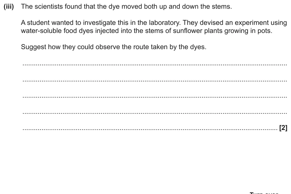
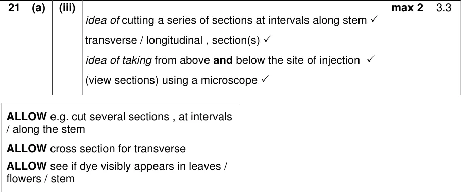
A level Paper 1 · June 2023 · Q21(a)(iii) · 2 marks · Medium
Suggest how to observe the route taken by a dye injected into a sunflower stem.
Show the mark scheme
Tap an image to open it full size.
From the examiners' report: Many answers just said 'use a microscope'. The marks went for cutting sections at intervals along the stem, transverse or longitudinal, above and below the injection site. Don't suggest a potometer or colorimeter here.
Question 4
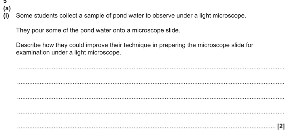
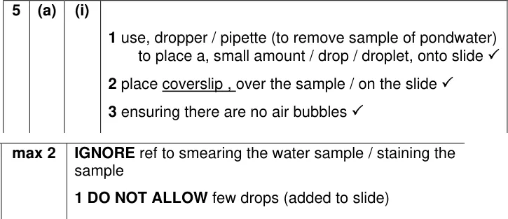
AS Paper 2 · June 2024 · Q5(a)(i) · 2 marks · Hard
Improve a technique where pond water is poured onto a microscope slide.
Show the mark scheme
Tap an image to open it full size.
From the examiners' report: Few students got both marks. A smear dries the sample, so it is no good for pond water. Name the cover slip, and use a pipette to add one drop.
Using a light microscope and a graticule
Question 5
A level Paper 1 · November 2020 · Q20(c)(ii) · 2 marks · Medium
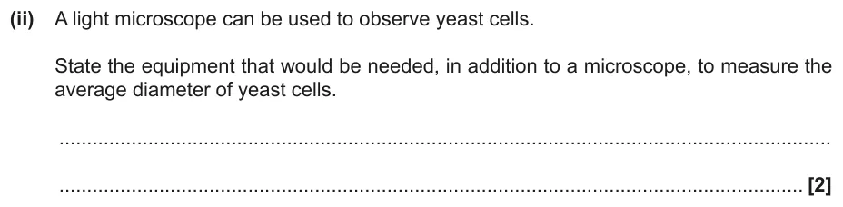
State the equipment needed, as well as a microscope, to measure the diameter of yeast cells.
Show the mark scheme

Tap an image to open it full size.
From the examiners' report: There is no examiners' comment for this part. Both marks need the full names: an eyepiece graticule to measure and a stage micrometer to calibrate it.
Question 6
AS Paper 1 · June 2018 · Q25(b) · 3 marks · Medium
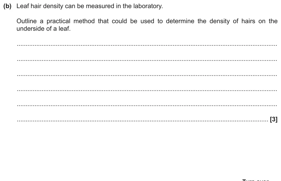
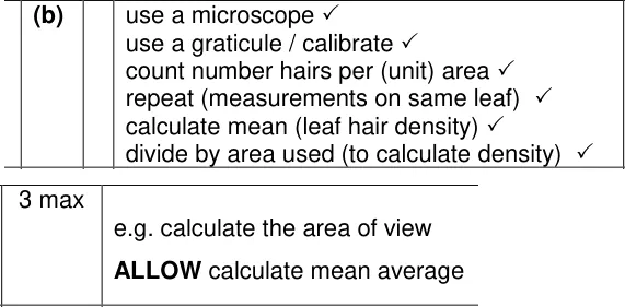
Outline a method to find the density of hairs on the underside of a leaf.
Show the mark scheme
Tap an image to open it full size.
From the examiners' report: Density here means hairs per unit area, not mass over volume. The best answers counted hairs in a measured area under the microscope and divided by that area; many tried to scale up to the whole leaf instead.
Question 7
AS Paper 1 · November 2021 · Q22(a) · 4 marks · Medium
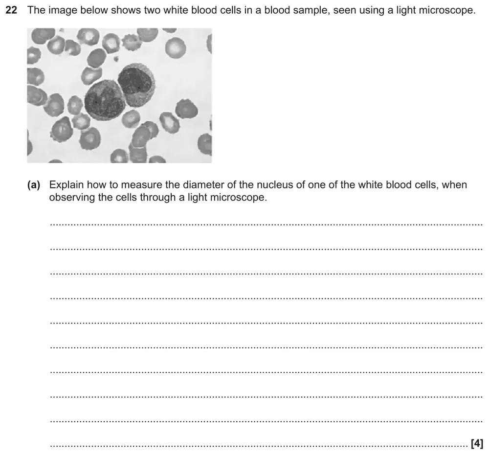
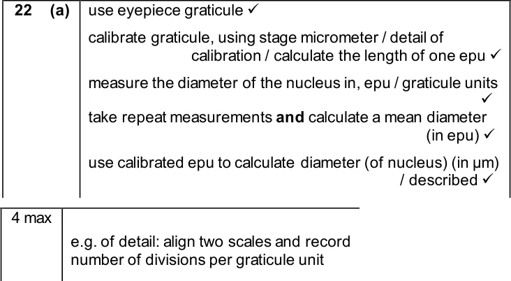
Explain how to measure the diameter of a white blood cell nucleus with a light microscope.
Show the mark scheme
Tap an image to open it full size.
From the examiners' report: There is no examiners' comment for this part. Four marks means four steps in order: graticule, calibrate with a stage micrometer, measure in eyepiece units and repeat, then convert to µm.
Question 8
AS Paper 2 · June 2024 · Q5(a)(ii) · 2 marks · Hard
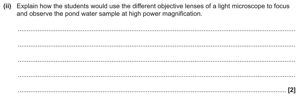
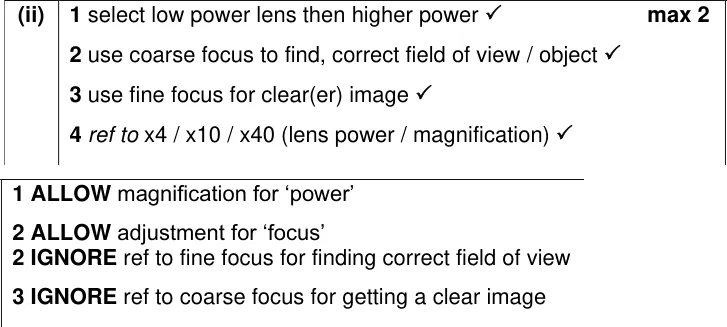
Explain how to use the objective lenses to focus on pond water at high power.
Show the mark scheme
Tap an image to open it full size.
From the examiners' report: Many students said to start on the lowest power lens but couldn't explain focusing. The lenses magnify; the coarse and fine focus knobs focus. Use coarse focus to find the cells, then fine focus to sharpen them.
Staining
Question 9
AS Paper 1 · June 2025 · Q26(b) · 2 marks · Medium
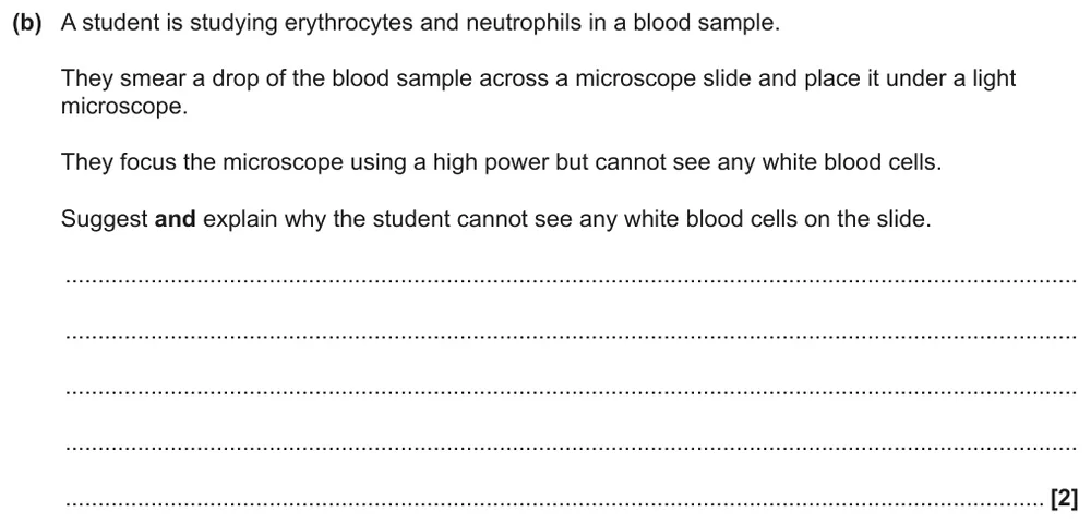
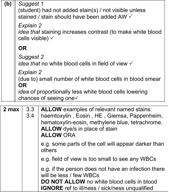
Suggest and explain why a student can't see any white blood cells in an unstained smear.
Show the mark scheme
Tap an image to open it full size.
From the examiners' report: Many said no stain had been added but didn't explain that a stain increases contrast. White blood cells are not too small for a light microscope, and saying the blood has 'no' white cells was not credited.
Question 10
A level Paper 3 · June 2022 · Q2(c) · 2 marks · Medium
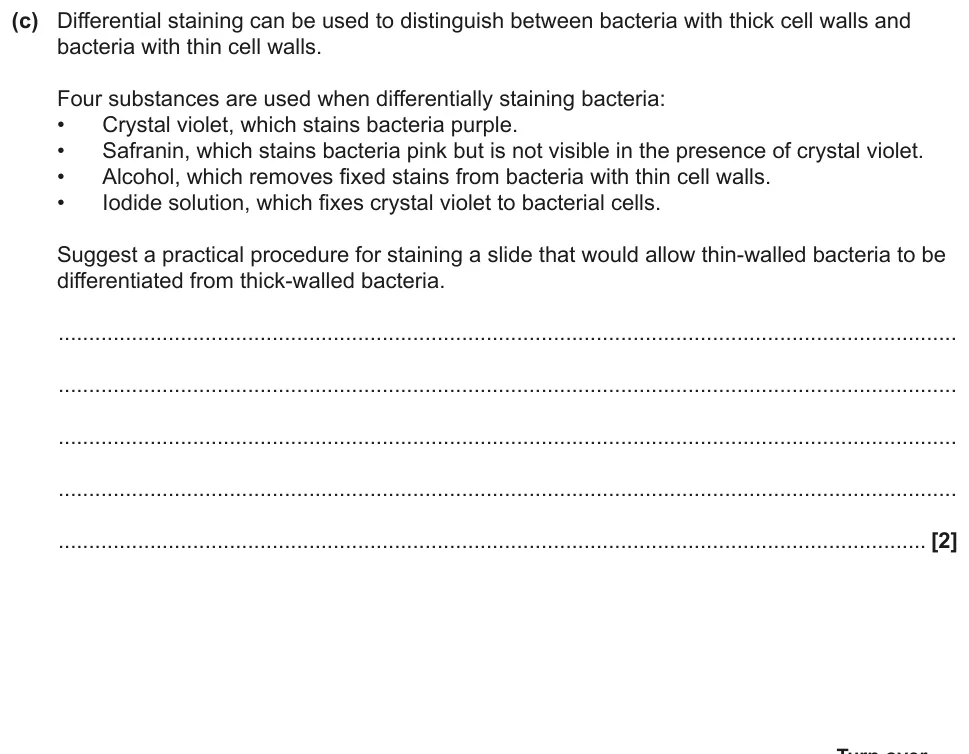
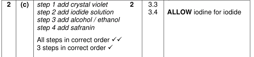
Put four substances in order to differentially stain thick- and thin-walled bacteria.
Show the mark scheme
Tap an image to open it full size.
From the examiners' report: Use every detail you are given. Add crystal violet and iodide one after the other, not together, and finish with safranin, or thin-walled bacteria won't be visible.
Question 11
A level Paper 2 · June 2025 · Q16(a)(iii) · 2 marks · Hard
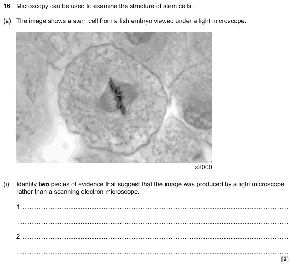
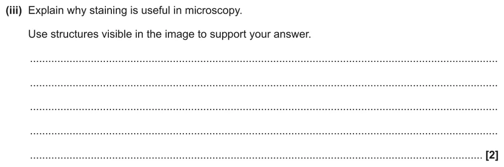
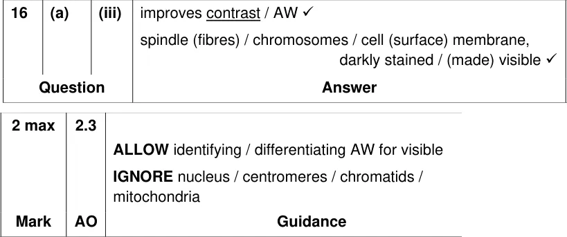
Explain why staining is useful, using structures visible in a light micrograph of a dividing cell.
Information from earlier in this question:
Show the mark scheme
Tap an image to open it full size.
From the examiners' report: Few students used the word contrast. Then name structures you can actually see in the image: there is no nucleus in a dividing cell, and an animal cell has a cell surface membrane, not a cell wall.
Light or electron microscope?
Question 12
AS Paper 2 · November 2020 · Q1(a)(i) · 3 marks · Medium
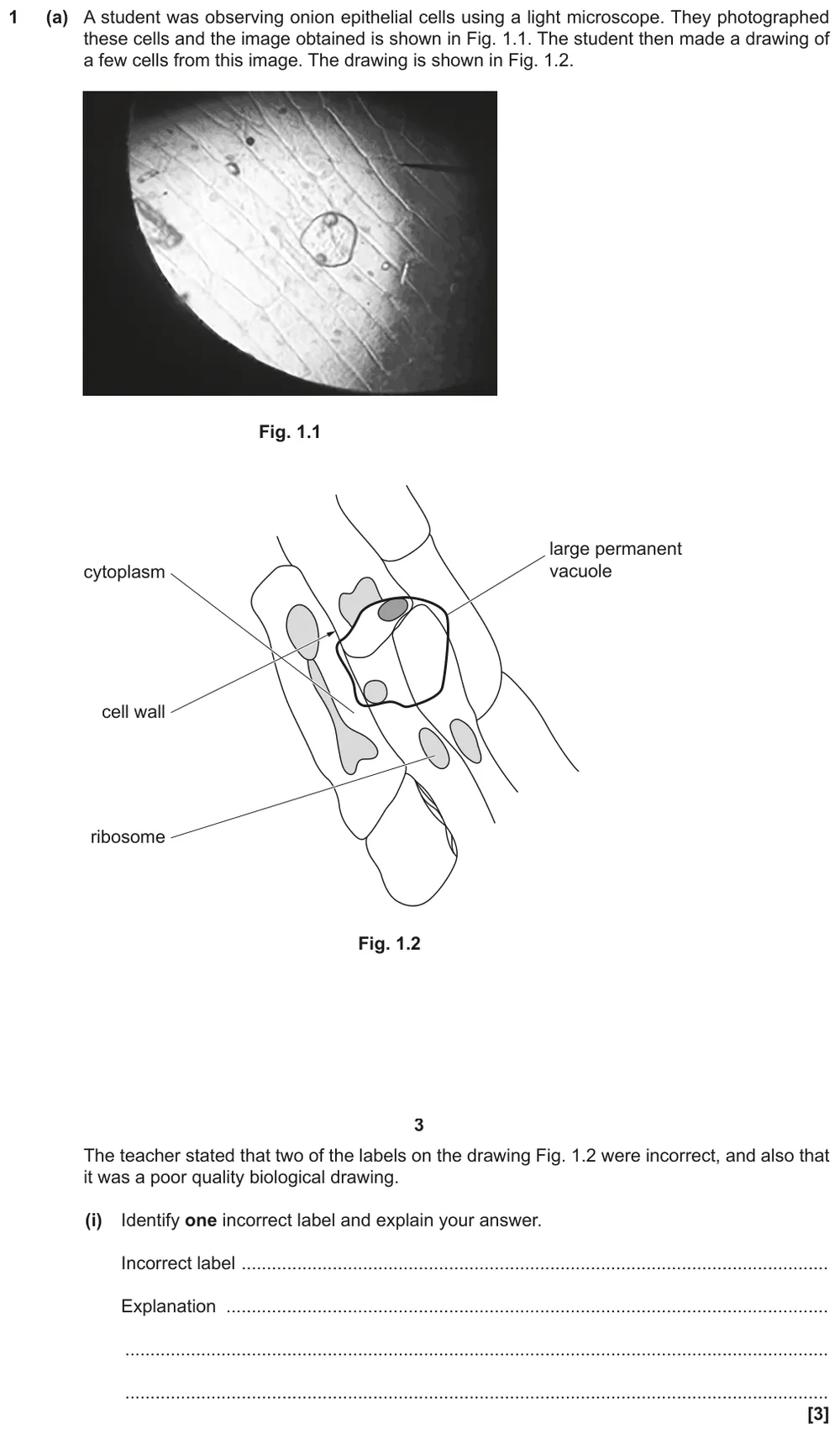
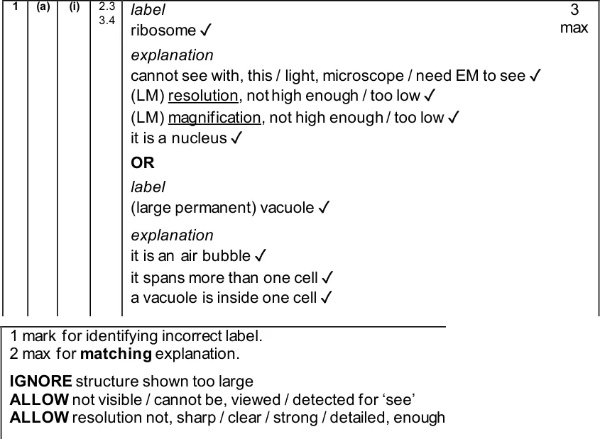
Identify an incorrect label on a drawing of onion cells and explain why it is wrong.
Show the mark scheme
Tap an image to open it full size.
From the examiners' report: The examiners' report has no specific comment on this part. For full marks name the wrong label, then explain it: a light microscope's resolution is too low to see ribosomes, so the structure is the nucleus.
Question 13
AS Paper 1 · June 2022 · Q25(b)(ii) · 3 marks · Medium
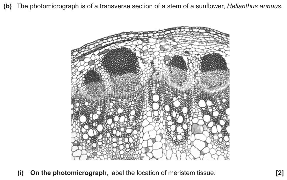
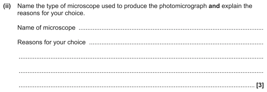
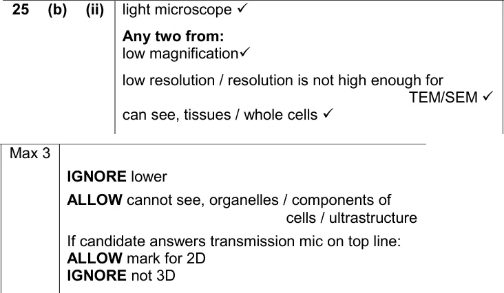
Name the microscope used for a photomicrograph of a sunflower stem and justify your choice.
Information from earlier in this question:
Show the mark scheme
Tap an image to open it full size.
From the examiners' report: Many students chose an electron microscope and claimed organelles were visible. Use the evidence in front of you: whole tissues and cells, low magnification and low resolution, and no organelles.
Scientific drawings
Question 14
AS Paper 2 · June 2024 · Q5(b) · 2 marks · Easy
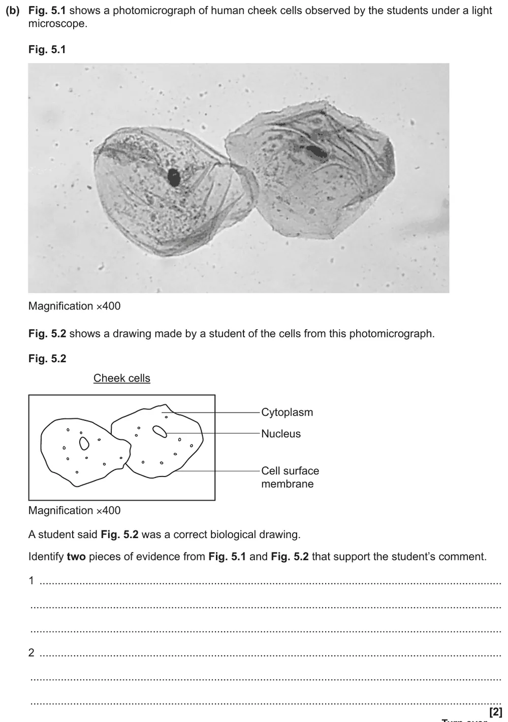
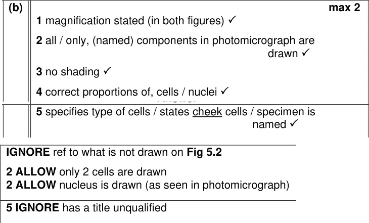
Give two pieces of evidence that a drawing of cheek cells is a correct biological drawing.
Show the mark scheme
Tap an image to open it full size.
From the examiners' report: Most students spotted no shading and the stated magnification. Use evidence from both figures, such as the proportions matching the photo, rather than listing drawing rules.
Question 15
A level Paper 2 · June 2024 · Q17(b)(ii) · 4 marks · Easy
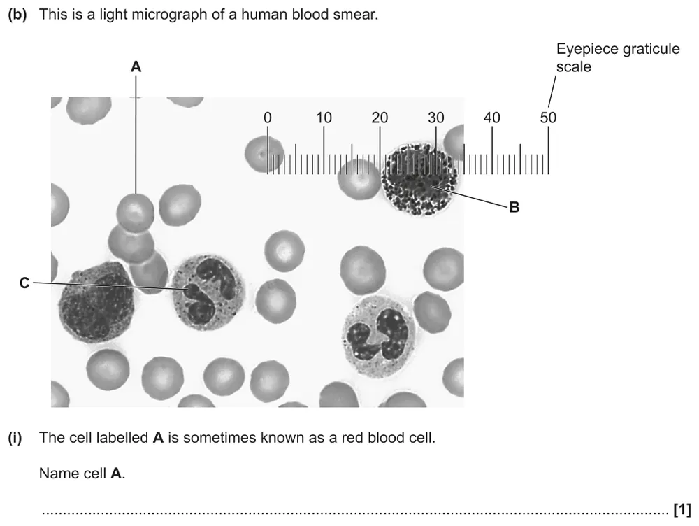
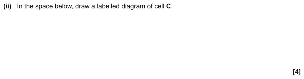
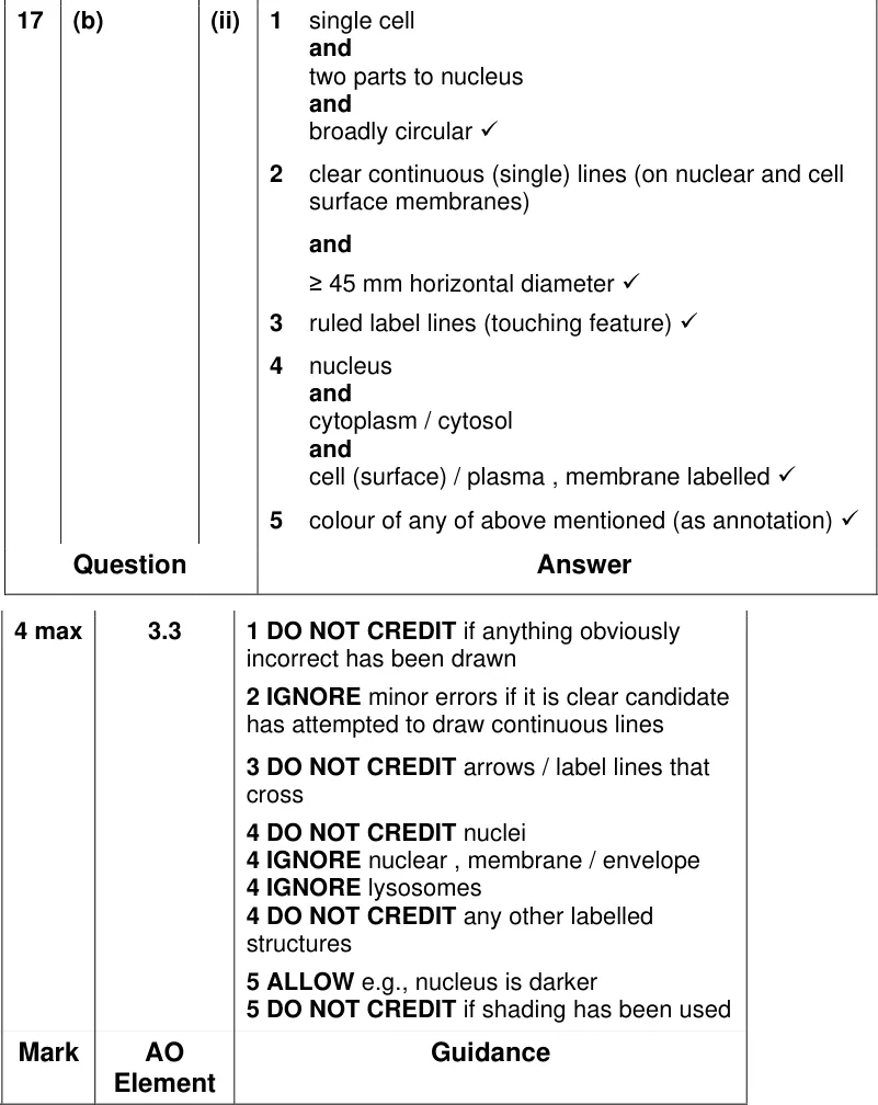
Draw a labelled diagram of a white blood cell from a blood smear.
Information from earlier in this question:
Show the mark scheme
Tap an image to open it full size.
From the examiners' report: Most drawings were a good size with clear single lines. Marks were lost for drawing only one lobe of the nucleus, labelling the lobes as 'nuclei', and adding mitochondria or ER that can't be seen.
Question 16
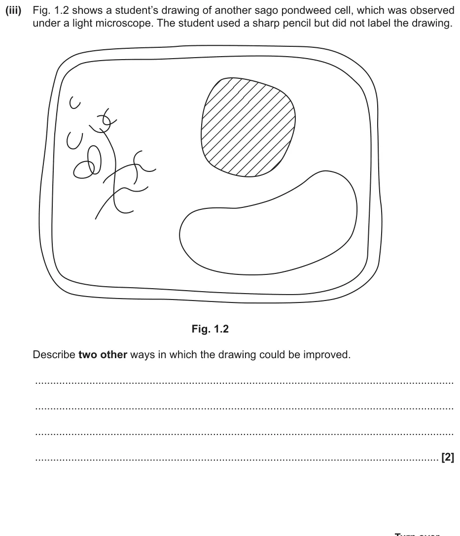
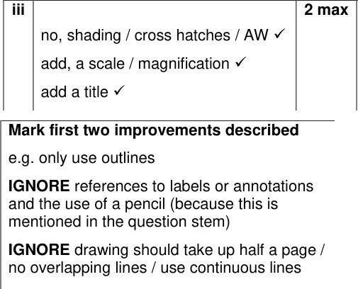
A level Paper 3 · June 2019 · Q1(a)(iii) · 2 marks · Medium
Describe two ways to improve a student's shaded drawing of a pondweed cell.
Show the mark scheme
Tap an image to open it full size.
From the examiners' report: Most students said to remove the shading or add a scale. Labels and a sharp pencil gained nothing because the question already mentions them, so read the stem first.
Question 17
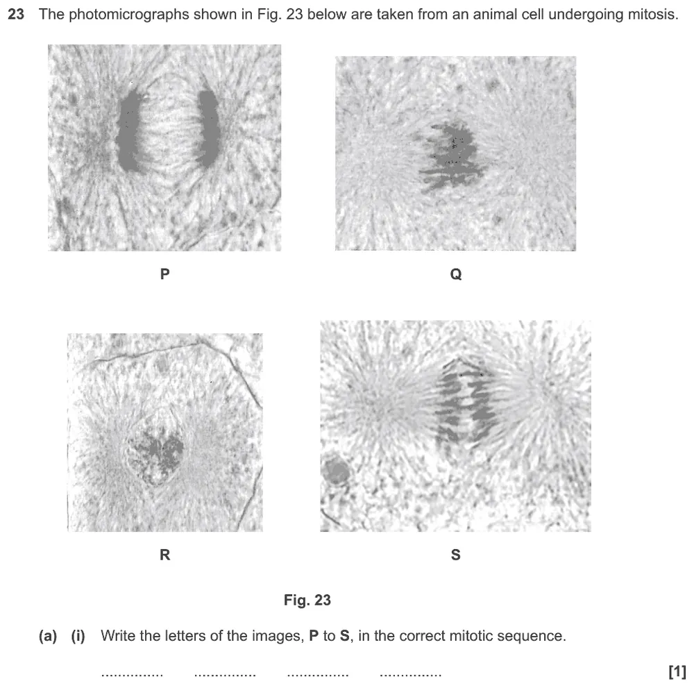
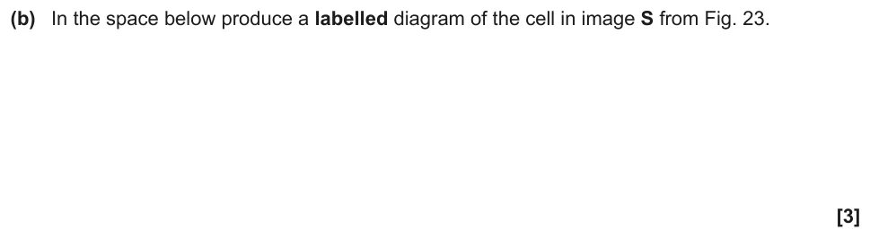
AS Paper 1 · June 2019 · Q23(b) · 3 marks · Hard
Produce a labelled diagram of an animal cell in anaphase from a photomicrograph.
Information from earlier in this question:
Show the mark scheme
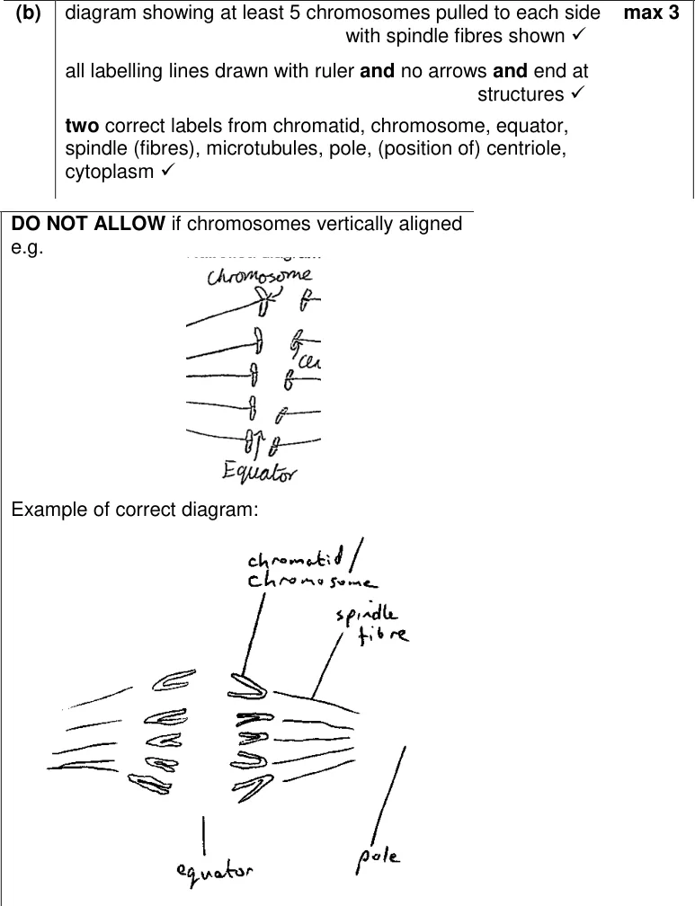
Tap an image to open it full size.
From the examiners' report: The most common error was drawing fewer than five chromosomes. Others drew label lines freehand or not touching the structure, or drew metaphase instead of anaphase. Draw what is in the photo.
Where students lose marks (from OCR examiners' reports and mark schemes)
Each point below is followed by wording that gained marks.
Lower the cover slip at an angle: Asked to improve a staining method, many wrote about gloves, aseptic technique or pressing on the cover slip to remove air bubbles.
✔ Gains credit: "Lower cover slip at an angle / use mounted needle" and "use blotting paper to remove excess stain / pull stain through".
One drop, then a cover slip: For pond water, students suggested a smear, which dries the sample, or described the cover slip as 'another slide'.
✔ Gains credit: "Use dropper / pipette to place a drop onto slide", "place coverslip over the sample", "ensuring there are no air bubbles".
Calibrate the eyepiece graticule: Measuring a cell needs two pieces of kit and a calibration step; answers that just say 'use a graticule' lose marks.
✔ Gains credit: "Eyepiece graticule" and "stage micrometer": "calibrate graticule using stage micrometer", "measure in epu", "take repeat measurements and calculate a mean".
The knobs focus, the lenses magnify: Many students said the objective lens focuses the image, or talked about changing the resolution.
✔ Gains credit: "Select low power lens then higher power", "use coarse focus to find the object", "use fine focus for clearer image".
A stain increases contrast: Students said a stain had not been added but didn't explain why it matters, and some said white blood cells are too small for a light microscope.
✔ Gains credit: "Staining increases contrast (to make white blood cells visible)"; "spindle fibres / chromosomes darkly stained".
Stains go on one at a time: In differential staining of bacteria, many applied crystal violet and iodine together, or left out safranin.
✔ Gains credit: "Add crystal violet, then iodide solution, then alcohol, then safranin", all in the correct order.
Use the evidence for a light microscope: Many claimed an electron microscope was used and that organelles were visible in a light photomicrograph.
✔ Gains credit: "Light microscope": "low magnification", "low resolution", "can see tissues / whole cells", organelles not visible.
Draw only what you can see: Drawings added mitochondria, ER or spindle fibres that were not visible, showed too few chromosomes, or used freehand label lines with arrowheads.
✔ Gains credit: "Clear continuous lines", "ruled label lines touching the feature", no shading, and only the structures visible in the image.
Download the questions
Download the PDF pack (17 questions, mark schemes at the end)
Keep practising
- OCR A magnification and microscopes quiz
- Arithmetic and Numerical Computation (M0): OCR A Level Biology exam questions by topic
- PAG 5 Colorimeter and Potometer: OCR A Level Biology exam questions by topic
- OCR A Level Biology practical skills quiz
- Cell Structure (2.1.1): OCR A Level Biology exam questions by topic
- OCR A Level Biology masterclasses
Questions, mark schemes and examiners' report extracts © OCR.