

This page puts together the last two topics of Option B, Human Musculoskeletal Anatomy: the structure and function of the skeleton (OB2) and joints (OB3). Each has only a handful of past questions, so they work best as one set. You need to name the bones of the axial and appendicular skeleton, describe vertebrae, fractures and postural deformities such as scoliosis, compare joint types, explain osteoarthritis and rheumatoid arthritis, and use levers and antagonistic muscles to explain movement.
These questions are short but exact. The examiners want both parts of fracture treatment (realign, then immobilise), the joint type named correctly (the elbow is a hinge, not a ball and socket), the immune system attacking antigens in the joint for rheumatoid arthritis, and the position of the fulcrum, effort and load when you name a lever.
I don't teach the Eduqas specification myself, so for one-to-one help I recommend Sam. He teaches Eduqas A level Biology at a top-rated sixth form college and is very popular with his students. You can ask about lessons with Sam on WhatsApp using the link at the bottom of this page.
What you need from GCSE and Year 12
Each question on this page assumes you're secure on these ideas. If any of them feels shaky, look it up before you start.
- The skeleton is made of bones linked at joints, with muscles attached by tendons.
- Muscles can only pull, so they work in pairs.
- Synovial joints have cartilage and fluid to reduce friction.
- Autoimmune diseases happen when the immune system attacks the body's own cells.
Key terms
These words come up in the questions and mark schemes. Examiners expect you to use them exactly.
- Axial skeleton: the skull, vertebral column and rib cage.
- Appendicular skeleton: the limbs and the pectoral and pelvic girdles.
- Simple (closed) fracture: the bone breaks but doesn't pierce the skin.
- Scoliosis: a sideways curve of the spine.
- Osteoarthritis: wear and tear of the articular cartilage, so bone rubs on bone.
- Rheumatoid arthritis: an autoimmune disease that inflames the synovial membrane.
- Hinge joint: allows movement in one plane, like the elbow and knee.
- Antagonistic muscles: a pair of muscles that pull in opposite directions, such as the biceps and triceps.
What these questions cover (Eduqas OB2 and OB3)
The questions test these statements from Option B (Human Musculoskeletal Anatomy) of the Eduqas A level Biology specification (A400). Option questions are in Component 3, and you answer only one of the three options.
- OB2(a) the structure of the appendicular and axial skeleton (pectoral and pelvic girdles, forelimb and hind limb)
- OB2(b) the types of fractures that can occur in the skeleton and their causes
- OB2(c) the structure and function of the vertebral column, general structure of a vertebra and the differences between cervical, thoracic and lumbar vertebrae and be able to rel
- OB2(d) postural deformities, including scoliosis, their causes and treatment
- OB2(e) the functions of the skeleton, including support, muscle attachment, protection, production of red blood cells and as a store of calcium
- OB3(a) the different types of joint in the human
- OB3(b) the cause and treatment of osteoarthritis
- OB3(c) the cause and treatment of rheumatoid arthritis
- OB3(d) the concept of joints acting as levers including examples of 1st order, 2nd order and 3rd order levers
- OB3(e) the structure of a typical synovial joint including the roles of cartilage, synovial membrane, synovial fluid, ligaments
- OB3(f) the antagonistic muscle action in the human forelimb including the role of tendons
How to use this page
Write your answer on paper before you open the mark scheme. Allow about 1.2 minutes per mark: the 20 questions are worth 38 marks, so about 46 minutes in total. Then mark your answer strictly against the mark scheme and read the examiners' tip underneath it. If you'd rather print the questions, download the PDF pack.
The skeleton
Question 1
A level Component 3 · June 2025 · Q8(a)(i) · 1 mark · Easy
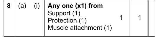
State one function of the axial skeleton.

Show the mark scheme
Tap an image to open it full size.
From the examiners' report: Give one clear function, such as support, protection or muscle attachment.
Question 2
A level Component 3 · June 2019 · Q9(a)(i) · 1 mark
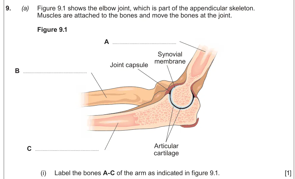
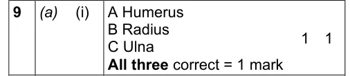
Label the humerus, radius and ulna on a diagram of the elbow.
Show the mark scheme
Tap an image to open it full size.
From the examiners' report: All three must be correct for the mark, so learn which forearm bone is on the thumb side (the radius).
Question 3
A level Component 3 · Autumn 2020 · Q9(a)(i) · 2 marks
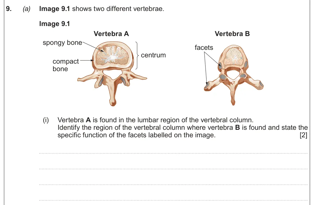
Identify which region of the spine a vertebra comes from and state the function of its facets.
Show the mark scheme

Tap an image to open it full size.
From the examiners' report: Most identified the thoracic vertebra; fewer saw that its facets are where the ribs articulate.
Question 4
A level Component 3 · Autumn 2020 · Q9(b)(iii) · 2 marks
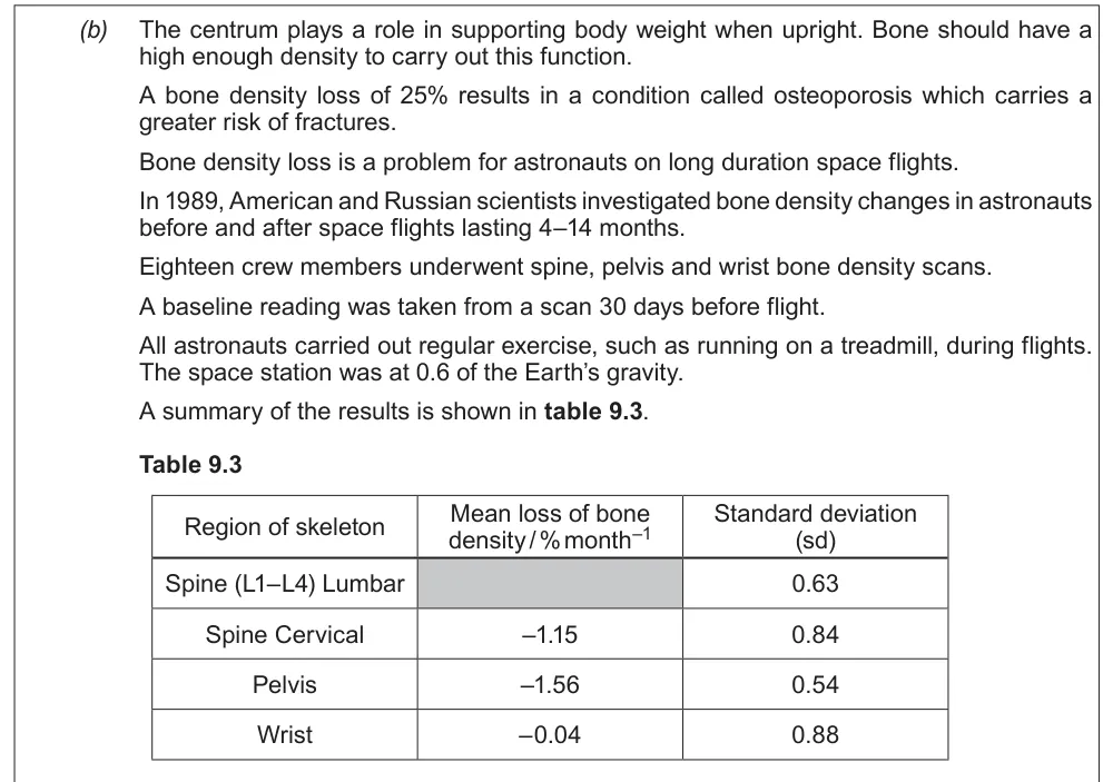
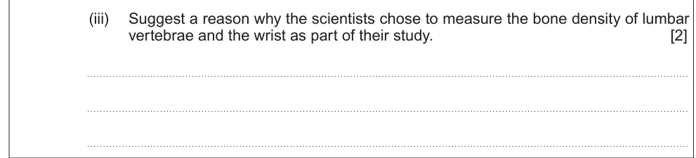
Suggest why scientists measured bone density in the lumbar vertebrae and the wrist.
Information from earlier in this question:
Show the mark scheme

Tap an image to open it full size.
From the examiners' report: Explain both choices: lumbar vertebrae bear the body's weight, while the wrist bears little and acts as a comparison.
Fractures and posture
Question 5
A level Component 3 · June 2017 · Q8(d)(i) · 1 mark
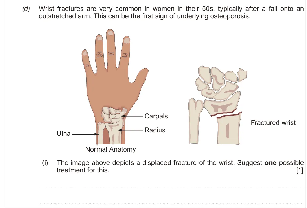
Suggest a treatment for a displaced fracture of the wrist.
Show the mark scheme

Tap an image to open it full size.
From the examiners' report: A displaced fracture needs two steps: realign the bone, then immobilise it in a cast or splint.
Question 6
A level Component 3 · June 2023 · Q8(b) · 3 marks · Hard
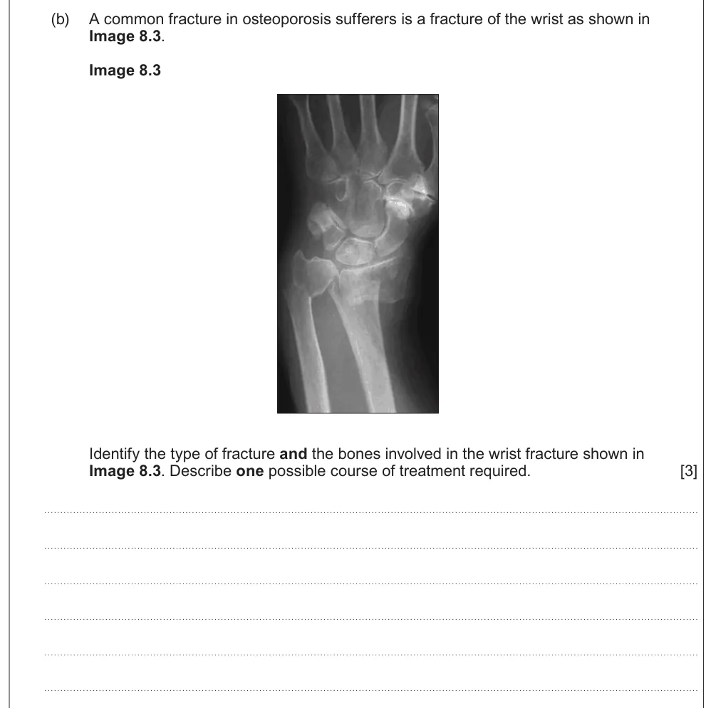
Identify a wrist fracture and the bones involved, and describe a treatment.
Show the mark scheme

Tap an image to open it full size.
From the examiners' report: Name the bones as well as the fracture type; that part caught some students out.
Question 7
A level Component 3 · June 2018 · Q9(c)(ii) · 1 mark · Hard
Suggest why surgery with screws or plates leads to faster recovery than bed rest.
Show the mark scheme

Tap an image to open it full size.
From the examiners' report: Being mobile sooner promotes healing and cuts complications such as blood clots and muscle wasting.
Question 8
A level Component 3 · June 2022 · Q8(d)(i) · 1 mark
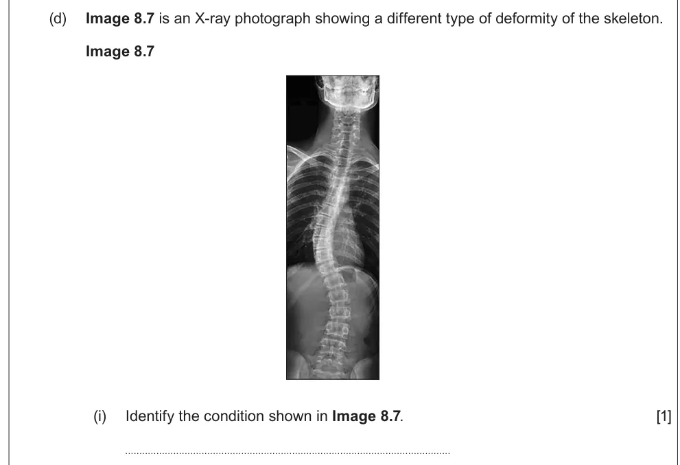
Identify the spinal deformity shown in an X-ray.
Show the mark scheme

Tap an image to open it full size.
From the examiners' report: This is recall: it's scoliosis.
Question 9
A level Component 3 · June 2022 · Q8(d)(ii) · 1 mark
State one potential cause of scoliosis.

Show the mark scheme

Tap an image to open it full size.
From the examiners' report: Make the cause clear, such as an inherited gene mutation or a problem with the muscles.
Question 10
A level Component 3 · June 2025 · Q8(a)(ii) · 2 marks
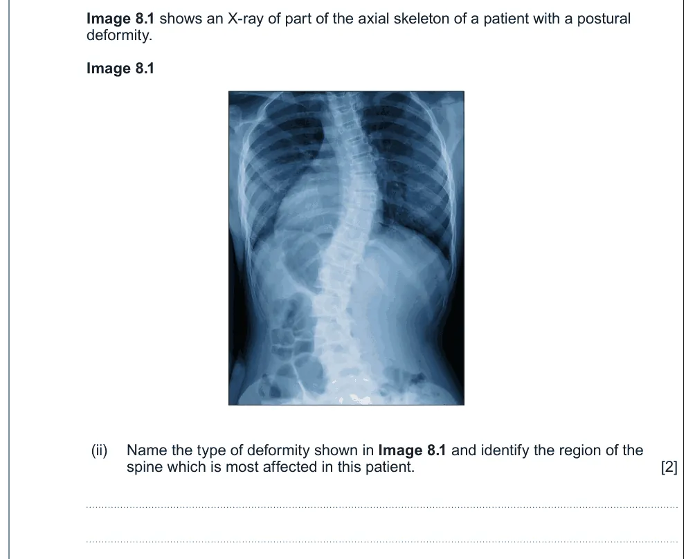
Name the deformity in an X-ray and the region of the spine most affected.
Show the mark scheme

Tap an image to open it full size.
From the examiners' report: Scoliosis, then use the X-ray to name the region of the spine.
Joints and arthritis
Question 11
A level Component 3 · June 2017 · Q8(e)(i) · 4 marks
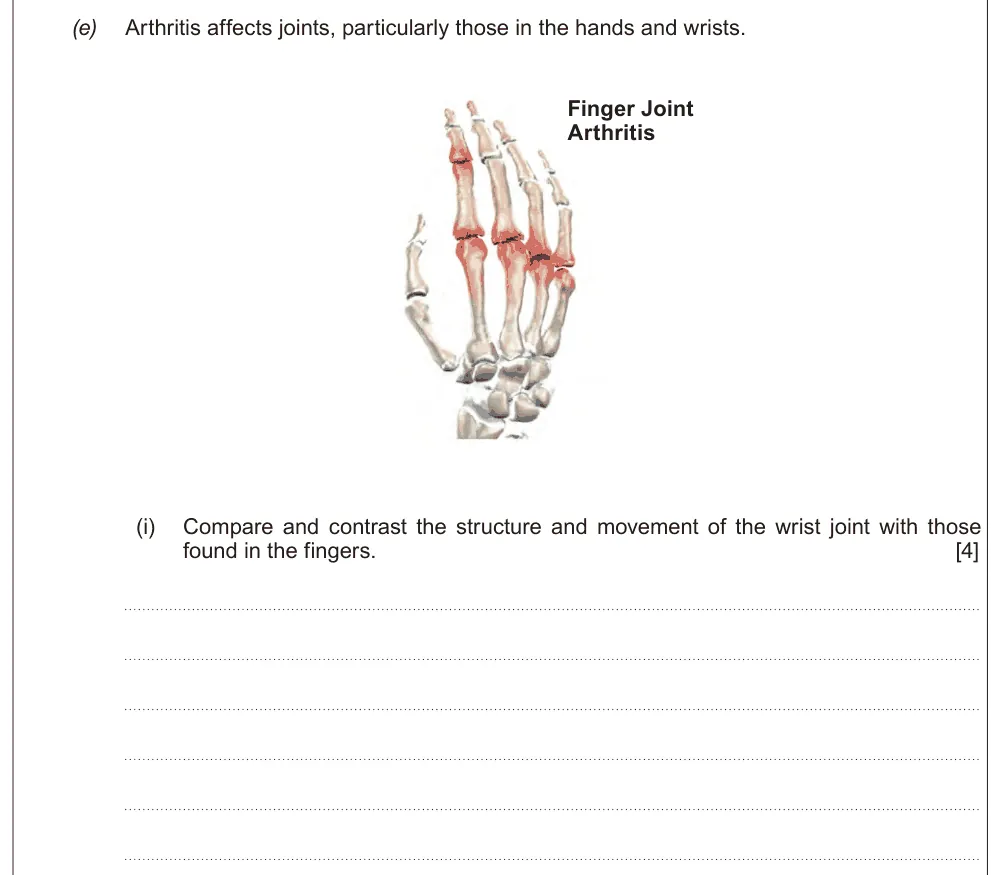
Compare the structure and movement of the wrist joint with the finger joints.
Show the mark scheme

Tap an image to open it full size.
From the examiners' report: Name both joint types and the movement each allows: the wrist is a gliding joint moving in more than one plane, fingers are hinge joints.
Question 12
A level Component 3 · June 2019 · Q9(a)(ii) · 2 marks
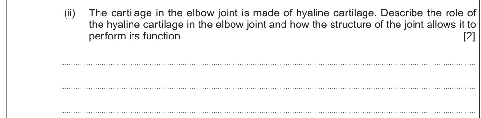
Describe the role of hyaline cartilage in the elbow and how the joint's structure allows its function.
Show the mark scheme

Tap an image to open it full size.
From the examiners' report: The elbow is a hinge joint moving in one plane. Many wrongly called it a ball and socket joint.
Question 13
A level Component 3 · Autumn 2021 · Q8(d)(i) · 2 marks
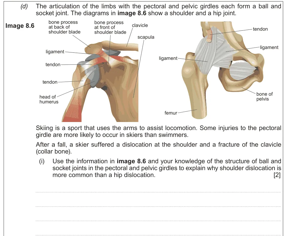
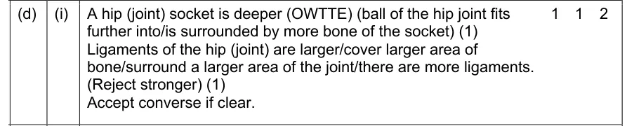
Explain why shoulder dislocation is more common than hip dislocation.
Show the mark scheme
Tap an image to open it full size.
Question 14
A level Component 3 · Autumn 2020 · Q9(c)(i) · 2 marks
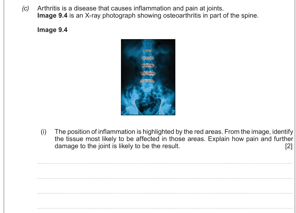
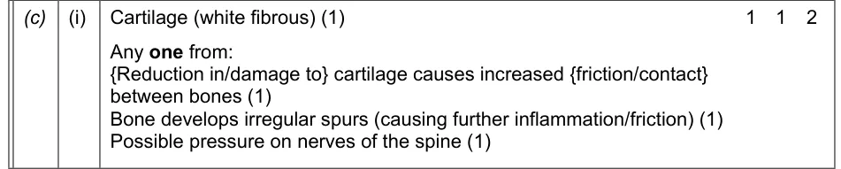
Identify the tissue affected by inflammation in a spine scan and explain how pain and further damage result.
Show the mark scheme
Tap an image to open it full size.
From the examiners' report: Name a tissue (cartilage), not a structure, then link cartilage loss to bone rubbing on bone.
Question 15
A level Component 3 · Autumn 2020 · Q9(c)(ii) · 1 mark
Explain the cause of inflammation in rheumatoid arthritis.

Show the mark scheme

Tap an image to open it full size.
From the examiners' report: Say that the immune system recognises proteins in the joint as antigens. "Recognises tissue as foreign" lacked the detail needed.
Question 16
A level Component 3 · June 2024 · Q8(c)(ii)II · 2 marks
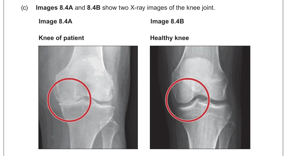
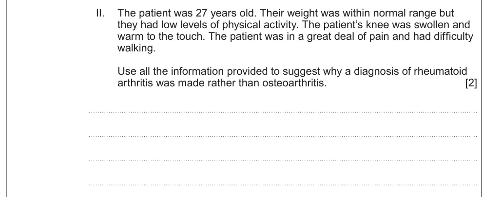
Use the patient's details to explain why rheumatoid arthritis was diagnosed instead of osteoarthritis.
Information from earlier in this question:
Show the mark scheme

Tap an image to open it full size.
From the examiners' report: Finish each point: the patient was young and inactive, so it's unlikely to be wear and tear, and warm swelling means inflammation.
Question 17
A level Component 3 · June 2025 · Q8(d)(ii) · 3 marks · Easy
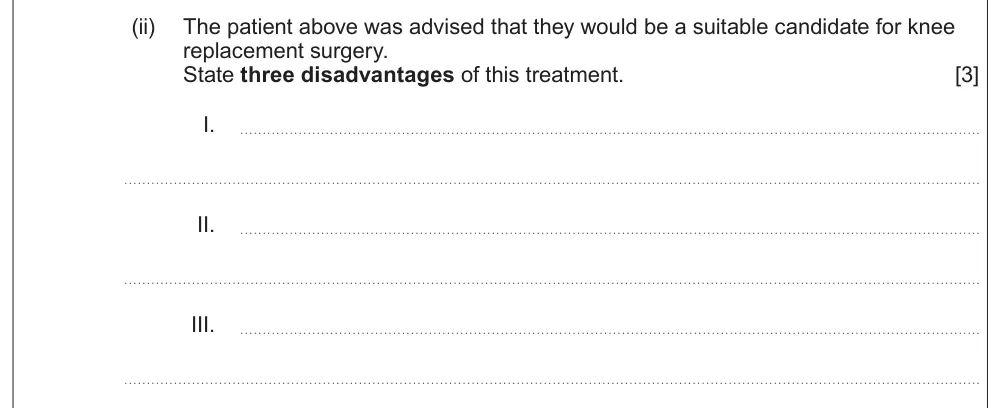
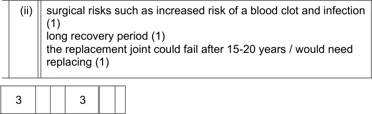
State three disadvantages of knee replacement surgery.
Show the mark scheme
Tap an image to open it full size.
From the examiners' report: Give three different points: surgical risks such as clots or infection, a long recovery, and the joint wearing out after 15–20 years.
Levers and muscles
Question 18
A level Component 3 · June 2018 · Q9(d)(i) · 1 mark
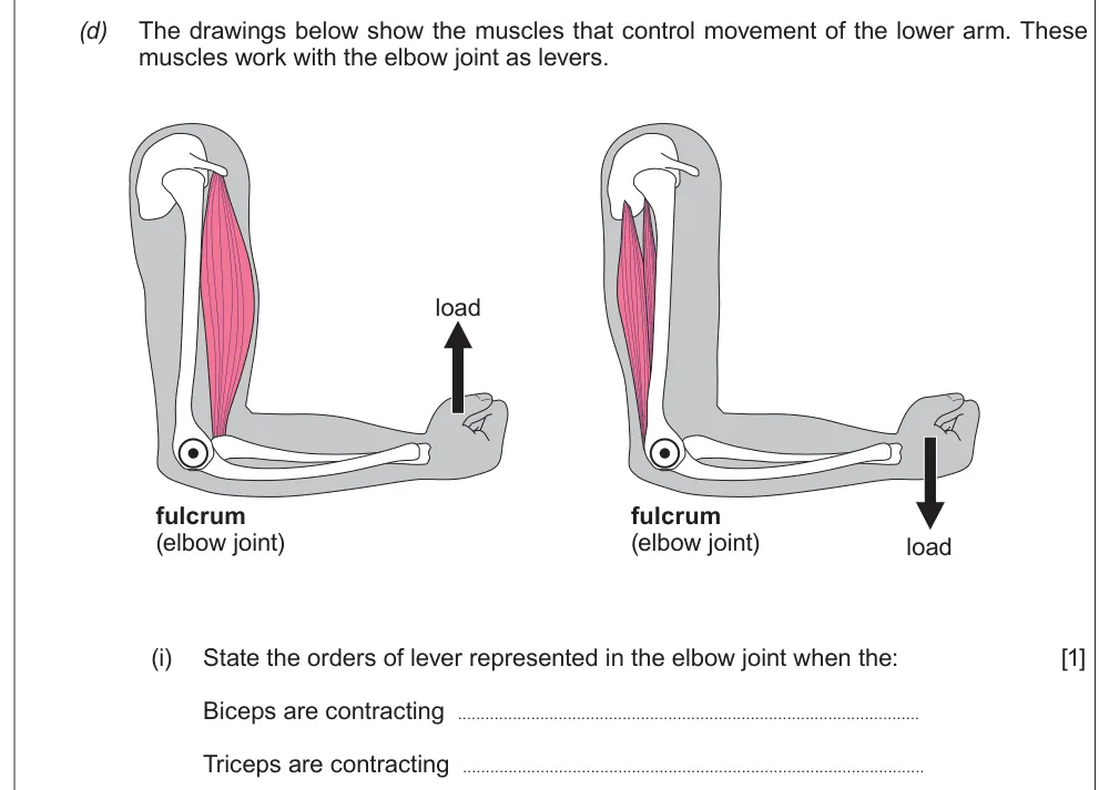
Name the order of lever when the biceps and when the triceps contract.
Show the mark scheme

Tap an image to open it full size.
From the examiners' report: Biceps flexing is a third order lever; triceps extending is a first order lever.
Question 19
A level Component 3 · June 2018 · Q9(d)(ii) · 2 marks
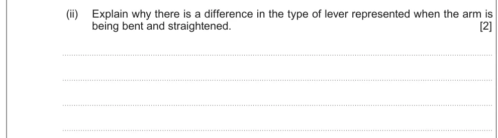
Explain why the lever type differs when the arm is bent and straightened.
Show the mark scheme

Tap an image to open it full size.
From the examiners' report: Say where each part sits: the effort is in the middle when the biceps contracts, the fulcrum when the triceps does.
Question 20
A level Component 3 · June 2023 · Q8(c)(iii) · 4 marks
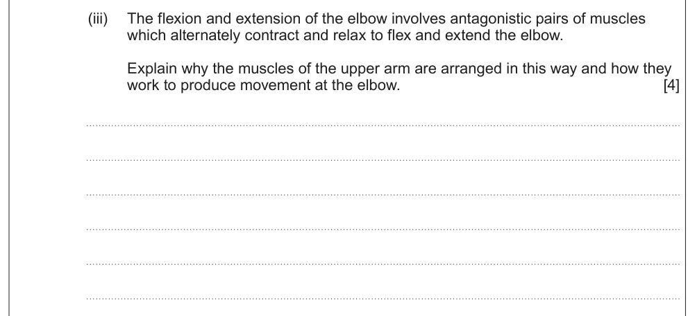
Explain why the upper arm muscles work as an antagonistic pair and how they move the elbow.
Show the mark scheme
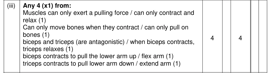
Tap an image to open it full size.
From the examiners' report: Start with the key fact that muscles can only pull, then say which muscle contracts and which relaxes for each movement.
Where students lose marks (from Eduqas examiners' reports and mark schemes)
Each point below is followed by wording that gained marks.
Realign, then immobilise: Many gave only the second part of the treatment or an unsuitable way of immobilising, such as nailing the bones together.
✔ Gains credit: "Realignment + immobilisation with cast or splint".
Moving early helps healing: Few students saw that getting the patient mobile aids recovery, although it's in the specification.
✔ Gains credit: "Patient will be mobile more quickly and this promotes healing / reduced complications with blood clots".
The elbow is a hinge: Many wrongly called the elbow a ball and socket joint.
✔ Gains credit: "It is a hinge joint", so it moves in one plane only (NOT one direction).
Antigens in the joint: Saying the immune system recognises tissue as foreign wasn't detailed enough.
✔ Gains credit: "Lymphocytes recognise proteins in the joint tissue as antigens".
Rib facets on a thoracic vertebra: Students identified the thoracic vertebra but missed what the facets are for.
✔ Gains credit: "Thoracic; articulation with ribs".
Name a tissue, not a structure: No marks were given for structures that aren't tissues.
✔ Gains credit: "Cartilage": damage "causes increased friction between bones".
Muscles only pull: Some couldn't clearly explain how the muscles bring about movement at the elbow.
✔ Gains credit: "Muscles can only exert a pulling force"; "when biceps contracts, triceps relaxes".
Download the questions
Download the PDF pack (20 questions, mark schemes at the end)
Keep practising
- Skeletal Tissues (Option B1): Eduqas A Level Biology exam questions by topic
- Eduqas A level Biology past papers, mark schemes and examiners' reports
- Eduqas A level Biology grade boundaries
- Ask about Eduqas A level Biology lessons with Sam on WhatsApp
Questions, mark schemes and examiners' report extracts © WJEC CBAC Ltd (Eduqas).