

Skeletal tissues is the first and largest topic in Option B, Human Musculoskeletal Anatomy, in Section B of Component 3. It covers the three types of cartilage, the make-up of compact bone and its Haversian systems, how osteoblasts and osteoclasts remodel bone, rickets, osteomalacia and osteoporosis, and then skeletal muscle: its structure, the sliding filament theory, fast and slow twitch fibres, and where the energy for contraction comes from.
Much of this option is precise recall, so the marks go to the exact word: chondrocytes not osteocytes, Haversian canal, hydroxyapatite and collagen. The examiners also want the reasoning a question asks for, such as why cartilage heals slowly (no blood vessels, so diffusion through the matrix), and they want you to use the graph or micrograph you're given rather than general knowledge.
I don't teach the Eduqas specification myself, so for one-to-one help I recommend Sam. He teaches Eduqas A level Biology at a top-rated sixth form college and is very popular with his students. You can ask about lessons with Sam on WhatsApp using the link at the bottom of this page.
What you need from GCSE and Year 12
Each question on this page assumes you're secure on these ideas. If any of them feels shaky, look it up before you start.
- The skeleton supports the body, protects organs and allows movement with muscles.
- Muscles contract when they receive impulses from motor neurones.
- Aerobic respiration releases more ATP than anaerobic respiration, which makes lactate in muscles.
- Calcium is needed for strong bones and vitamin D for absorbing it.
Key terms
These words come up in the questions and mark schemes. Examiners expect you to use them exactly.
- Chondrocyte: a cartilage cell, sitting in a space in the matrix.
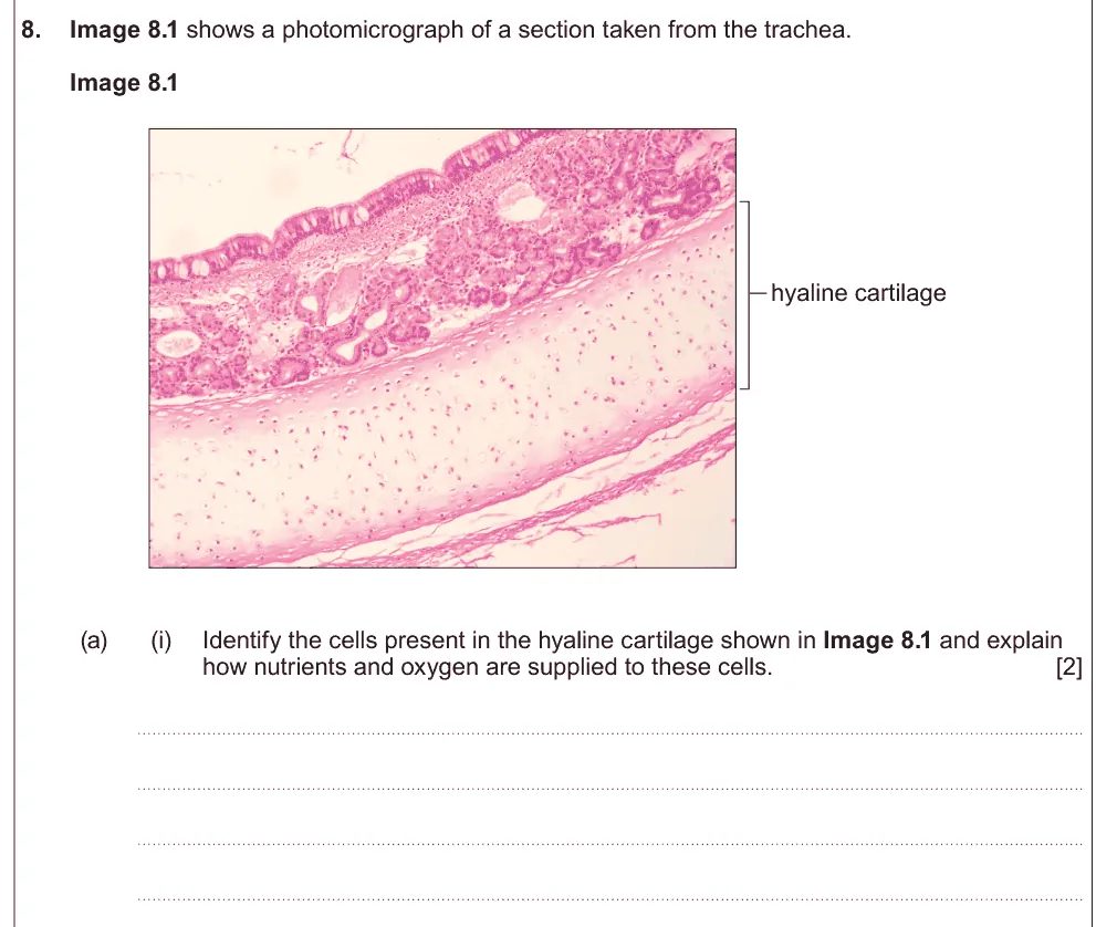
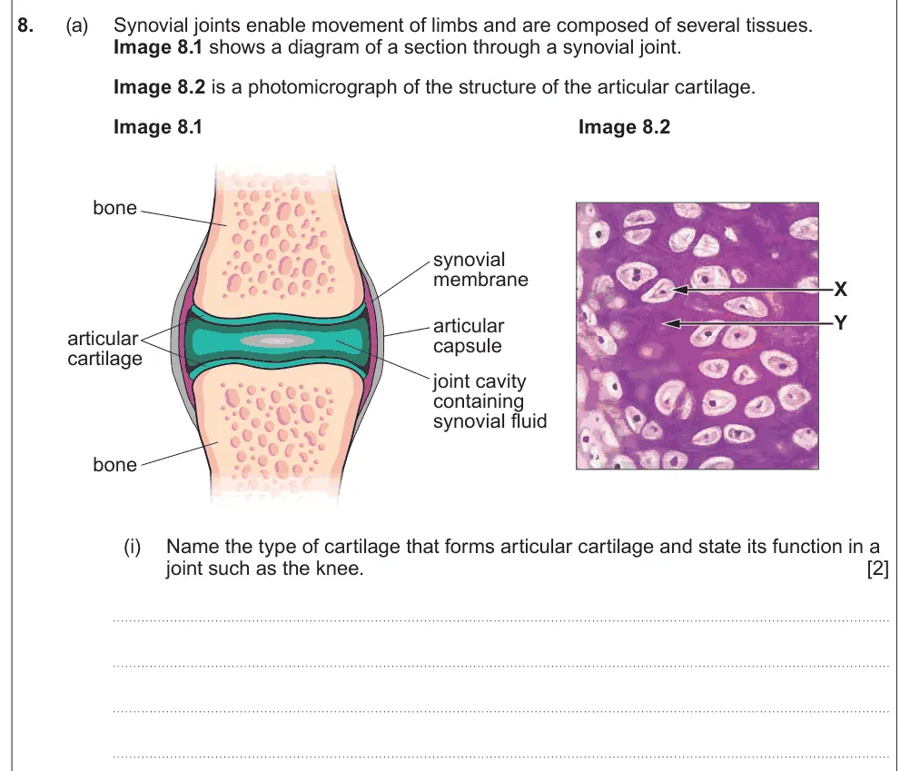
- Hyaline cartilage: firm cartilage with collagen fibres, found on joint surfaces and in the trachea.
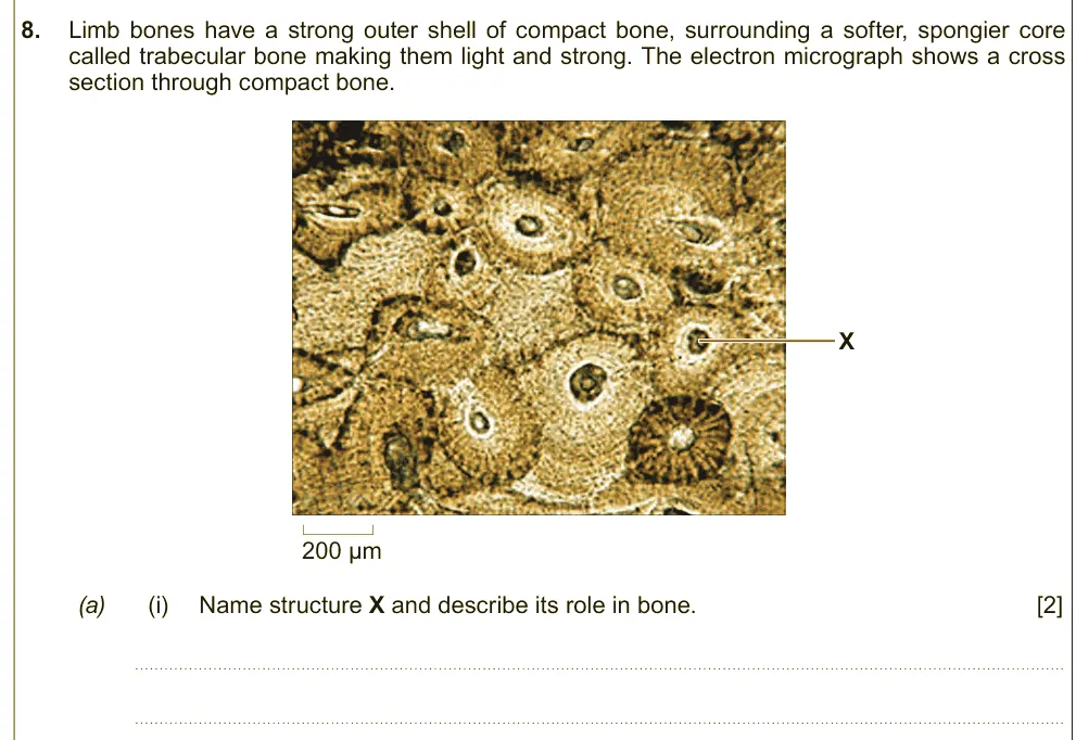
- Haversian system (osteon): a unit of compact bone: rings of matrix around a central Haversian canal.
- Osteoblast: a cell that lays down new bone matrix.
- Osteoclast: a cell that breaks bone down (resorption).
- Sarcomere: the unit of a myofibril between two Z lines.
- Troponin and tropomyosin: proteins on actin that block the myosin binding sites until calcium ions arrive.
- Creatine phosphate: a store in muscle that rapidly regenerates ATP from ADP.
What these questions cover (Eduqas OB1)
The questions test these statements from Option B (Human Musculoskeletal Anatomy) of the Eduqas A level Biology specification (A400). Option questions are in Component 3, and you answer only one of the three options.
- (a) the structure of cartilage including hyaline cartilage, yellow elastic cartilage, white fibrous cartilage
- (b) the components of compact bone, a matrix which is 30% organic (mainly the protein collagen) and 70% inorganic (the main component being hydroxyapatite containing calcium
- (c) the functions of the organic and inorganic components of compact bone (no past question tests this on its own; the bone composition questions use it)
- (d) the functions of osteoblasts and osteoclasts
- (e) the structure and function of the Haversian systems
- (f) rickets and osteomalacia as disorders of bone caused by a calcium or vitamin D deficiency
- (g) the causes, symptoms and treatment of osteoporosis and brittle bone disease
- (h) the structure and ultra-structure of skeletal muscle
- (i) the sliding filament theory to include structure of the thin filaments (actin with two accessory proteins, tropomyosin and troponin) and thick filaments (myosin)
- (j) the differences between ‘fast twitch’ and ‘slow twitch’ muscles
- (k) the effects of anaerobic conditions including the role of creatinine phosphate and the build-up of lactic acid in muscles
- (l) the use of glycogen and protein as the main sources of energy during muscle contraction
How to use this page
Write your answer on paper before you open the mark scheme. Allow about 1.2 minutes per mark: the 20 questions are worth 46 marks, so about 55 minutes in total. Then mark your answer strictly against the mark scheme and read the examiners' tip underneath it. If you'd rather print the questions, download the PDF pack.
Cartilage
Question 1
A level Component 3 · June 2024 · Q8(a)(i) · 2 marks · Easy
Identify the cells in hyaline cartilage and explain how they get nutrients and oxygen.
Show the mark scheme

Tap an image to open it full size.
From the examiners' report: Name the cells as chondrocytes, not osteocytes. Cartilage has no blood vessels, so finish by saying that diffusion happens through the matrix.
Question 2
A level Component 3 · June 2022 · Q8(a)(i) · 2 marks
Name the cartilage that forms articular cartilage and state its function in the knee.
Show the mark scheme

Tap an image to open it full size.
From the examiners' report: Give one function only: it reduces friction so the bones move smoothly. Listing several functions in the hope one is right doesn't work.
Question 3
A level Component 3 · June 2019 · Q9(a)(iii) · 3 marks
Compare hyaline and yellow elastic cartilage and explain the benefit of each.
Show the mark scheme

Tap an image to open it full size.
From the examiners' report: Compare the fibres (collagen in hyaline, elastin in yellow elastic), then link each to its job: firm in joints, flexible in the ear.
Bone
Question 4
A level Component 3 · Autumn 2020 · Q9(a)(iii) · 2 marks
Name one organic and one inorganic compound in bone matrix.

Show the mark scheme

Tap an image to open it full size.
From the examiners' report: Give compounds, not ions or elements: collagen and hydroxyapatite (calcium phosphate).
Question 5
A level Component 3 · June 2017 · Q8(a)(i) · 2 marks · Easy
Name structure X in compact bone and describe its role.
Show the mark scheme

Tap an image to open it full size.
From the examiners' report: It's the Haversian canal, and it carries blood vessels that deliver oxygen and nutrients to the bone cells.
Question 6
A level Component 3 · June 2017 · Q8(b) · 4 marks
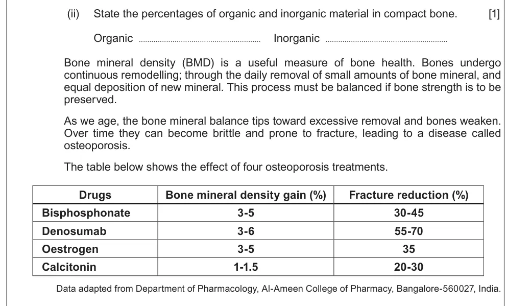
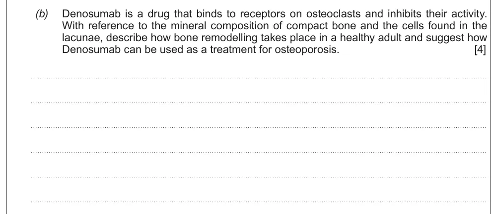
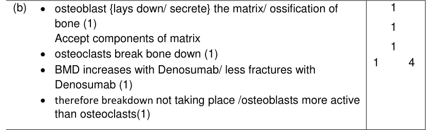
Describe bone remodelling and suggest how denosumab could treat osteoporosis.
Information from earlier in this question:
Show the mark scheme
Tap an image to open it full size.
From the examiners' report: Give the job of both cells, osteoblasts laying down matrix and osteoclasts breaking it down, then use the data on denosumab.
Question 7
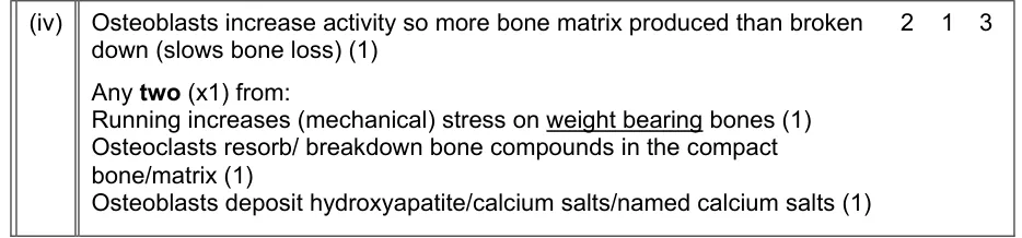
A level Component 3 · Autumn 2020 · Q9(b)(iv) · 3 marks
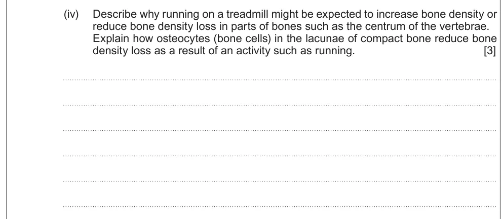
Explain how bone cells reduce bone density loss when someone runs.
Show the mark scheme
Tap an image to open it full size.
From the examiners' report: Bone density depends on the balance of building and breakdown. Link running's mechanical stress to osteoblasts making more matrix than osteoclasts remove.
Question 8
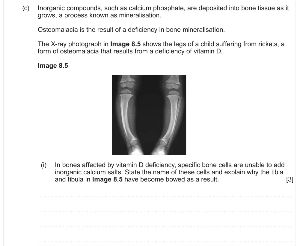
A level Component 3 · June 2022 · Q8(c)(i) · 3 marks
Name the cells affected by vitamin D deficiency and explain why the leg bones become bowed.
Show the mark scheme

Tap an image to open it full size.
From the examiners' report: Osteoblasts can't add calcium salts, so the bones are too soft to support the body's weight.
Question 9
A level Component 3 · June 2019 · Q9(b)(iv) · 1 mark
Name the adult form of rickets and explain why it is less serious in adults.

Show the mark scheme

Tap an image to open it full size.
From the examiners' report: Osteomalacia; adult bones have stopped growing, so they aren't deformed in the same way.
Question 10
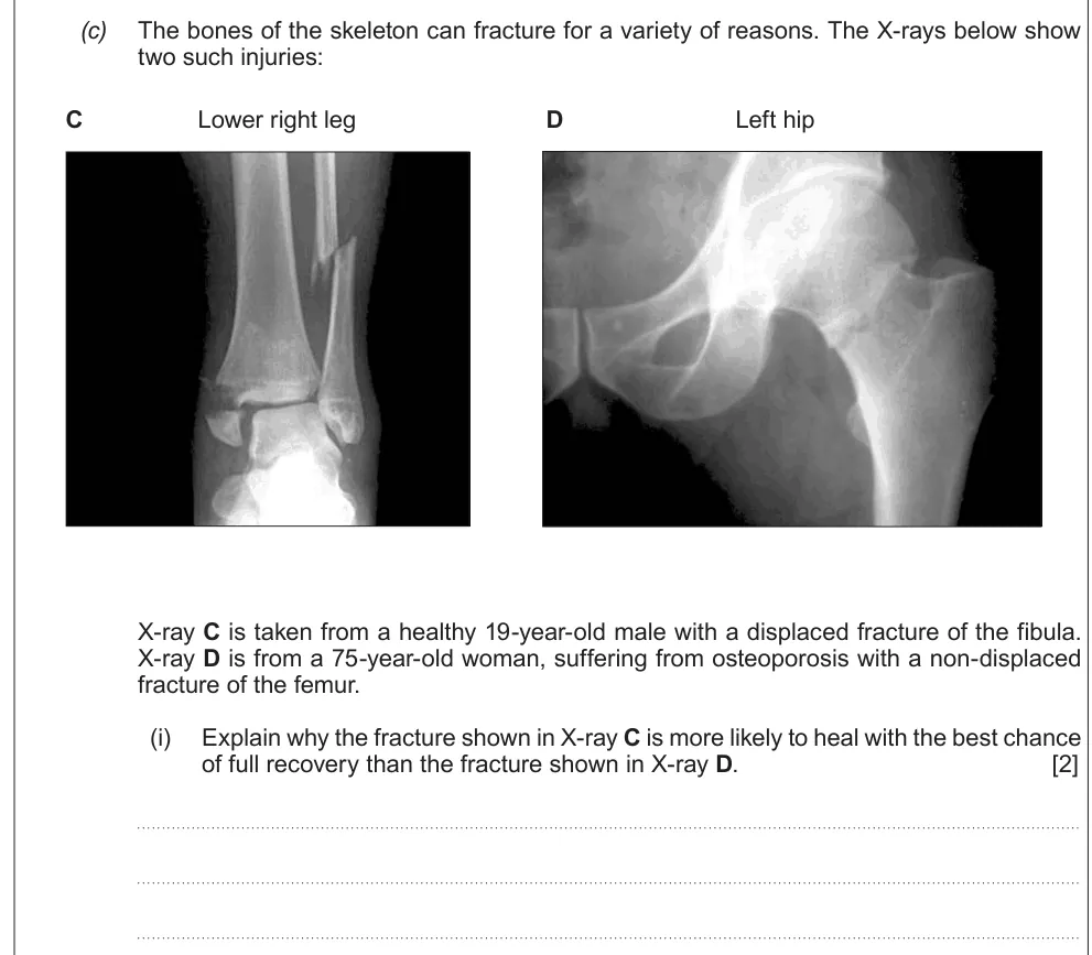
A level Component 3 · June 2018 · Q9(c)(i) · 2 marks
Use two X-rays to explain why one fracture is more likely to heal fully than the other.
Show the mark scheme

Tap an image to open it full size.
From the examiners' report: Use the information given and talk about cell activity: in osteoporosis osteoblasts are less active, so new bone forms more slowly.
Muscle structure and contraction
Question 11
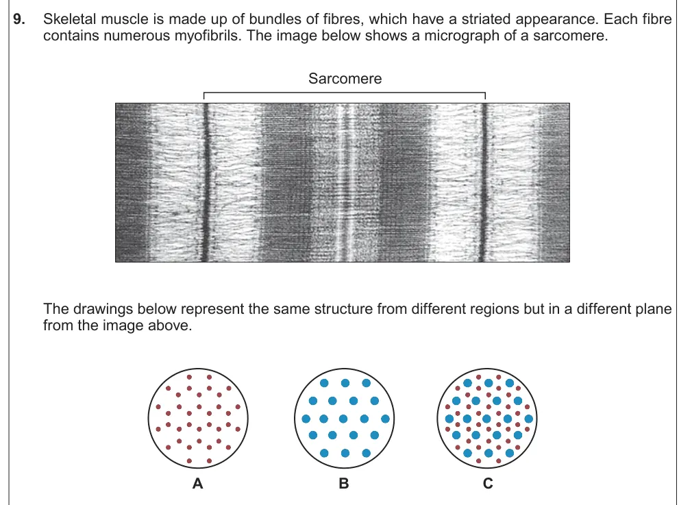
A level Component 3 · June 2018 · Q9(a)(ii) · 3 marks
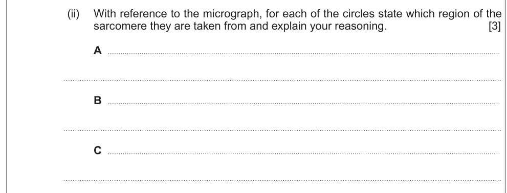
Identify which region of a sarcomere each cross-section comes from and give your reasons.
Information from earlier in this question:
Show the mark scheme

Tap an image to open it full size.
From the examiners' report: The question asks for reasons, so explain each one: actin only, myosin only, or overlapping filaments.
Question 12
A level Component 3 · Autumn 2020 · Q9(c)(iv) · 3 marks
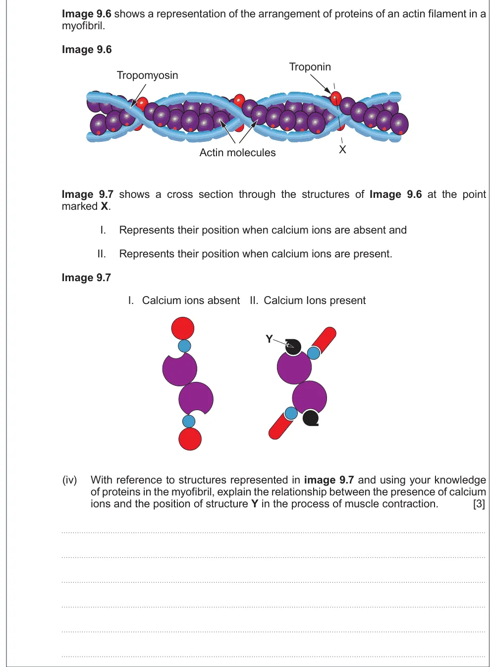
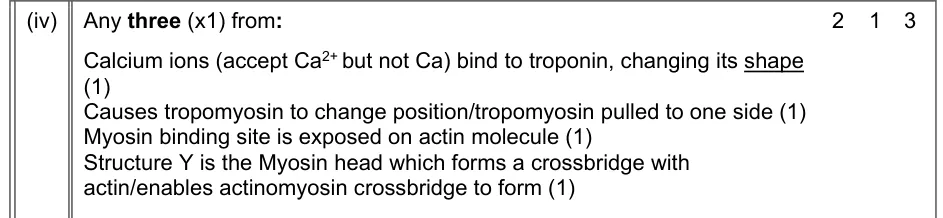
Explain the link between calcium ions and the position of the myosin head during contraction.
Show the mark scheme
Tap an image to open it full size.
From the examiners' report: Use the terms in order: calcium binds troponin, tropomyosin moves, binding sites on actin are exposed and the myosin head binds.
Question 13
A level Component 3 · June 2022 · Q8(b)(i) · 2 marks
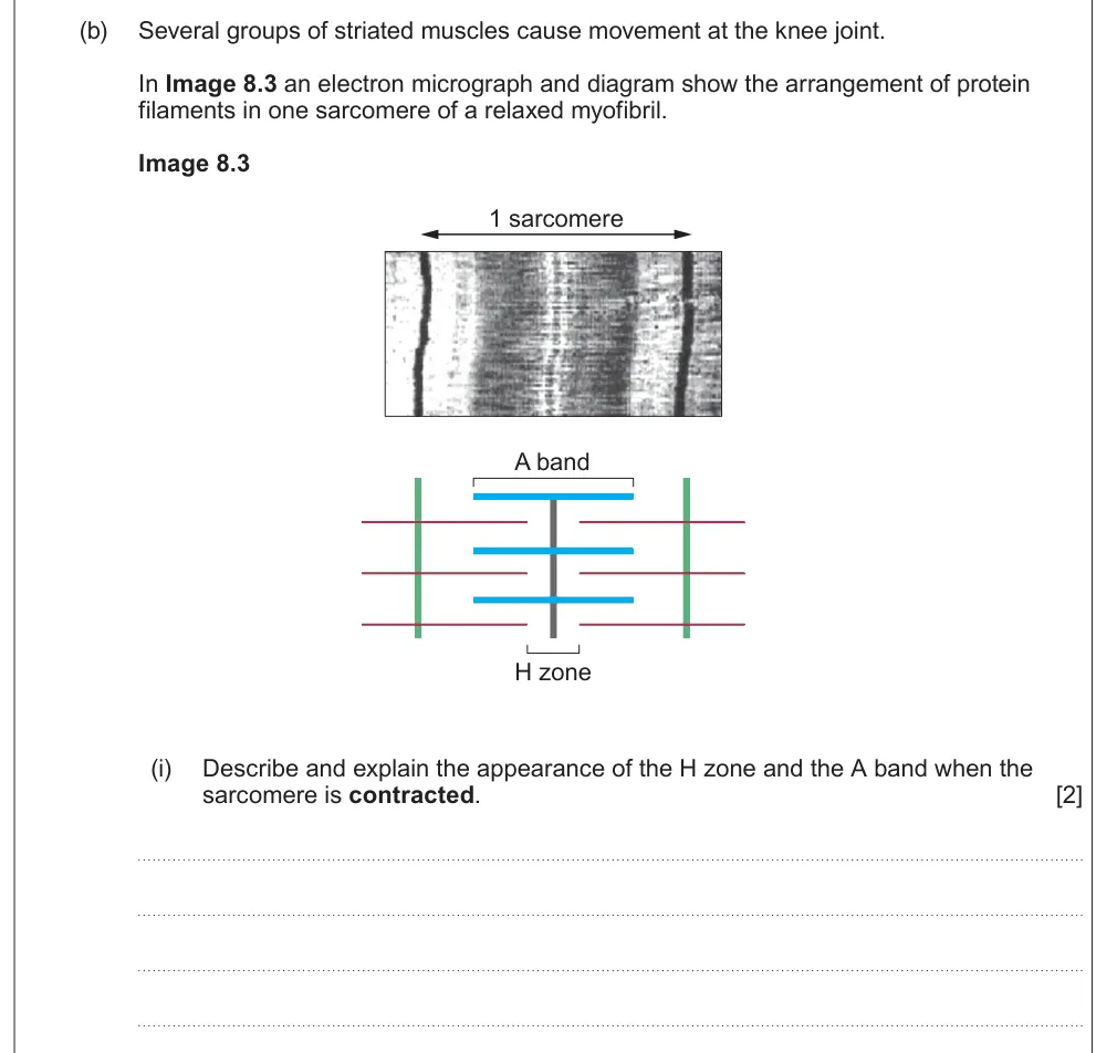
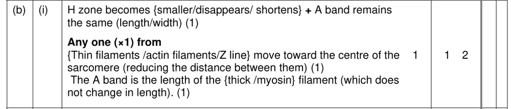
Describe and explain how the H zone and A band change when a sarcomere contracts.
Show the mark scheme
Tap an image to open it full size.
From the examiners' report: The A band stays the same length because it is the length of the myosin; the H zone shortens as actin slides towards the centre.
Question 14
A level Component 3 · June 2018 · Q9(b)(ii) · 2 marks
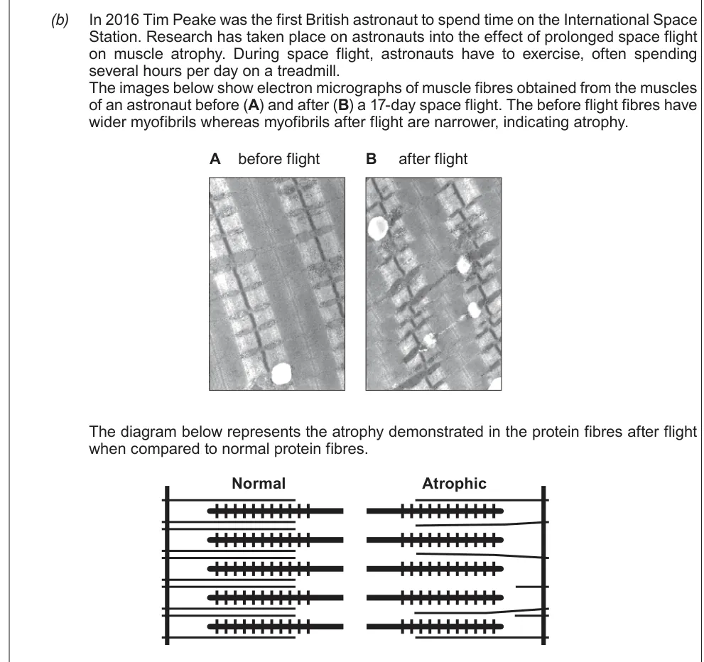
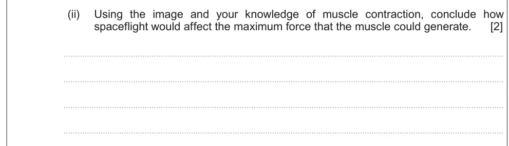
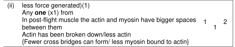
Use micrographs to conclude how spaceflight affects the maximum force a muscle can generate.
Information from earlier in this question:
Show the mark scheme
Tap an image to open it full size.
From the examiners' report: Look at the image: there is less actin, so fewer cross bridges can form and the force is lower.
Question 15
A level Component 3 · June 2025 · Q8(b)(ii) · 2 marks
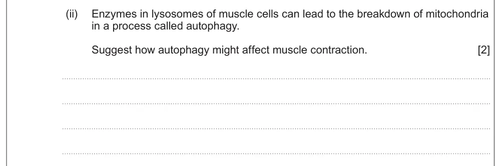
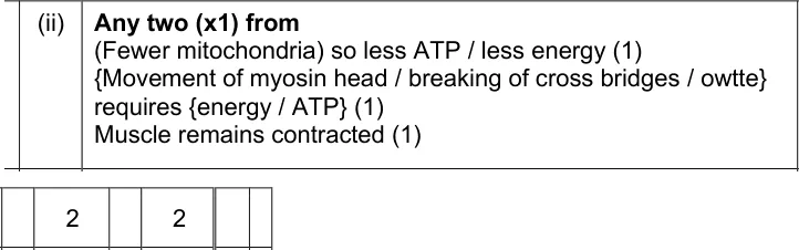
Suggest how the breakdown of mitochondria by autophagy might affect muscle contraction.
Show the mark scheme
Tap an image to open it full size.
From the examiners' report: Fewer mitochondria means less ATP, and ATP is needed to move the myosin head and break cross bridges.
Fibre types and energy
Question 16
A level Component 3 · Autumn 2021 · Q8(b)(i) · 2 marks
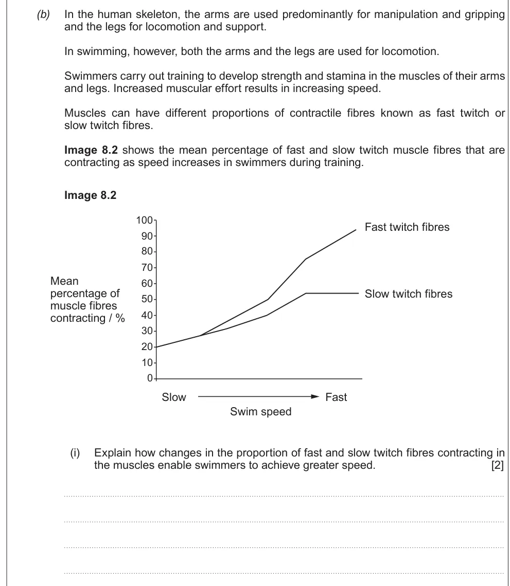
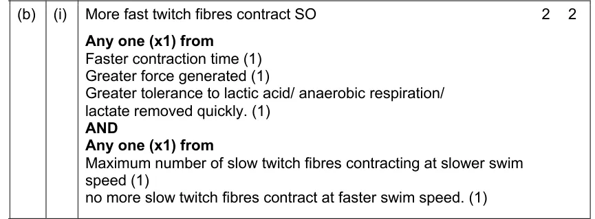
Explain how changing the proportion of fast and slow twitch fibres lets swimmers go faster.
Show the mark scheme
Tap an image to open it full size.
Question 17
A level Component 3 · June 2025 · Q8(c)(i) · 1 mark · Easy
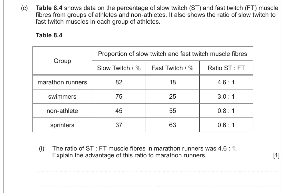
Explain the advantage of a 4.6 : 1 ratio of slow to fast twitch fibres for marathon runners.
Show the mark scheme

Tap an image to open it full size.
From the examiners' report: Be exact: slow twitch fibres fatigue slowly, with less lactic acid build-up. Vague wording lost marks.
Question 18
A level Component 3 · June 2019 · Q9(c)(i) · 3 marks
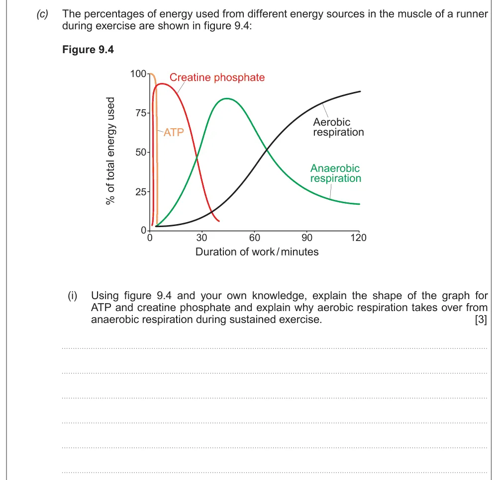
Explain the shapes of the ATP and creatine phosphate graphs and why aerobic respiration takes over.
Show the mark scheme

Tap an image to open it full size.
From the examiners' report: ATP is used up first; creatine phosphate then rephosphorylates ADP; aerobic respiration takes over as lactate builds up.
Question 19
A level Component 3 · June 2024 · Q8(d)(i)I · 2 marks · Hard
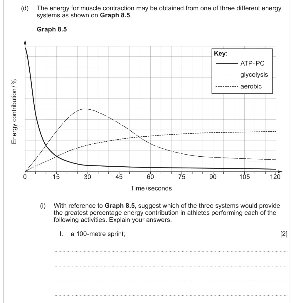
Use a graph of energy systems to suggest which one powers a 100 m sprint.
Show the mark scheme

Tap an image to open it full size.
From the examiners' report: Use the evidence on the graph: a sprint lasts about 10 seconds, when the ATP-PC system releases the most energy.
Question 20
A level Component 3 · June 2024 · Q8(d)(iii) · 2 marks
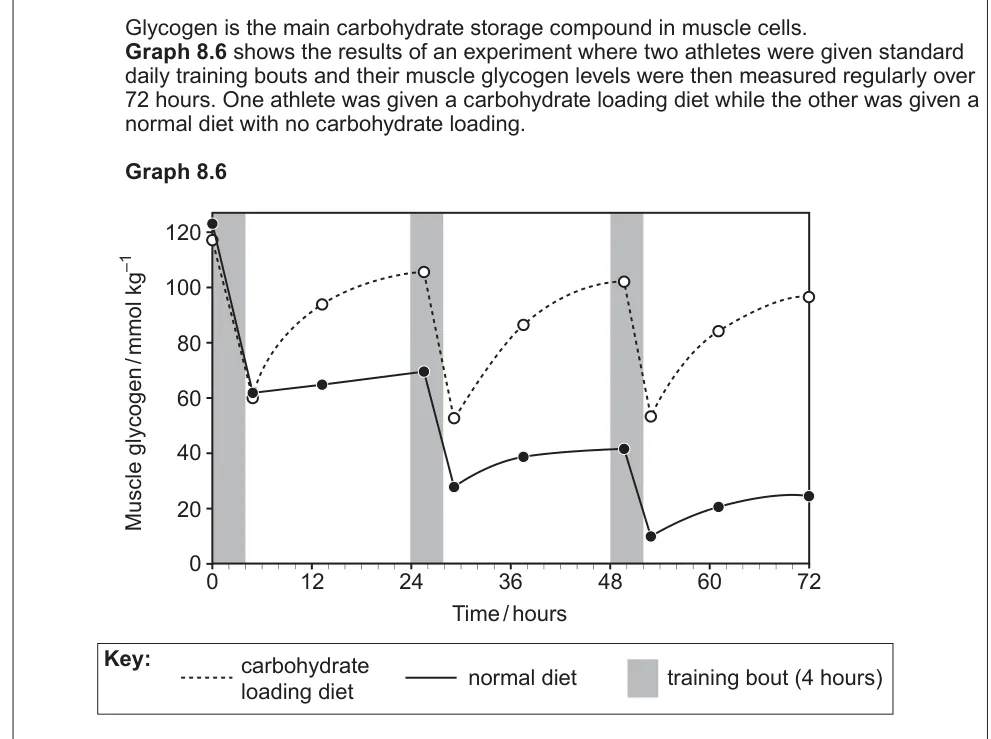
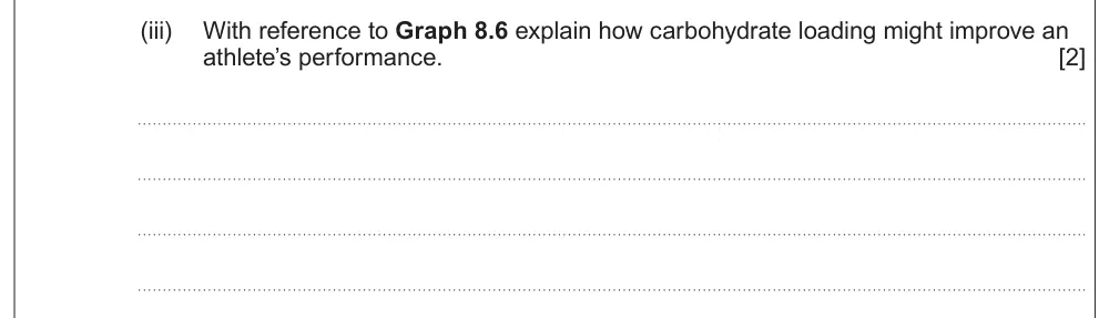
Use a graph to explain how carbohydrate loading might improve an athlete's performance.
Information from earlier in this question:
Show the mark scheme

Tap an image to open it full size.
From the examiners' report: Quote what the graph shows: muscle glycogen is restored to a higher level, so the athlete can train harder or for longer.
Where students lose marks (from Eduqas examiners' reports and mark schemes)
Each point below is followed by wording that gained marks.
Chondrocytes, then diffusion through the matrix: Many knew cartilage lacks blood vessels but didn't say diffusion happens through the matrix; weaker answers called the cells osteocytes.
✔ Gains credit: "Chondrocyte" and "by diffusion through the matrix".
Compounds, not ions: Answers named ions or elements instead of compounds, and organic compounds were harder to recall.
✔ Gains credit: "Organic – collagen; Inorganic – hydroxyapatite/calcium phosphate".
Both cells decide bone density: The idea of bone breakdown was missed by many; few saw that density depends on the balance of both cells.
✔ Gains credit: "Osteoblasts increase activity so more bone matrix produced than broken down".
Haversian canal carries the blood supply: Students confused the name of the Haversian canal and its role in transport.
✔ Gains credit: "Haversian canal": "O2 delivery / CO2 removal / nutrients".
Give the reasons you're asked for: Many identified the regions of the sarcomere but lost marks for not explaining their reasoning.
✔ Gains credit: "A – I band, as it only contains thin filaments".
The A band doesn't change: Many couldn't explain how the H zone shortens.
✔ Gains credit: "H zone becomes smaller + A band remains the same length", because "thin filaments move toward the centre".
A sprint is about 10 seconds: Many claimed a 100 m sprint lasts over 30 seconds and chose glycolysis, without using the graph.
✔ Gains credit: "ATP PC": a short activity, with more energy released than by the other systems in the first 10 seconds.
Download the questions
Download the PDF pack (20 questions, mark schemes at the end)
Keep practising
- Skeleton and Joints (Option B2 and B3): Eduqas A Level Biology exam questions by topic
- Eduqas A level Biology past papers, mark schemes and examiners' reports
- Eduqas A level Biology grade boundaries
- Ask about Eduqas A level Biology lessons with Sam on WhatsApp
Questions, mark schemes and examiners' report extracts © WJEC CBAC Ltd (Eduqas).