

Neuroscience is the second topic of Option C, Neurobiology and Behaviour, in Section B of Component 3. It covers ways of studying the brain without surgery (CT, MRI, fMRI, PET and EEG), the critical periods when the brain develops vision, hearing and language, neuroplasticity after injury or with new skills, and how childhood experiences can change gene expression and raise the risk of mental illness later in life.
The marks here usually go to a comparison or a link. When you compare scans, give both sides: what each one shows and how it is made. When you explain a critical period, link synapse formation and pruning to the timing in the graph. When you explain childhood stress, follow the cortisol pathway through to the loss of negative feedback.
I don't teach the Eduqas specification myself, so for one-to-one help I recommend Sam. He teaches Eduqas A level Biology at a top-rated sixth form college and is very popular with his students. You can ask about lessons with Sam on WhatsApp using the link at the bottom of this page.
What you need from GCSE and Year 12
Each question on this page assumes you're secure on these ideas. If any of them feels shaky, look it up before you start.
- Neurones connect at synapses, and the brain has billions of them.
- Genes are switched on and off; methylation of DNA can stop a gene being transcribed.
- Negative feedback keeps hormone levels steady.
- X-rays and magnetic fields can make images of the inside of the body.
Key terms
These words come up in the questions and mark schemes. Examiners expect you to use them exactly.
- CT scan: uses X-rays to make cross-sectional images of the brain's structure.
- MRI: uses magnetic fields and radio waves to make detailed images of soft tissue, without X-rays.
- fMRI: functional MRI: shows active brain areas by detecting extra oxygenated blood flow.
- PET scan: detects a radioactive tracer taken up most by the most active cells.
- Critical period: a window in early development when a sense or skill must be stimulated to develop normally.
- Synaptic pruning: the removal of synapses that aren't used.
- Neuroplasticity: the brain's ability to change its neural pathways, for example after injury.
What these questions cover (Eduqas OC2)
The questions test these statements from Option C (Neurobiology and Behaviour) of the Eduqas A level Biology specification (A400). Option questions are in Component 3, and you answer only one of the three options.
- (a) the different techniques used for studying the brain without invasive neurosurgery including functional magnetic resonance imaging (fMRI), computerised tomography (CT), p
- (b) how the brain develops and that there are critical periods for certain aspects of human learning and language acquisition
- (c) neuroplasticity
- (d) how the expression of genes can affect brain development and the impact this may have on an individual’s behaviour (no past question tests this on its own)
- (e) how altered gene expression in childhood could predispose adults to an increased risk of mental illness
How to use this page
Write your answer on paper before you open the mark scheme. Allow about 1.2 minutes per mark: the 15 questions are worth 36 marks, so about 43 minutes in total. Then mark your answer strictly against the mark scheme and read the examiners' tip underneath it. If you'd rather print the questions, download the PDF pack.
Studying the brain
Question 1
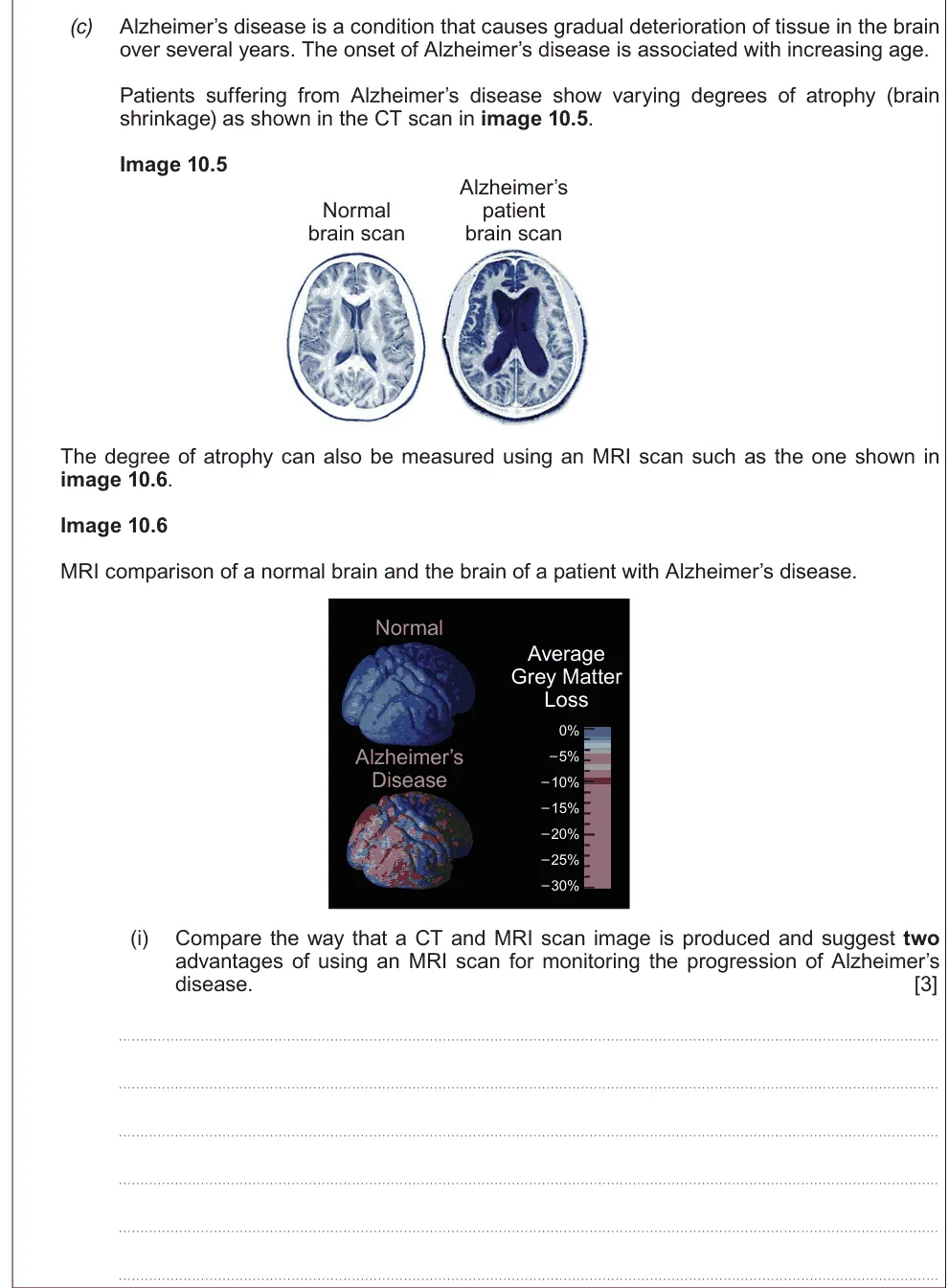
A level Component 3 · Autumn 2020 · Q10(c)(i) · 3 marks
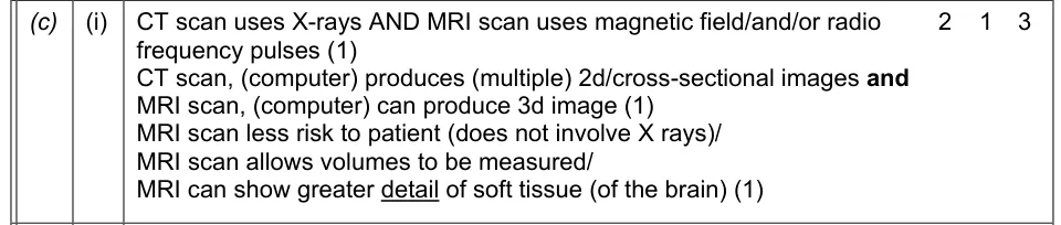
Compare how CT and MRI images are made and suggest two advantages of MRI for monitoring Alzheimer's disease.
Show the mark scheme
Tap an image to open it full size.
From the examiners' report: Make every point a comparison: CT uses X-rays and gives 2D slices, MRI uses magnetic fields and can give 3D images. One-sided points didn't score.
Question 2
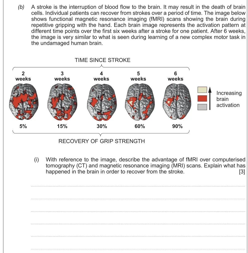
A level Component 3 · June 2018 · Q10(b)(i) · 3 marks · Easy
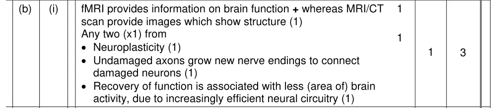
Describe the advantage of fMRI over CT and MRI and explain how the brain recovered from a stroke.
Show the mark scheme
Tap an image to open it full size.
From the examiners' report: Compare the scans: fMRI shows function, while CT and MRI show structure. Then explain recovery with neuroplasticity.
Question 3
A level Component 3 · June 2017 · Q9(b)(iii) · 2 marks
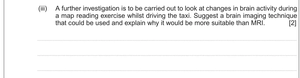
Suggest a brain imaging technique for a taxi driver who is driving, and why it suits better than MRI.
Show the mark scheme

Tap an image to open it full size.
From the examiners' report: Think about the situation: it must be portable, so EEG. Many chose fMRI, CT or PET, and some even ECG.
Question 4
A level Component 3 · June 2022 · Q9(d)(i) · 3 marks
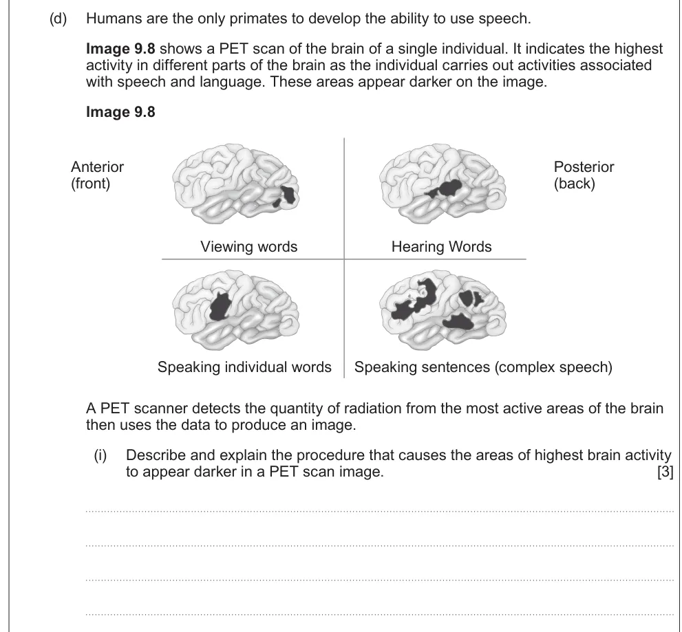
Describe and explain why the most active areas of the brain appear darker on a PET scan.
Show the mark scheme

Tap an image to open it full size.
From the examiners' report: Keep the procedure brief and explain the key link: the most active cells take up more of the radioactive glucose tracer.
Question 5
A level Component 3 · June 2025 · Q9(b)(i) · 2 marks · Easy
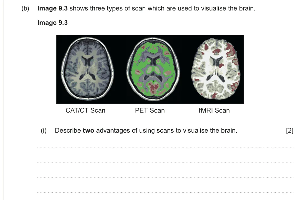
Describe two advantages of using scans to see the brain.
Show the mark scheme

Tap an image to open it full size.
From the examiners' report: Scans are non-invasive, so no surgery, and they show detail of the whole brain or its soft tissue.
Question 6
A level Component 3 · June 2025 · Q9(b)(iii) · 2 marks · Easy
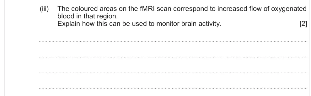
Explain how increased oxygenated blood flow on an fMRI scan shows brain activity.
Show the mark scheme

Tap an image to open it full size.
From the examiners' report: More active neurones need more ATP from aerobic respiration, so more oxygen is delivered to them.
Critical periods and neuroplasticity
Question 7
A level Component 3 · Autumn 2020 · Q10(b)(i) · 1 mark · Easy
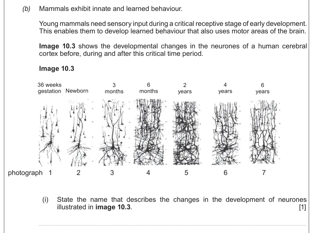
Name the changes in neurone development shown in a series of photographs.
Show the mark scheme

Tap an image to open it full size.
From the examiners' report: Most recognised it: neuroplasticity.
Question 8
A level Component 3 · Autumn 2020 · Q10(b)(ii) · 3 marks
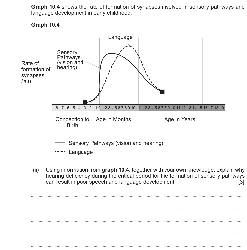
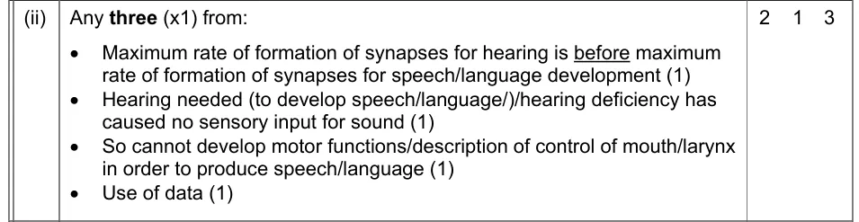
Use a graph to explain why hearing loss during the critical period harms speech and language.
Show the mark scheme
Tap an image to open it full size.
From the examiners' report: Use synapse formation from the graph, not vague "nervous pathways", and get the order right: hearing synapses peak before language synapses.
Question 9
A level Component 3 · Autumn 2020 · Q10(b)(iii) · 1 mark
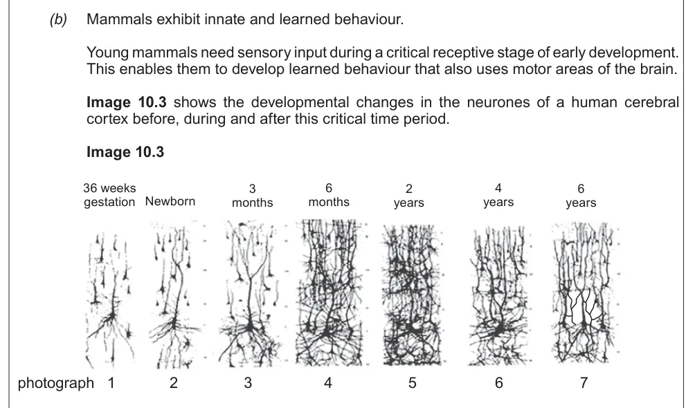
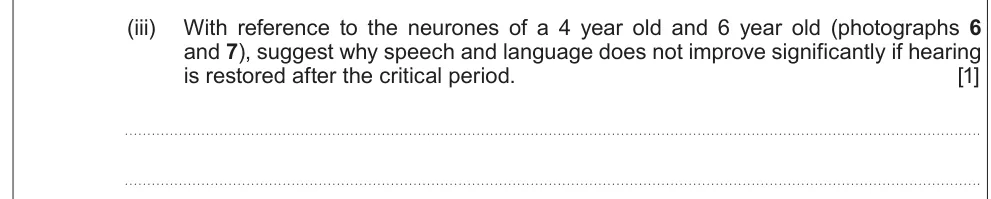
Suggest why speech doesn't improve much if hearing is restored after the critical period.
Information from earlier in this question:
Show the mark scheme

Tap an image to open it full size.
From the examiners' report: Link synaptic pruning to timing: the unused synapses have already been lost by the time hearing is restored.
Question 10
A level Component 3 · June 2023 · Q9(b)(iv) · 4 marks
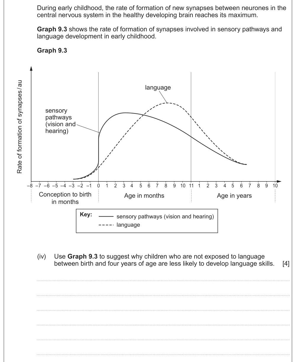
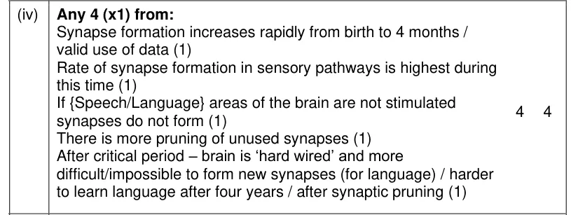
Use a graph to suggest why children without language before age four may not develop language skills.
Show the mark scheme
Tap an image to open it full size.
From the examiners' report: Quote the graph, then explain: if the language areas aren't stimulated during peak synapse formation, the synapses don't form or are pruned.
Question 11
A level Component 3 · June 2017 · Q9(b)(ii) · 4 marks
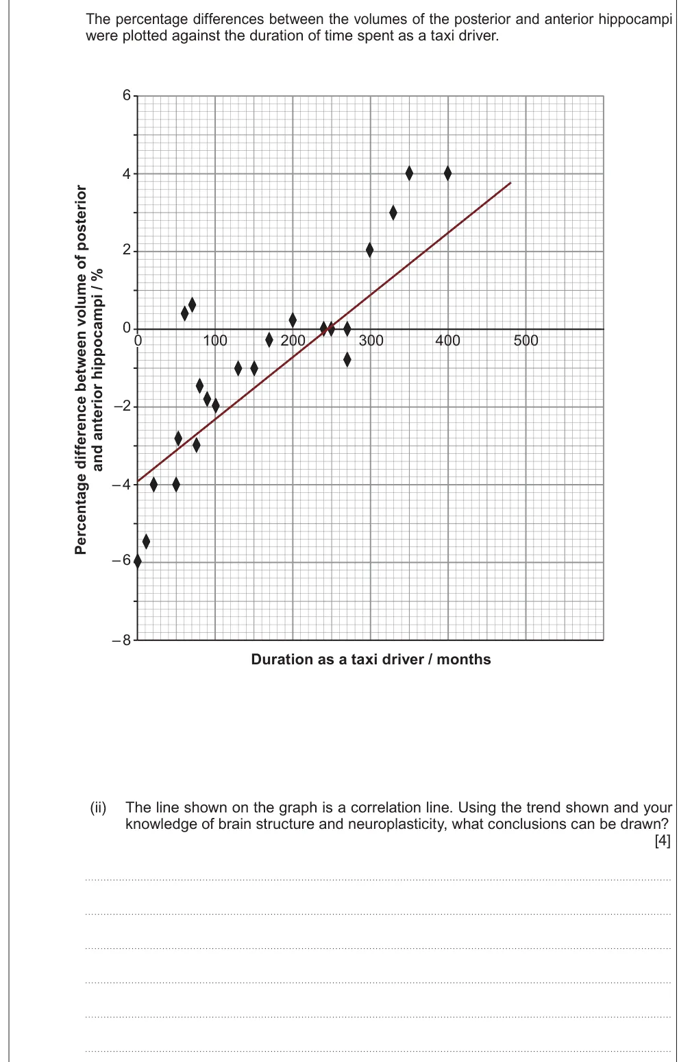
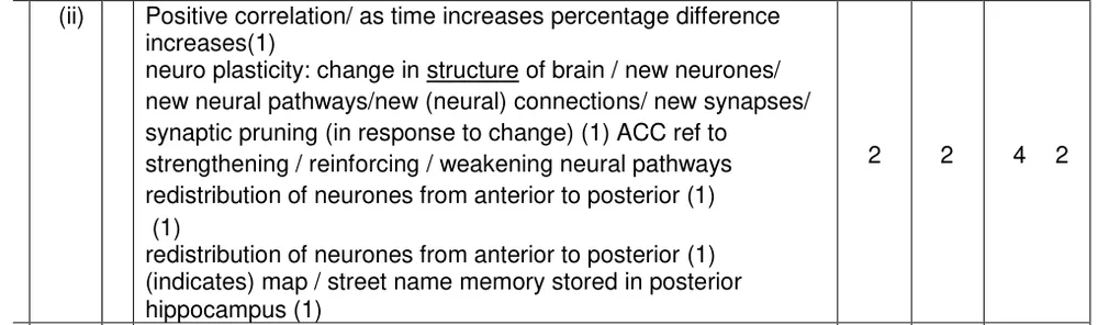
Draw conclusions from a correlation between years as a taxi driver and hippocampus size.
Show the mark scheme
Tap an image to open it full size.
From the examiners' report: State the positive correlation, then link it to neuroplasticity: new neural connections form with use.
Question 12
A level Component 3 · June 2023 · Q9(b)(ii) · 2 marks
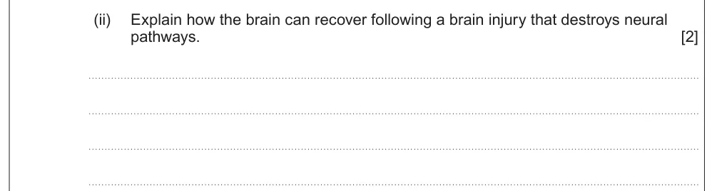
Explain how the brain can recover after an injury destroys neural pathways.
Show the mark scheme

Tap an image to open it full size.
From the examiners' report: Name neuroplasticity, then say what happens: neurones grow new connections that bypass the damaged area.
Genes, stress and mental illness
Question 13
A level Component 3 · Autumn 2021 · Q9(b)(ii) · 3 marks
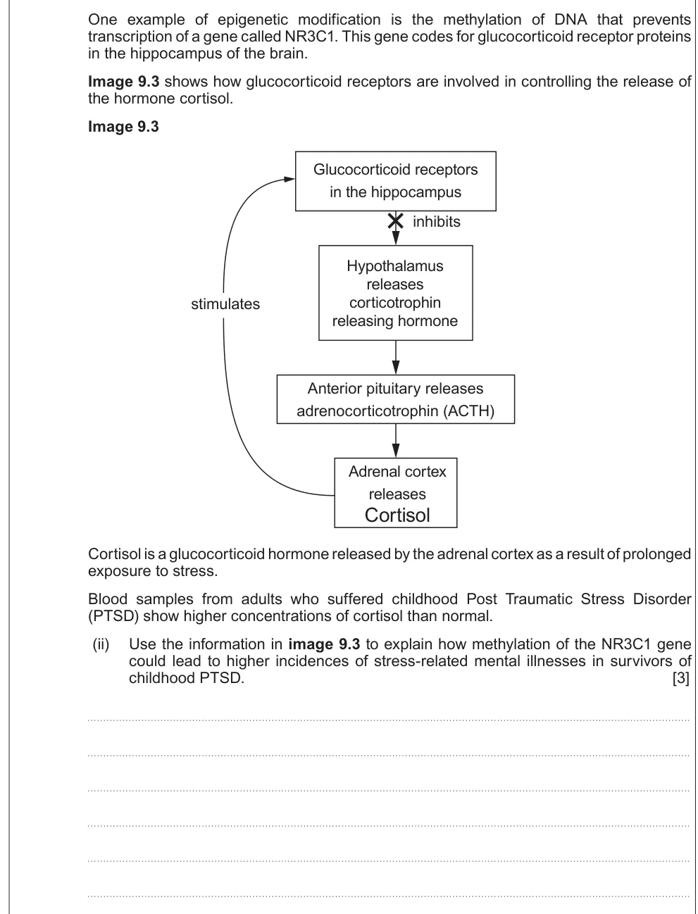
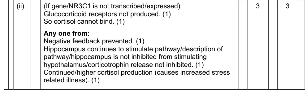
Explain how methylation of the NR3C1 gene could lead to more stress-related mental illness after childhood PTSD.
Show the mark scheme
Tap an image to open it full size.
Question 14
A level Component 3 · June 2019 · Q10(c)(ii) · 1 mark
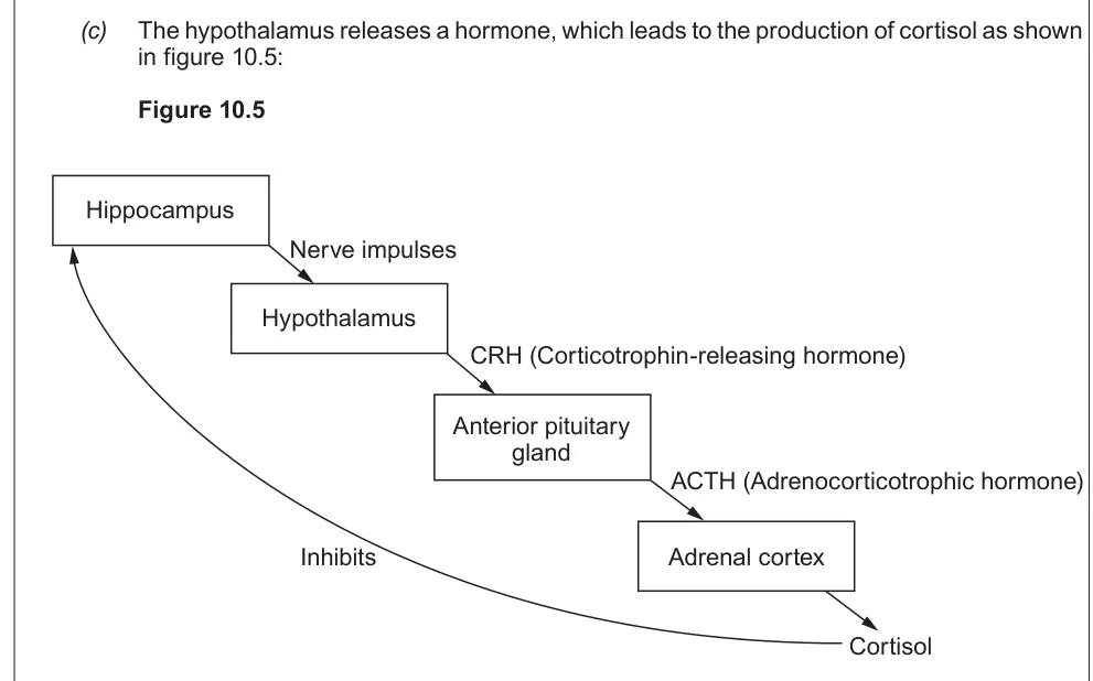
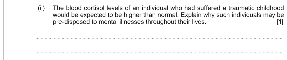
Explain why people with a traumatic childhood may be predisposed to mental illness.
Information from earlier in this question:
Show the mark scheme

Tap an image to open it full size.
From the examiners' report: Raised cortisol means negative feedback isn't working, which increases vulnerability to mental illness.
Question 15
A level Component 3 · June 2024 · Q9(c)(iii) · 2 marks · Easy
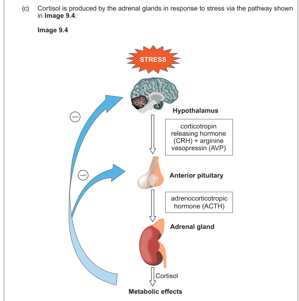
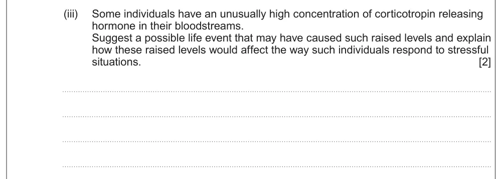
Suggest a life event that could raise CRH levels and explain how it affects responses to stress.
Information from earlier in this question:
Show the mark scheme

Tap an image to open it full size.
From the examiners' report: Childhood trauma; then follow the pathway to more ACTH and cortisol, so the person becomes over-stressed.
Where students lose marks (from Eduqas examiners' reports and mark schemes)
Each point below is followed by wording that gained marks.
Compare both scans: Marks were only given when the comparison of both types of scan was clear; several gave only one valid point.
✔ Gains credit: "CT scan uses X-rays AND MRI scan uses magnetic field/radio frequency pulses".
Function versus structure: Most knew what fMRI is for but didn't compare it with CT and MRI.
✔ Gains credit: "fMRI provides information on brain function, whereas MRI/CT provide images which show structure".
Portable means EEG: Very few identified EEG; many said fMRI, CT or PET, and some suggested an ECG.
✔ Gains credit: "Electroencephalography/EEG": portable, whereas MRI isn't.
Explain why active areas show up: Descriptions of the PET procedure were over-detailed, and few explained why more activity showed.
✔ Gains credit: "More radioactivity taken up by cells that are most active / have highest glucose uptake".
Synapses, not "nervous pathways": Good answers used synapse formation from the graph rather than vague references to nervous pathways.
✔ Gains credit: "Maximum rate of formation of synapses for hearing is before maximum rate of formation of synapses for speech".
Pruning before hearing returns: Answers described synaptic pruning but didn't link it to when hearing was restored.
✔ Gains credit: "Many synapses lost / synaptic pruning before hearing is restored".
Download the questions
Download the PDF pack (15 questions, mark schemes at the end)
Keep practising
- The Brain (Option C1): Eduqas A Level Biology exam questions by topic
- Behaviour (Option C3): Eduqas A Level Biology exam questions by topic
- Eduqas A level Biology past papers, mark schemes and examiners' reports
- Eduqas A level Biology grade boundaries
- Ask about Eduqas A level Biology lessons with Sam on WhatsApp
Questions, mark schemes and examiners' report extracts © WJEC CBAC Ltd (Eduqas).