

The Microscope in Cell Studies (1.1): Cambridge (CIE) A Level Biology 9700 Exam Questions by Topic
The microscope in cell studies is sub-topic 1.1 of Topic 1, Cell structure, in the Cambridge International AS & A Level Biology syllabus (9700). You need to calculate magnifications and actual sizes from drawings, photomicrographs and electron micrographs, use an eyepiece graticule and stage micrometer scale to make measurements in millimetres, micrometres and nanometres, and define resolution and magnification with reference to light and electron microscopy. The questions on this page start at outcome 1.1.3: the question bank has no Paper 2 or Paper 4 parts on making temporary slides or drawing cells (1.1.1 and 1.1.2). This is AS content, examined on Papers 1 and 2. A level students meet it again on Paper 4.
The examiners' reports return to the same points. In calculations, most marks are lost on units: a line measured in centimetres has to be converted to millimetres before it is multiplied by 1000 to give micrometres. Round the answer as instructed and do not give a unit with a magnification. It is the magnification of the image that matters, not the magnification of the microscope. When you compare microscopes, write about resolution, give the values in nanometres and name a structure, such as a ribosome, that can be seen. 'Clearer' and 'more detail' on their own do not gain credit.
Studying Cambridge International A level Biology and want more help? Tom teaches CIE students one-to-one online, including students in the UAE, Saudi Arabia, Hong Kong, India and Indonesia, and every lesson is recorded. You can book a one-to-one lesson or a free 15-minute discovery chat, or register your interest in a CIE group class starting in September 2027, using the links at the bottom of this page.
What you need from IGCSE
Each question on this page assumes you're secure on these ideas. If any of them feels shaky, look it up before you start.
- Magnification = image size ÷ actual size.
- There are 1000 micrometres (µm) in 1 millimetre (mm).
- A light microscope shows structures such as the nucleus, cell wall, chloroplasts and vacuole; smaller structures such as ribosomes can only be seen with an electron microscope.
- How to round an answer to a stated number of significant figures.
Key terms
These words come up in the questions and mark schemes. Examiners expect you to use them exactly.
- Magnification: the number of times larger an image is than the actual size of the object: image size ÷ actual size.
- Resolution: the ability to distinguish between two points that are close together. It is about 200 nm for a light microscope and about 0.5 nm for an electron microscope.
- Micrometre (µm): one thousandth of a millimetre: 1 mm = 1000 µm.
- Nanometre (nm): one thousandth of a micrometre: 1 µm = 1000 nm.
- Eyepiece graticule: a scale in the eyepiece of a light microscope, used to measure specimens once it has been calibrated.
- Stage micrometer: a microscope slide with an accurate scale on it, used to calibrate the eyepiece graticule.
- Transmission electron micrograph: an image made with electrons that have passed through a very thin section, so it shows the structures inside a cell.
- Scanning electron micrograph: an image made with electrons from the surface of a specimen, so it shows surface contours and looks three-dimensional.
What these questions cover (Cambridge 9700 Topic 1: Cell structure)
The questions test these learning outcomes from sub-topic 1.1 of the Cambridge International AS & A Level Biology syllabus (9700) for 2025 to 2027.
- 1.1.3 calculate magnifications of images and actual sizes of specimens from drawings, photomicrographs and electron micrographs
- 1.1.4 use an eyepiece graticule and stage micrometer scale to make measurements and use the appropriate units
- 1.1.5 define resolution and magnification and explain the differences between these terms, with reference to light microscopy and electron microscopy
How to use this page
Write your answer on paper before you open the mark scheme. Allow about 1.2 minutes per mark: the 22 questions are worth 38 marks, so about 46 minutes in total. Then mark your answer strictly against the mark scheme and read the examiners' tip underneath it. If you'd rather print the questions, download the PDF pack.
Calculating magnification and actual size (1.1.3)
Question 1
Paper 2 · February/March 2025 (22) · Q1(b) · 1 mark · Easy
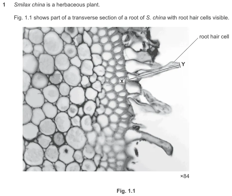
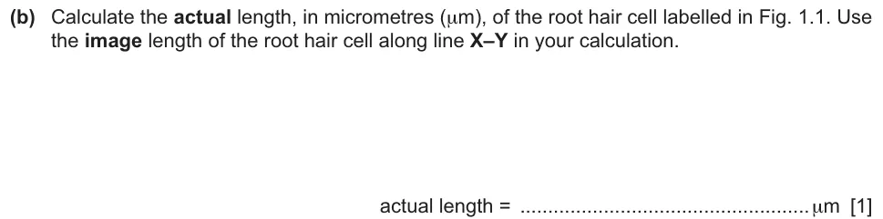
Calculate the actual length, in micrometres, of a root hair cell from the image and its magnification.
Information from earlier in this question:
Show the mark scheme

Tap an image to open it full size.
From the examiners' report: Most students measured the line correctly and knew which calculation to do. Marks were lost through errors in converting units or in rounding.
Question 2
Paper 2 · October/November 2025 (24) · Q2(c)(i) · 2 marks
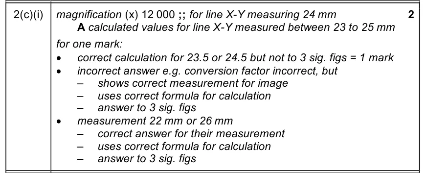
Calculate the magnification of a scanning electron micrograph from an actual diameter of 2 µm, to three significant figures.
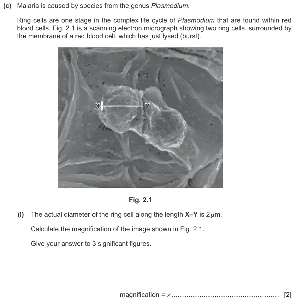
Show the mark scheme
Tap an image to open it full size.
Question 3
Paper 2 · May/June 2024 (23) · Q1(b)(i) · 2 marks
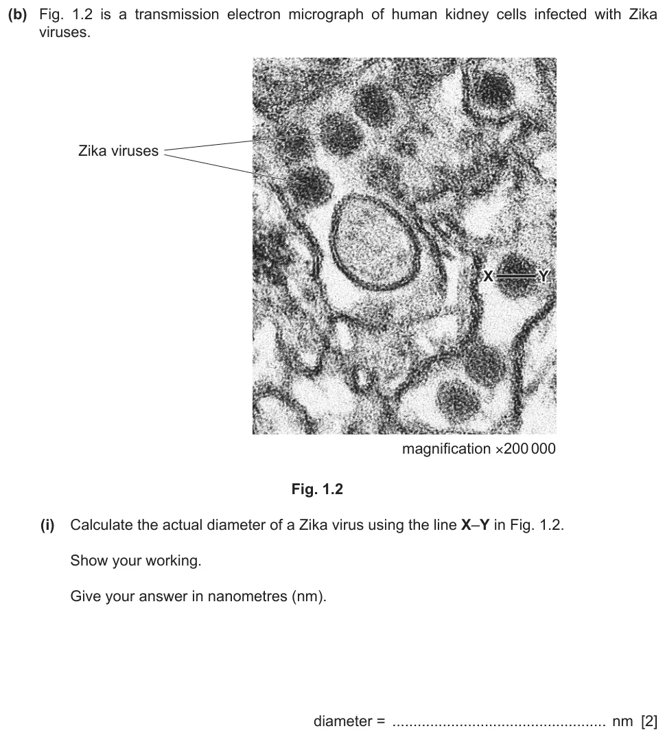
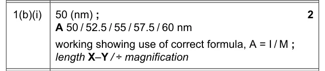
Calculate the actual diameter of a Zika virus in nanometres from a transmission electron micrograph.
Show the mark scheme
Tap an image to open it full size.
From the examiners' report: Most measured X–Y correctly and used a correct formula. Many gave 5 instead of 50 because they measured 1 cm and did not change it to millimetres before converting. A correct formula, clearly used, gained partial credit when the final value was wrong.
Question 4
Paper 2 · May/June 2022 (22) · Q1(b) · 2 marks · Easy
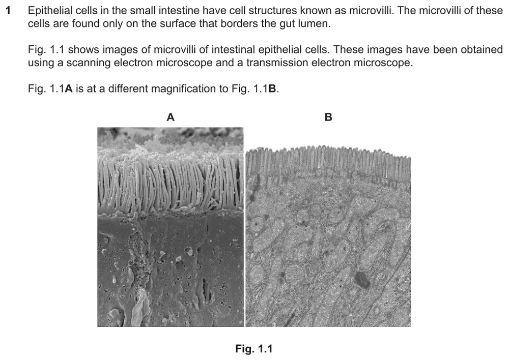
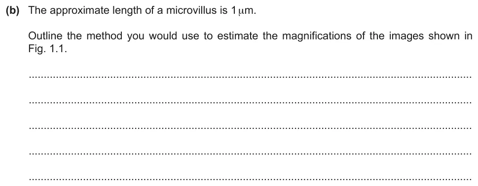
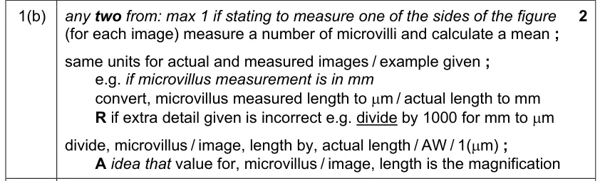
Outline how you would estimate the magnification of an image when the actual length of a microvillus is known.
Information from earlier in this question:
Show the mark scheme
Tap an image to open it full size.
From the examiners' report: The most detailed answers measured several microvilli in the image and calculated a mean length. Students who gained only one mark knew the formula but gave a wrong conversion factor for mm or cm, or did not say that the two lengths must be in the same units.
Question 5
Paper 2 · February/March 2022 (22) · Q4(b) · 2 marks · Easy
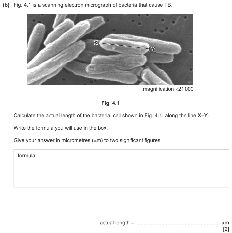
Write the formula and calculate the actual length of a bacterial cell in micrometres, to two significant figures.
Show the mark scheme

Tap an image to open it full size.
From the examiners' report: Most used the correct formula. Some put the wrong figures into it, and some did not give the answer to two significant figures as the question asked.
Question 6
Paper 2 · May/June 2022 (21) · Q5(b) · 2 marks
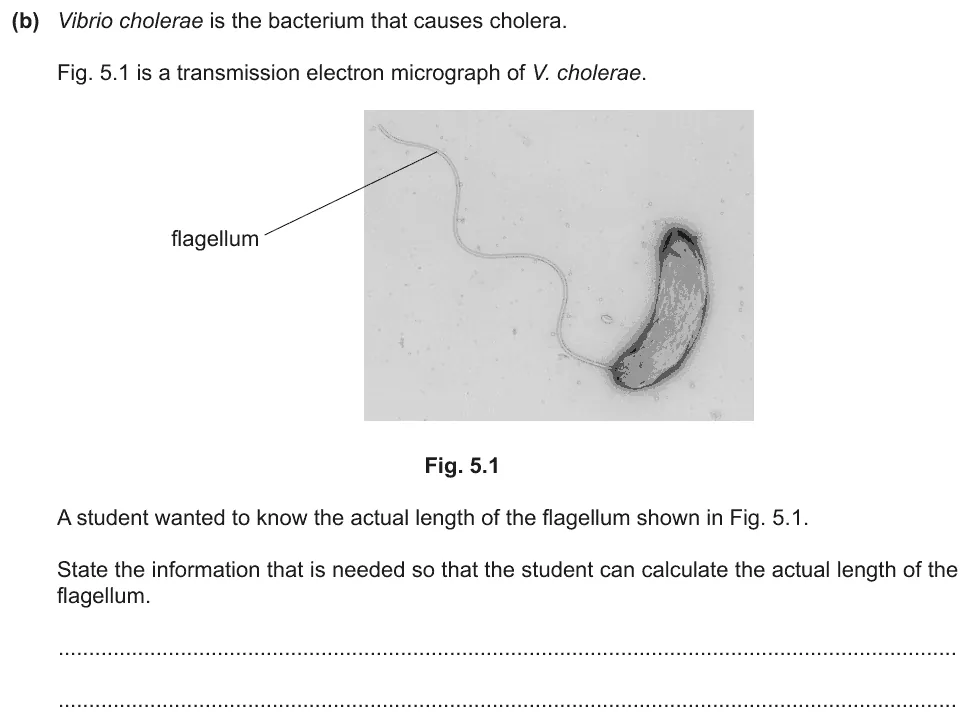
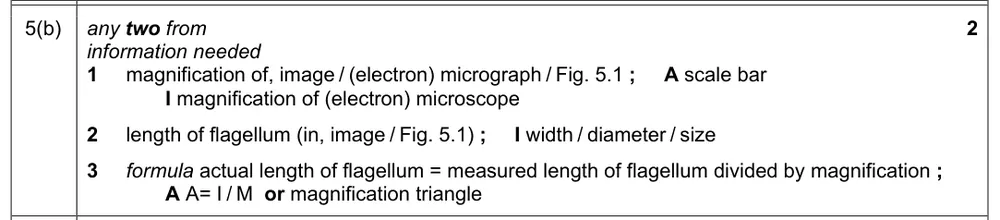
State the information a student needs to calculate the actual length of a flagellum in an electron micrograph.
Show the mark scheme
Tap an image to open it full size.
From the examiners' report: It is the magnification of the image (the electron micrograph) that is needed, not the magnification of the microscope worked out from its eyepiece and objective lenses. Only some remembered that the measured length of the flagellum in the image is needed as well.
Question 7
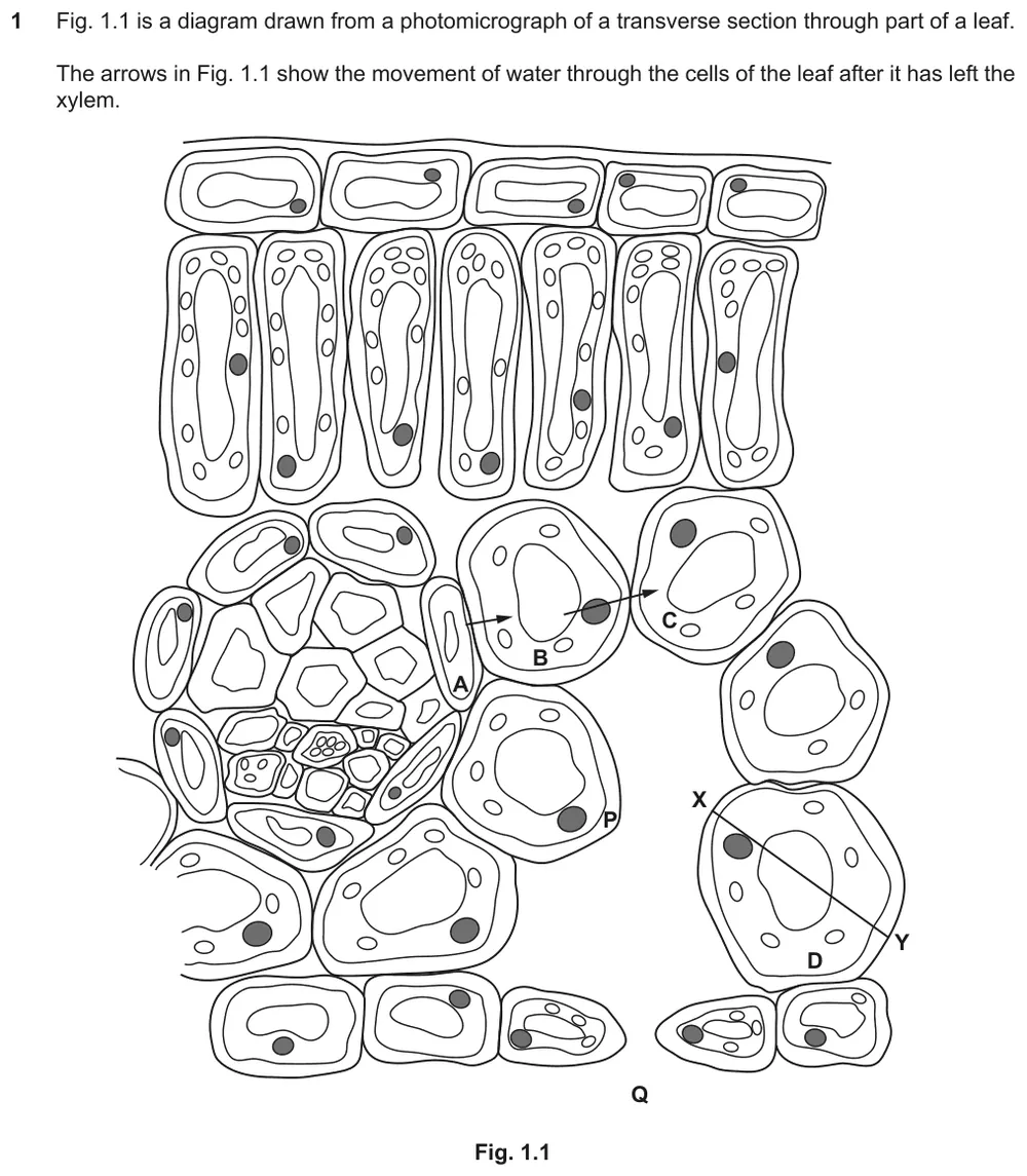
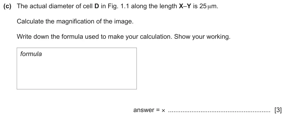
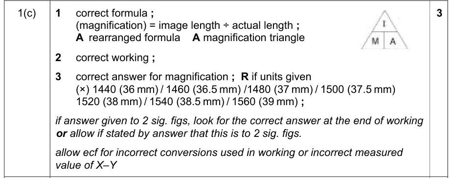
Paper 2 · October/November 2020 (22) · Q1(c) · 3 marks · Easy
Write the formula and calculate the magnification of an image from the actual diameter of a cell.
Information from earlier in this question:
Show the mark scheme
Tap an image to open it full size.
From the examiners' report: The formula was well known. Students who lost marks measured in centimetres and multiplied straight by 1000 to get micrometres, forgetting to multiply by 10 first to get millimetres. Some made errors with standard form and ended up with an unrealistic magnification.
Question 8
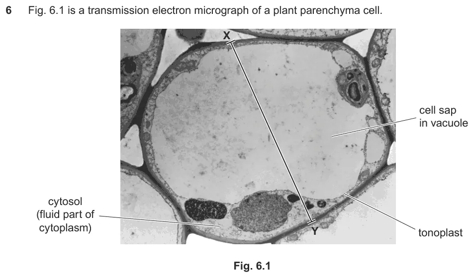
Paper 2 · February/March 2020 (22) · Q6(b)(ii) · 2 marks · Easy
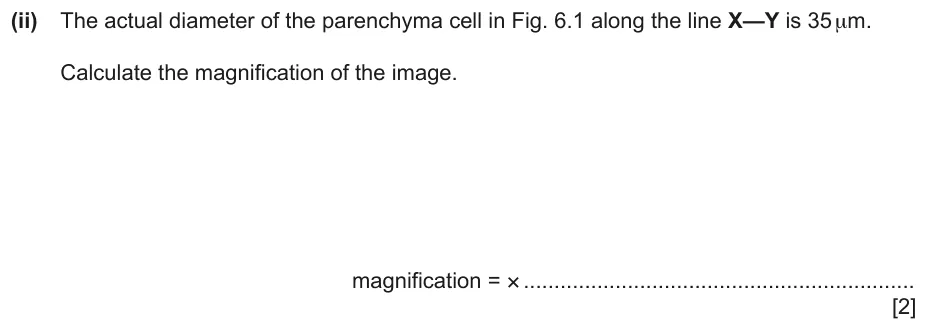
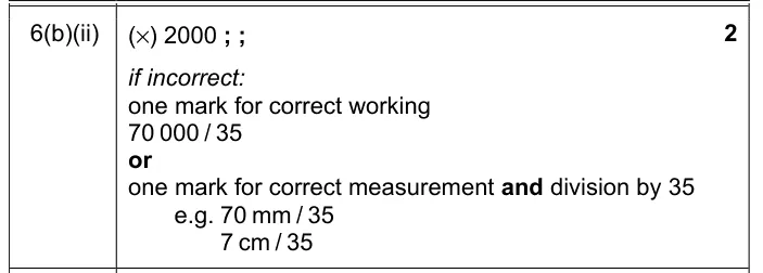
Calculate the magnification of an image of a parenchyma cell from its actual diameter of 35 µm.
Information from earlier in this question:

Show the mark scheme
Tap an image to open it full size.
From the examiners' report: Most measured the line correctly and did the right calculation. Some wrongly gave a unit with the magnification.
Question 9
Paper 2 · May/June 2018 (22) · Q1(b) · 2 marks
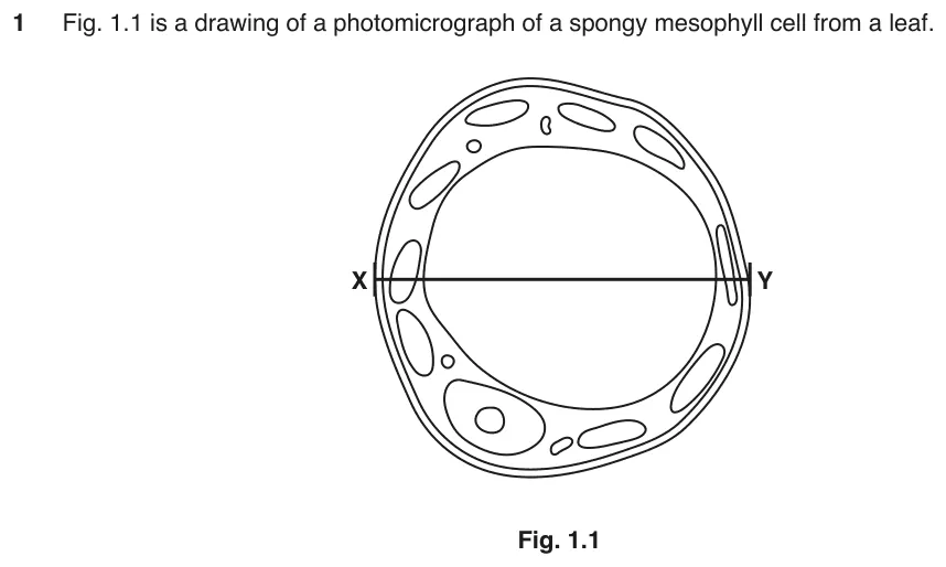
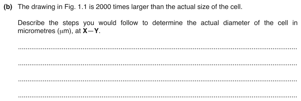
Describe the steps you would follow to find the actual diameter of a cell, in micrometres, from a drawing.
Information from earlier in this question:
Show the mark scheme

Tap an image to open it full size.
From the examiners' report: No calculation was needed. Good answers made it clear that the ruler measurement has to be converted to micrometres and then divided by the magnification. A common error was to multiply a centimetre measurement by 1000 instead of 10 000, and 'measure in micrometres' is not correct.
Question 10
Paper 2 · May/June 2017 (22) · Q3(d) · 3 marks
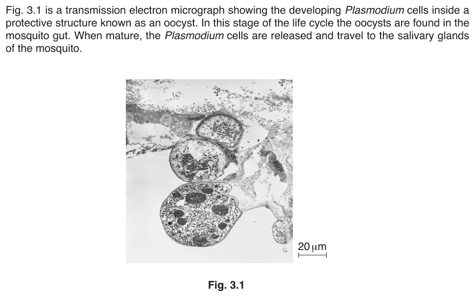
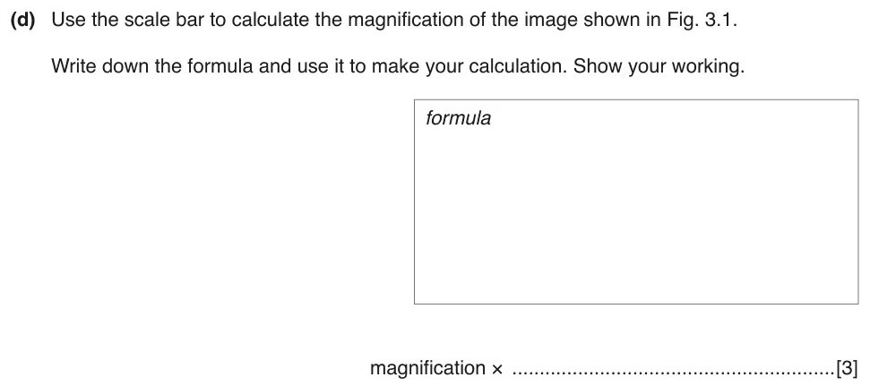
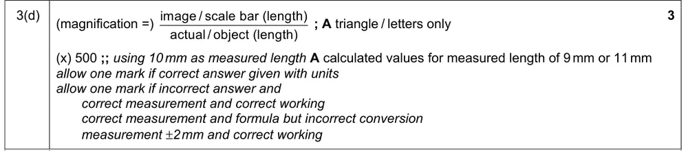
Use the scale bar to calculate the magnification of a transmission electron micrograph.
Information from earlier in this question:
Show the mark scheme
Tap an image to open it full size.
From the examiners' report: Use only the measured length of the scale bar and the actual length written below it. Some reached the right answer but tried to use the image itself, which was not necessary.
Eyepiece graticule, stage micrometer and units (1.1.4)
Question 11
Paper 2 · May/June 2024 (22) · Q1(b)(i) · 1 mark
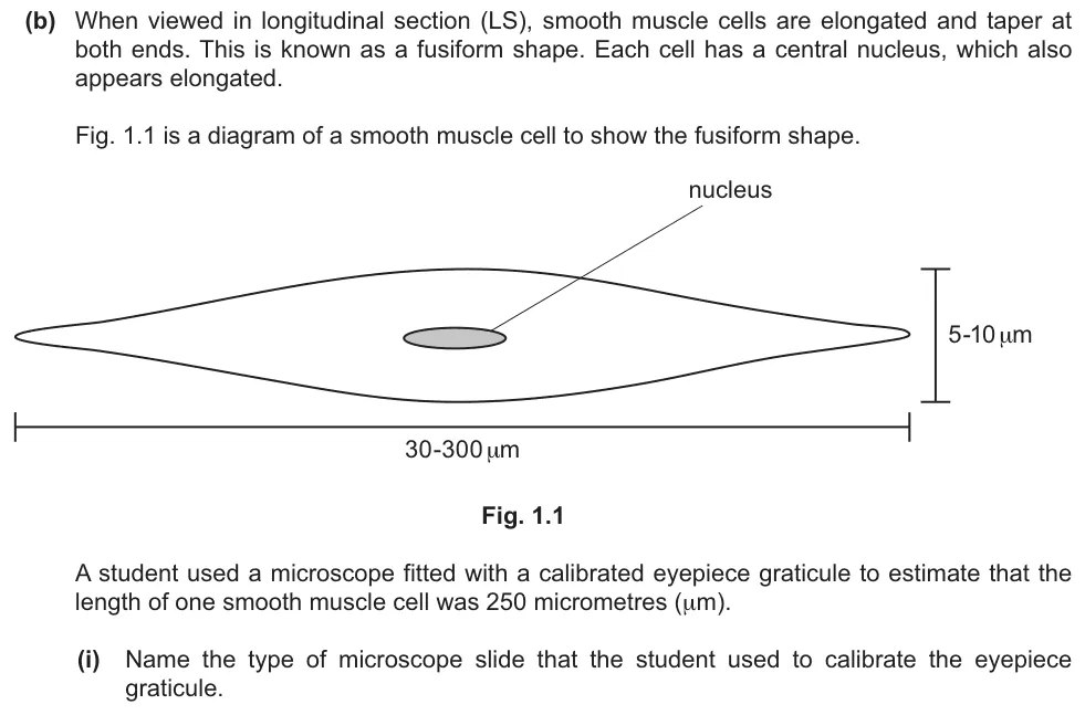
Name the type of microscope slide used to calibrate an eyepiece graticule.
Show the mark scheme

Tap an image to open it full size.
From the examiners' report: Most who gave the correct answer, stage micrometer, also spelt it correctly. Many wrongly named a type of microscope, such as 'light microscope' or 'electron microscope'. 'Microscope slide' and 'micrometer slide' were also incorrect.
Question 12
Paper 2 · May/June 2024 (22) · Q1(b)(ii) · 1 mark
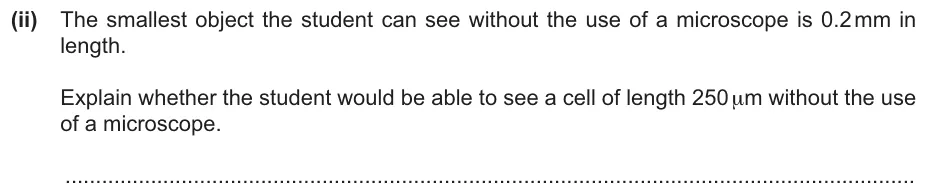
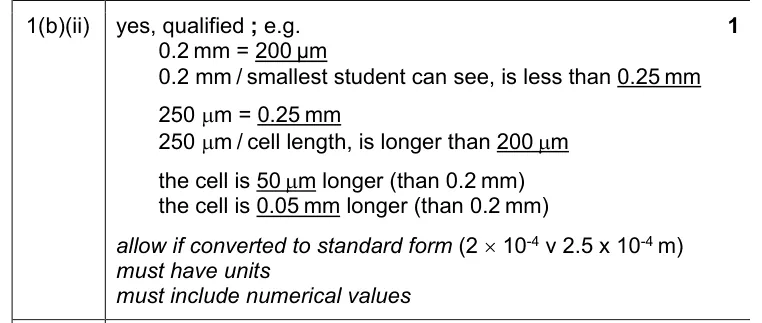
Explain whether a cell 250 µm long can be seen without a microscope by someone who can see objects of 0.2 mm.
Show the mark scheme
Tap an image to open it full size.
From the examiners' report: There are two steps: a correct conversion, then a decision. Some converted correctly but still said the cell could not be seen. Others wrote 'yes' but needed to explain it with calculated values and units.
Question 13
Paper 2 · October/November 2019 (22) · Q1(c)(ii) · 1 mark
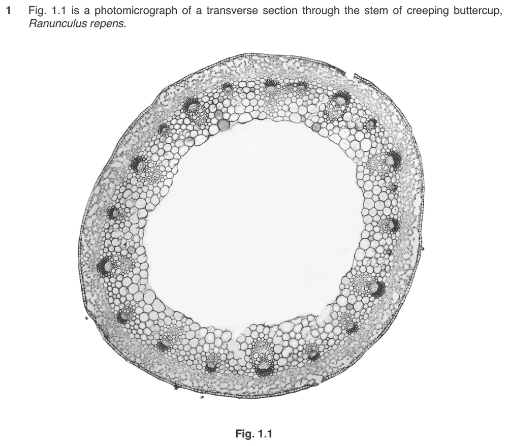
State the piece of equipment used in a light microscope to work out the correct proportions of tissues.
Information from earlier in this question:


Show the mark scheme

Tap an image to open it full size.
From the examiners' report: Only an eyepiece graticule is needed to work out the proportions of tissues, and many students knew this.
Question 14
Paper 2 · February/March 2019 (22) · Q6(d)(ii) · 1 mark · Easy
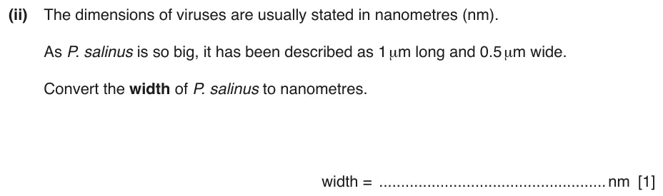
Convert the width of a giant virus from micrometres to nanometres.
Information from earlier in this question:

Show the mark scheme

Tap an image to open it full size.
Resolution, magnification and types of microscope (1.1.5)
Question 15
Paper 2 · February/March 2025 (22) · Q1(a) · 1 mark
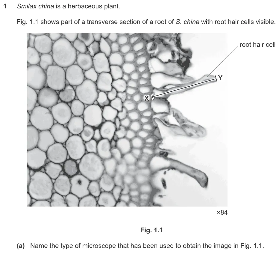
Name the type of microscope used to obtain an image of a section of a root.
Show the mark scheme

Tap an image to open it full size.
From the examiners' report: Many students wrongly thought that the image had been obtained with an electron microscope.
Question 16
Paper 2 · February/March 2024 (22) · Q5(a)(i) · 1 mark
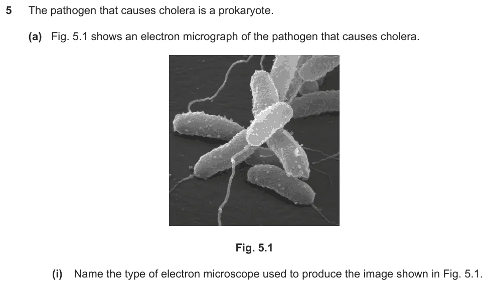
Name the type of electron microscope used to produce an image of a bacterium.
Show the mark scheme

Tap an image to open it full size.
Question 17
Paper 2 · February/March 2018 (22) · Q1(a) · 1 mark
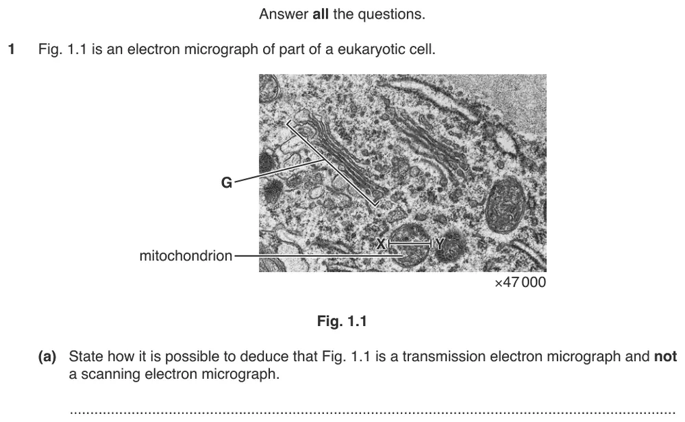
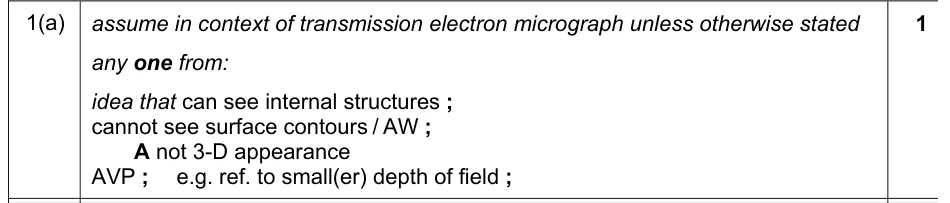
State how you can tell that an image is a transmission electron micrograph and not a scanning electron micrograph.
Show the mark scheme
Tap an image to open it full size.
From the examiners' report: There were two ways to gain the mark. Many gave a feature that can be seen in the image, such as the organelles. Others gave a feature of scanning electron micrographs that is absent, such as surface contours.
Question 18
Paper 2 · February/March 2020 (22) · Q6(b)(i) · 1 mark · Easy
Explain what is meant by the term magnification.
Information from earlier in this question:
Show the mark scheme

Tap an image to open it full size.
From the examiners' report: Some answers were too vague, such as 'zooming in to see more detail'. Others wrongly implied that magnification is an increase in the size of the actual object.
Question 19
Paper 2 · October/November 2020 (22) · Q4(a)(ii) · 2 marks
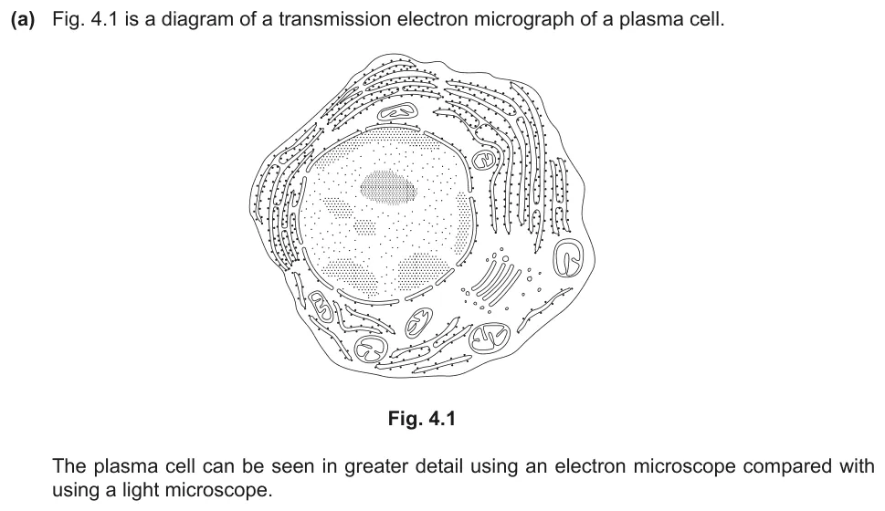
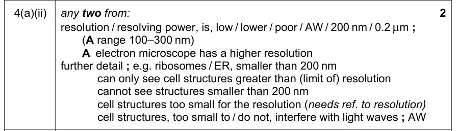
Explain why ribosomes and endoplasmic reticulum cannot be seen using a light microscope.
Information from earlier in this question:
Show the mark scheme
Tap an image to open it full size.
From the examiners' report: Link the small size of the structures to the limit of resolution. 'The structures are too small for the resolution of the light microscope' gains credit; 'the structures are small' on its own does not. Weaker answers gave 200 µm, gave the wavelength of light as the resolution, or wrote only about magnification.
Question 20
Paper 2 · October/November 2022 (21) · Q1(b) · 3 marks · Easy
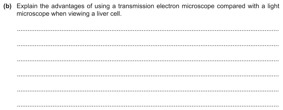
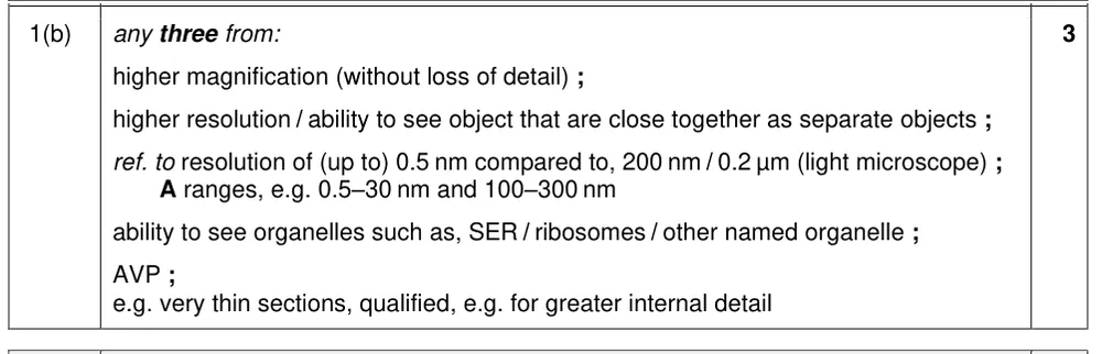
Explain the advantages of a transmission electron microscope over a light microscope for viewing a liver cell.
Show the mark scheme
Tap an image to open it full size.
From the examiners' report: 'Greater detail' needed to be related to the greater resolution of the microscope. 'More organelles can be seen' needed the organelles to be named.
Question 21
Paper 2 · October/November 2019 (22) · Q3(a) · 2 marks
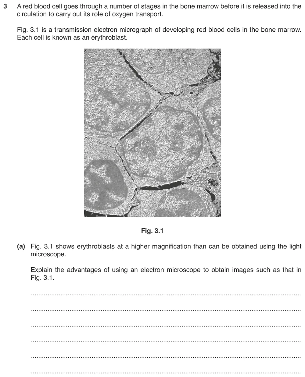
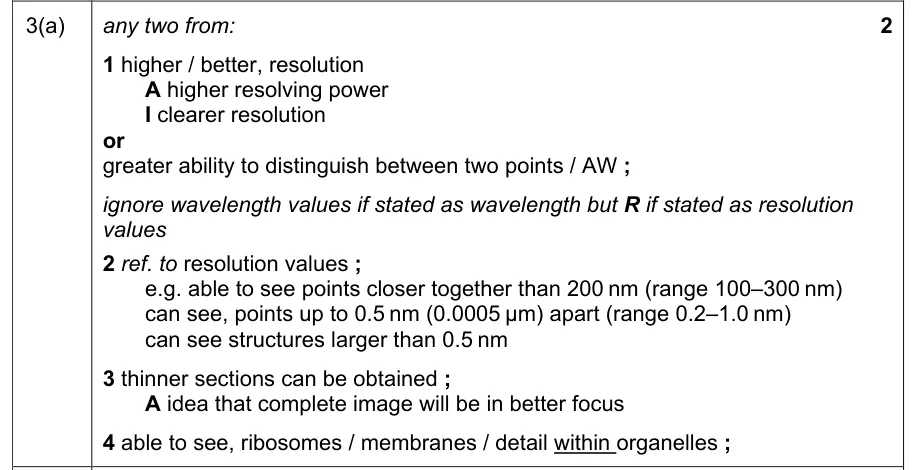
Explain the advantages of using an electron microscope to obtain an image of developing red blood cells.
Show the mark scheme
Tap an image to open it full size.
From the examiners' report: Write comparative sentences: 'electron microscopes have a high resolution' is not a comparison with the light microscope. Higher magnification was given in the question, so repeating it gained no credit. 'More detail' needed an example such as ribosomes, and some wrote 200 µm when they meant 200 nm.
Question 22
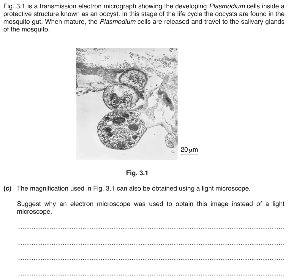
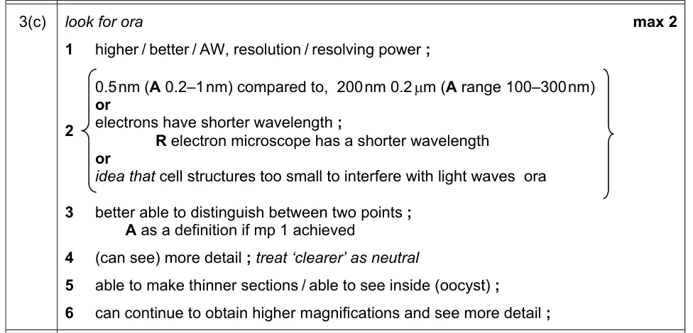
Paper 2 · May/June 2017 (22) · Q3(c) · 2 marks · Easy
Suggest why an electron microscope was used when a light microscope could give the same magnification.
Show the mark scheme
Tap an image to open it full size.
From the examiners' report: 'The image is clearer' was not enough. It is the electrons that have a shorter wavelength than light, not the microscope. The question said that a light microscope could give the same magnification, so higher magnification was not a valid answer.
Where students lose marks (from Cambridge examiners' reports and mark schemes)
Each point below comes from the examiners' reports and mark schemes, and is followed by wording that gained marks.
Convert centimetres to millimetres first. Students measured the line in centimetres and multiplied straight by 1000, so the magnification was out by a factor of 10.
✔ Gains credit: '(magnification) = image length ÷ actual length', with the line in micrometres: a line of '36 mm' gives '(×) 1440'.
Magnification has no units. Students measured and calculated correctly and then gave a unit with the magnification.
✔ Gains credit: '(×) 2000', from 'correct working 70 000 / 35'.
Round as the question asks. Students used the correct formula but did not give the answer to two significant figures.
✔ Gains credit: 'length X–Y (in mm) × 1000 ÷ 21 000', giving '1.7–1.8 (μm) (to 2 significant figures)'.
Magnification of the image, not the microscope. Students said that the magnification of the microscope is needed and explained how to find it from the eyepiece and objective lenses.
✔ Gains credit: the 'magnification of image' or a 'scale bar', the 'length of flagellum' in the image, and 'actual length of flagellum = measured length of flagellum divided by magnification'.
Stage micrometer is a slide. Students named a type of microscope, or wrote 'microscope slide' or 'micrometer slide'.
✔ Gains credit: 'stage micrometer' or 'stage micrometer scale'.
Too small for the resolution. Students wrote that the structures are small without mentioning resolution, gave 200 µm, or wrote only about magnification.
✔ Gains credit: 'resolution' is 'low' or '200 nm', so you 'cannot see structures smaller than 200 nm': 'cell structures too small for the resolution'.
Magnification describes the image. Students wrote 'zooming in to see more detail', or implied that the actual object gets bigger.
✔ Gains credit: 'number of times an image is larger than' its 'actual' size, or 'image size ÷ actual size'.
Compare the two microscopes. Students stated only that electron microscopes have a high resolution, repeated the higher magnification given in the question, or wrote 'can see more detail in the cell'.
✔ Gains credit: 'higher' or 'better' 'resolution', 'able to see points closer together than 200 nm', and 'able to see ribosomes'.
Download the questions
Download the PDF pack (22 questions, mark schemes at the end)
Keep practising
- Cells as the Basic Units of Living Organisms (1.2): Cambridge (CIE) exam questions by topic
- Cambridge (CIE) A level Biology 9700 past papers, mark schemes and examiners' reports
- Cambridge (CIE) A level Biology grade thresholds
- Book a one-to-one CIE lesson or a free 15-minute discovery chat with Tom
- Register your interest in a CIE A level Biology group class
Questions, mark schemes and examiners' report extracts © Cambridge University Press & Assessment.