

Cells as the Basic Units of Living Organisms (1.2): Cambridge (CIE) A Level Biology 9700 Exam Questions by Topic
Cells as the basic units of living organisms is sub-topic 1.2 of Topic 1, Cell structure, in the Cambridge International AS & A Level Biology syllabus (9700). You need to recognise the organelles of eukaryotic cells and outline their structures and functions, interpret photomicrographs, electron micrographs and drawings, compare plant and animal cells, and state that cells use ATP from respiration. You also need the key features of a prokaryotic cell, how it compares with eukaryotic cells, and the structure of viruses. This is AS content, examined on Papers 1 and 2. A level students meet it again on Paper 4; questions marked A level on this page come from Paper 4.
The examiners' reports return to the same points. Name structures in full: 'rough endoplasmic reticulum' and not RER, 'nuclear envelope' and not nuclear membrane, and 'nucleolus' when the question is about making ribosomal subunits. Do not confuse cristae with cisternae, or describe the capsid of a virus as a membrane or cell wall. On an electron micrograph, think about the relative sizes of organelles before you name them, and add labels when the question tells you to. Mitochondria produce ATP; they do not 'produce energy'. When you draw a bacterium, show the circular DNA as a closed loop and leave out the nucleus and mitochondria.
Studying Cambridge International A level Biology and want more help? Tom teaches CIE students one-to-one online, including students in the UAE, Saudi Arabia, Hong Kong, India and Indonesia, and every lesson is recorded. You can book a one-to-one lesson or a free 15-minute discovery chat, or register your interest in a CIE group class starting in September 2027, using the links at the bottom of this page.
What you need from IGCSE
Each question on this page assumes you're secure on these ideas. If any of them feels shaky, look it up before you start.
- Plant and animal cells both have a cell membrane, cytoplasm, a nucleus, ribosomes and mitochondria.
- Plant cells also have a cell wall, a large vacuole and often chloroplasts.
- Bacterial cells have a cell wall, cell membrane, cytoplasm, ribosomes and circular DNA, but no nucleus.
- Aerobic respiration takes place in mitochondria and releases energy.
Key terms
These words come up in the questions and mark schemes. Examiners expect you to use them exactly.
- Nucleolus: the structure inside the nucleus that makes ribosomal subunits from proteins and ribosomal RNA.
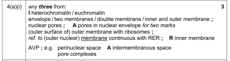
- Nuclear envelope: the two membranes around the nucleus. It has nuclear pores, and its outer membrane is continuous with the rough endoplasmic reticulum.
- Smooth endoplasmic reticulum: tubular membranes with no ribosomes, where triglycerides and other lipids are synthesised.
- Cristae: the folds of the inner membrane of a mitochondrion.
- Tonoplast: the partially permeable membrane around the large permanent vacuole of a plant cell.
- Peptidoglycan (murein): the main component of the cell wall of a prokaryotic cell.
- 70S ribosomes: the smaller ribosomes of prokaryotic cells, also found in mitochondria and chloroplasts. The cytoplasm of eukaryotic cells has larger 80S ribosomes.
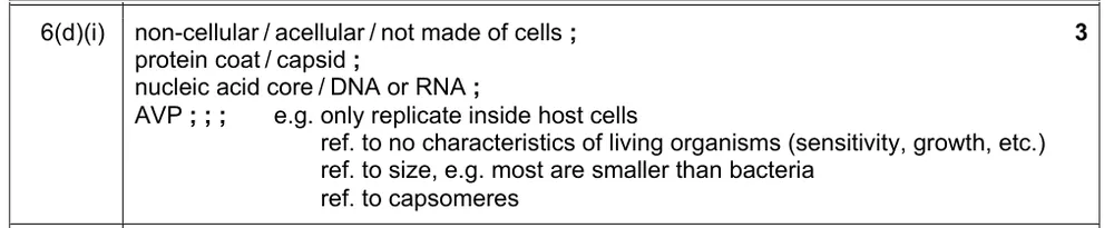
- Capsid: the protein coat of a virus, around its nucleic acid core.
What these questions cover (Cambridge 9700 Topic 1: Cell structure)
The questions test these learning outcomes from sub-topic 1.2 of the Cambridge International AS & A Level Biology syllabus (9700) for 2025 to 2027.
- 1.2.1 recognise organelles and other cell structures found in eukaryotic cells and outline their structures and functions
- 1.2.2 describe and interpret photomicrographs, electron micrographs and drawings of typical plant and animal cells
- 1.2.3 compare the structure of typical plant and animal cells
- 1.2.4 state that cells use ATP from respiration for energy-requiring processes
- 1.2.5 outline key structural features of a prokaryotic cell as found in a typical bacterium
- 1.2.6 compare the structure of a prokaryotic cell as found in a typical bacterium with the structures of typical eukaryotic cells in plants and animals
- 1.2.7 state that all viruses are non-cellular structures with a nucleic acid core (either DNA or RNA) and a capsid made of protein, and that some viruses have an outer envelope made of phospholipids
How to use this page
Write your answer on paper before you open the mark scheme. Allow about 1.2 minutes per mark: the 22 questions are worth 53 marks, so about 64 minutes in total. Then mark your answer strictly against the mark scheme and read the examiners' tip underneath it. If you'd rather print the questions, download the PDF pack.
Organelles: structure and function (1.2.1)
Question 1
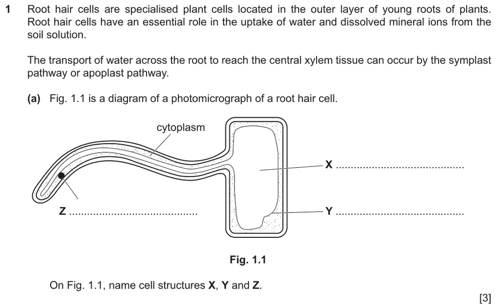
Paper 2 · May/June 2025 (22) · Q1(a) · 3 marks
Name cell structures X, Y and Z on a diagram of a root hair cell.
Show the mark scheme

Tap an image to open it full size.
Question 2
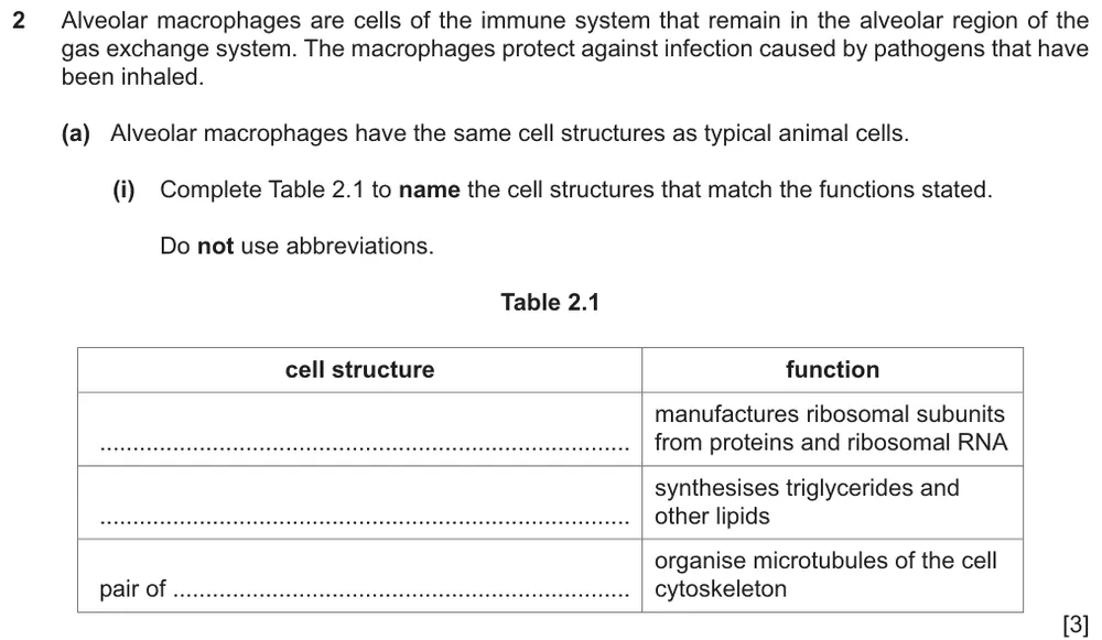
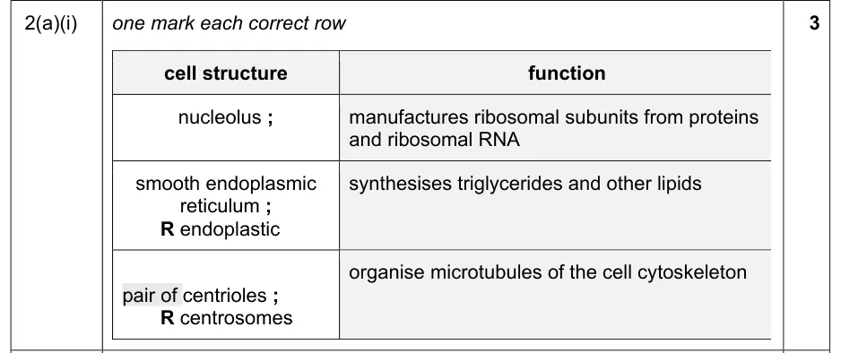
Paper 2 · May/June 2024 (22) · Q2(a)(i) · 3 marks
Complete the table to name the cell structures that match three functions.
Show the mark scheme
Tap an image to open it full size.
From the examiners' report: The most common wrong answers for the nucleolus were the nucleus, the ribosome and the rough endoplasmic reticulum. The Golgi body and rough endoplasmic reticulum were often given in place of the smooth endoplasmic reticulum. 'Centrosomes' was the most frequent wrong answer for the pair of centrioles.
Question 3
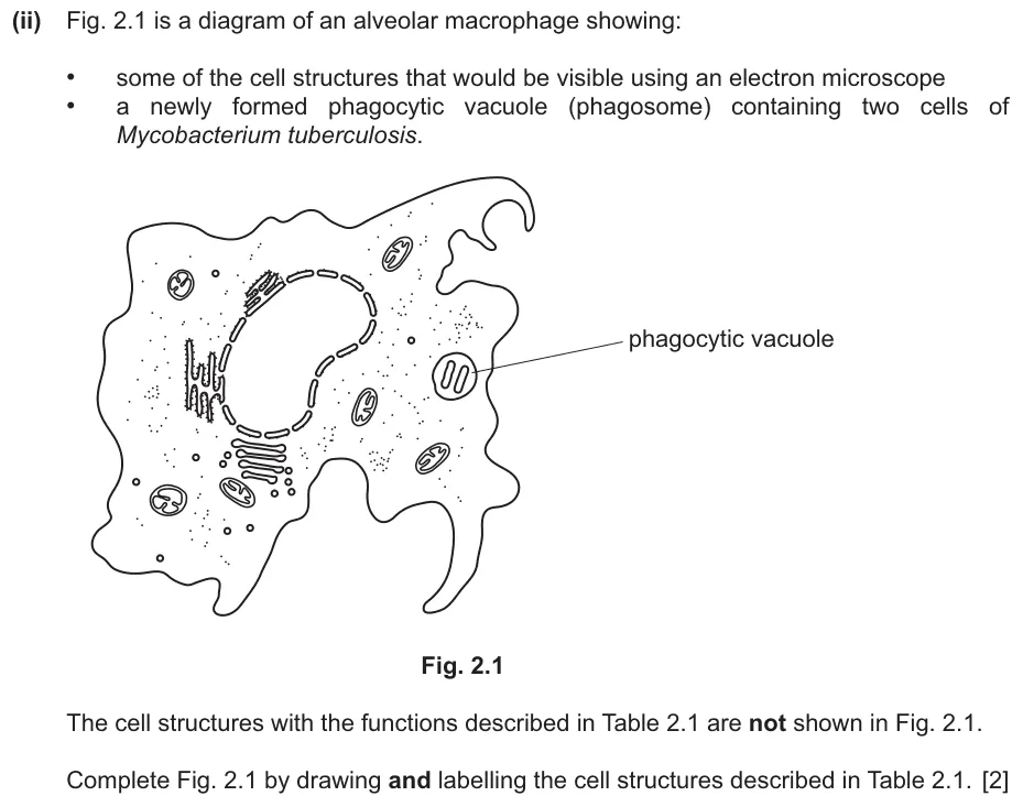
Paper 2 · May/June 2024 (22) · Q2(a)(ii) · 2 marks
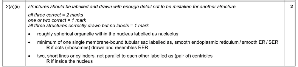
Draw and label the three cell structures from the table on a diagram of a macrophage.
Show the mark scheme
Tap an image to open it full size.
From the examiners' report: There were many very good drawings. The pair of centrioles gained credit least often: some drew them as two ovals, or as sticks parallel to each other and not at right angles.
Question 4
Paper 2 · October/November 2020 (22) · Q4(a)(i) · 3 marks
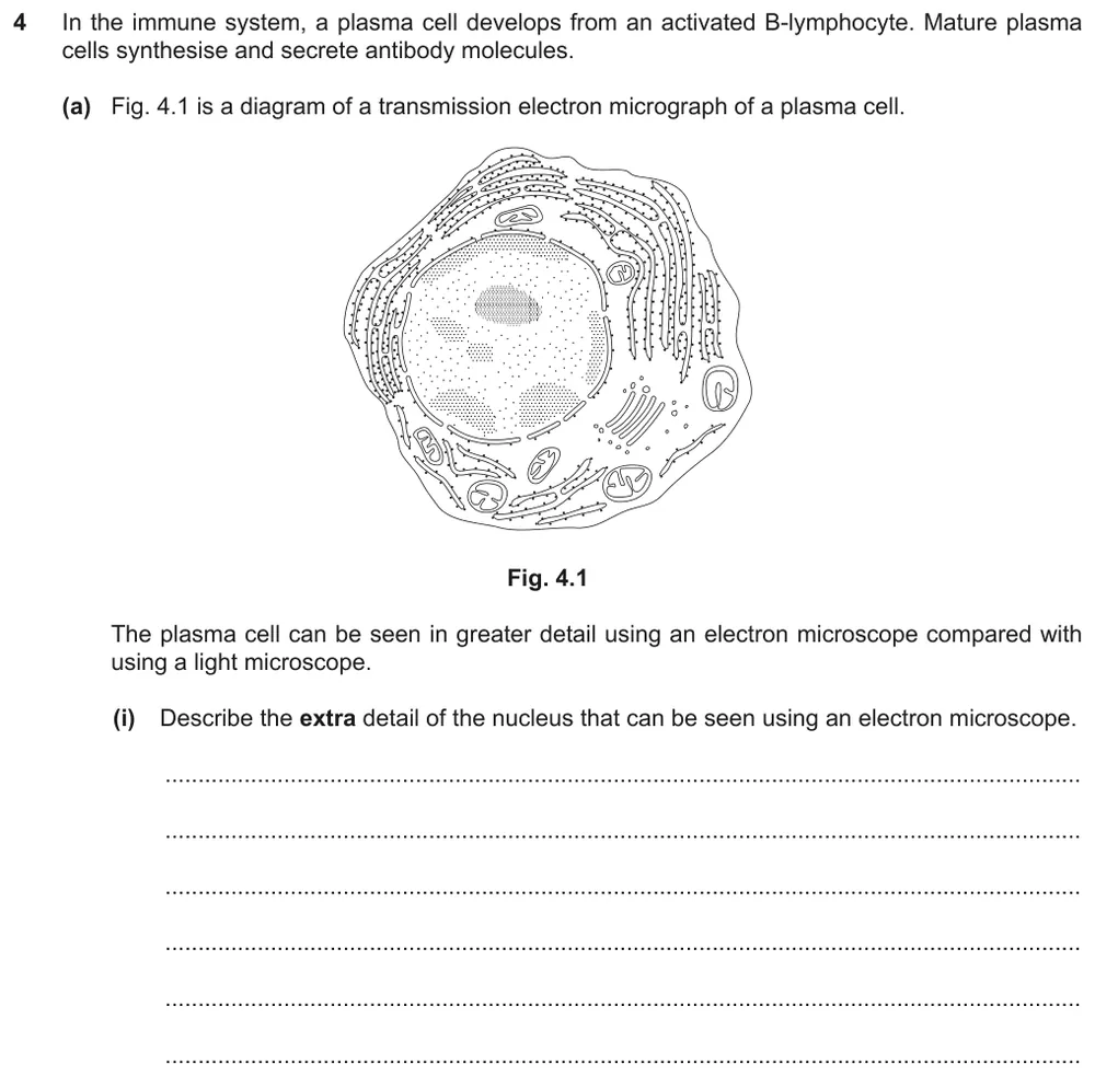
Describe the extra detail of the nucleus that can be seen using an electron microscope.
Show the mark scheme
Tap an image to open it full size.
From the examiners' report: Many wrote 'nuclear membrane' for the nuclear envelope and did not gain credit. Only the strongest answers placed ribosomes on the surface of the outer membrane, or said that the outer membrane is continuous with the rough endoplasmic reticulum. Some described the extra detail of the whole plasma cell, which did not answer the question.
Question 5
Paper 2 · October/November 2018 (22) · Q6(a) · 3 marks · Easy
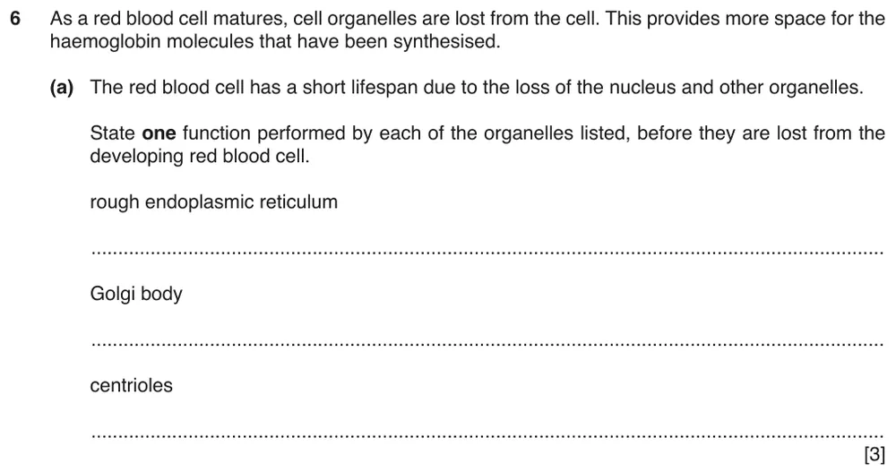
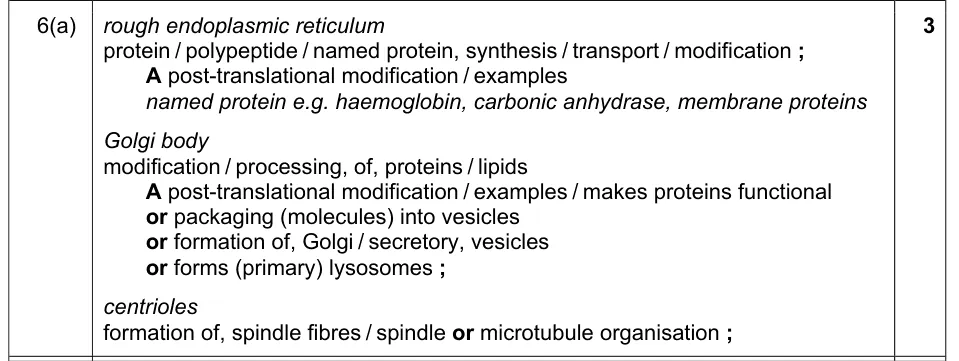
State one function of the rough endoplasmic reticulum, the Golgi body and the centrioles.
Show the mark scheme
Tap an image to open it full size.
From the examiners' report: For the Golgi body, more detail than 'forms vesicles' was needed. For centrioles, saying that they are involved in mitosis was too vague: refer to organising the microtubules of the spindle, or to forming spindle fibres.
Question 6
Paper 2 · May/June 2023 (22) · Q4(a)(ii) · 2 marks
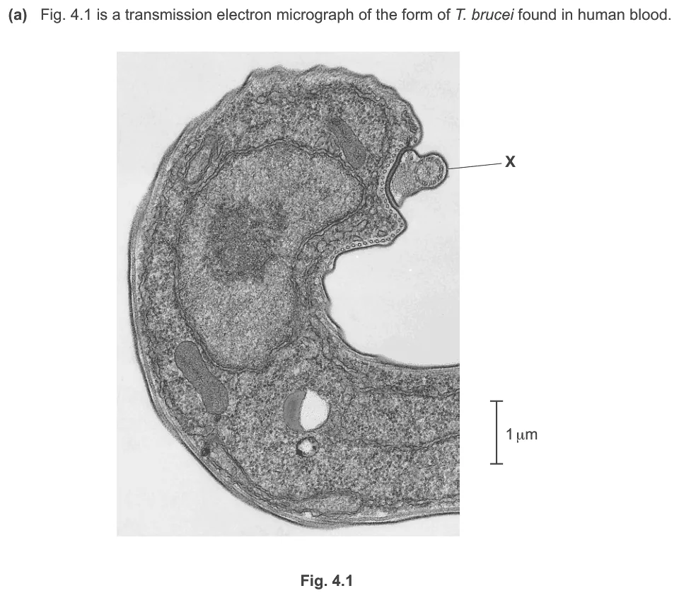
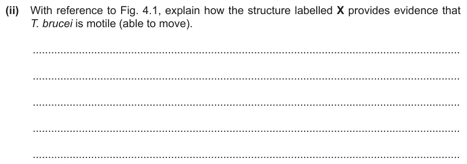
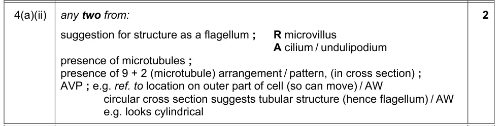
Explain how structure X on the electron micrograph is evidence that the organism can move.
Information from earlier in this question:

Show the mark scheme
Tap an image to open it full size.
From the examiners' report: Look closely at structure X: it resembles a transverse section of a cilium, so refer to microtubules and the 9 + 2 pattern. Many did not mention the microtubules and linked movement to endocytosis or phagocytosis.
Question 7
Paper 4 (A level) · May/June 2024 (43) · Q8(c) · 2 marks · Hard
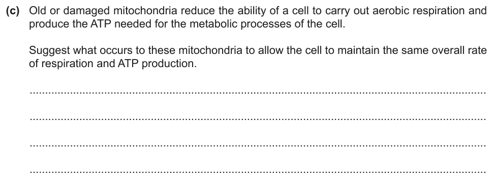
Suggest what happens to old or damaged mitochondria so that the cell keeps up its rate of ATP production.
Show the mark scheme

Tap an image to open it full size.
From the examiners' report: Only a minority said that the mitochondria are broken down by lysosomes. A few added that the products of the breakdown are used to make new mitochondria.
Interpreting electron micrographs (1.2.2)
Question 8
Paper 2 · October/November 2023 (22) · Q3(c)(i) · 3 marks
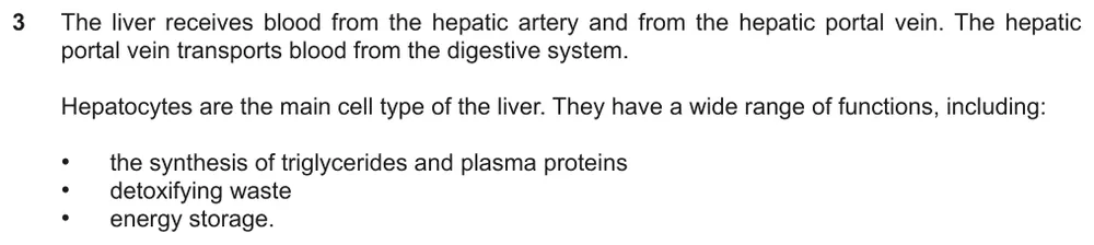
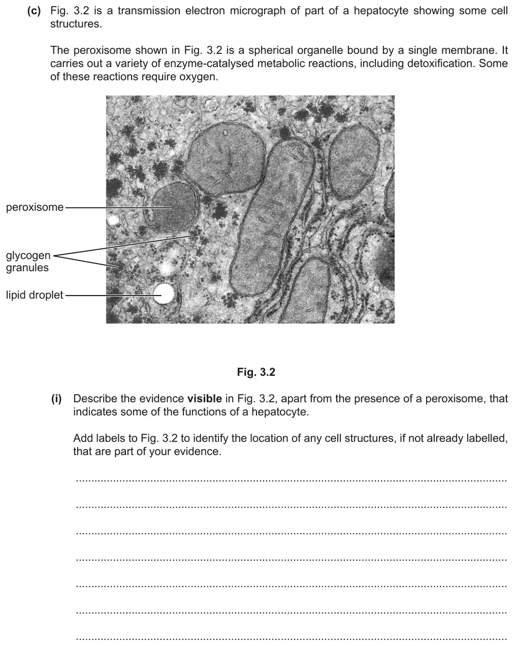
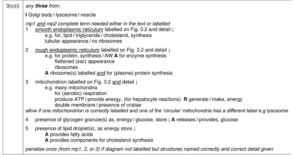
Describe the evidence in an electron micrograph of a liver cell that shows some of the cell's functions, and label the structures you use.
Information from earlier in this question:
Show the mark scheme
Tap an image to open it full size.
From the examiners' report: Credit was only given when the organelles were also correctly labelled on the micrograph, which some students missed. A common error was to label rough endoplasmic reticulum as the Golgi body. Mitochondria do not 'produce energy' and are not a store of ATP.
Question 9
Paper 2 · October/November 2023 (22) · Q3(c)(ii) · 1 mark
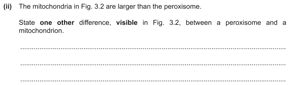
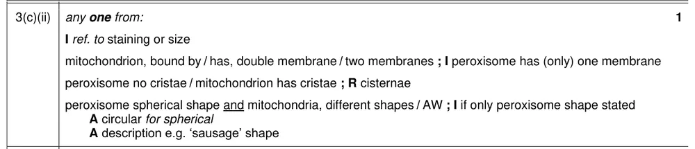
State one visible difference, other than size, between a peroxisome and a mitochondrion.
Show the mark scheme
Tap an image to open it full size.
From the examiners' report: Most gave the double membrane of the mitochondrion; fewer noted the cristae. A common error was to call the cristae 'cisternae'.
Question 10
Paper 2 · October/November 2022 (21) · Q1(a)(i) · 2 marks · Hard
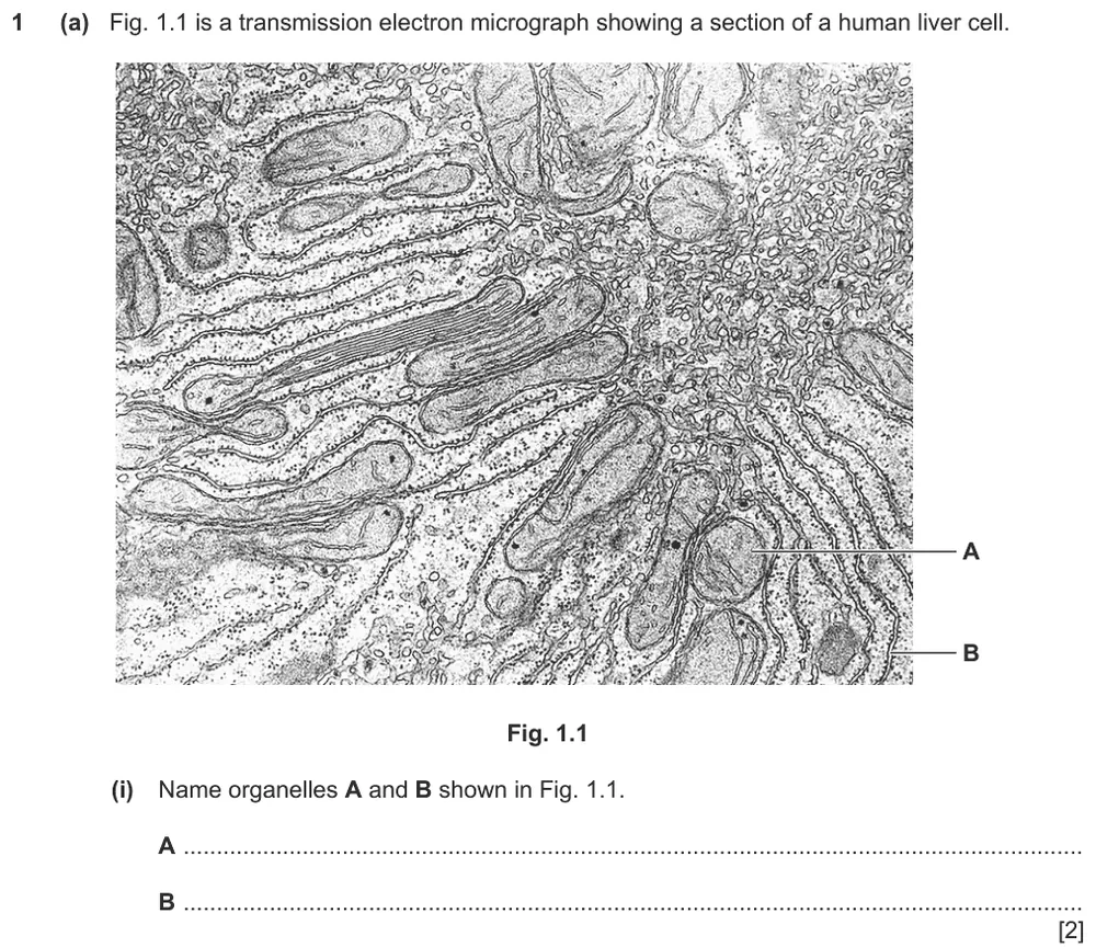
Name organelles A and B on a transmission electron micrograph of a liver cell.
Show the mark scheme

Tap an image to open it full size.
From the examiners' report: Many called A the nucleus. Think about scale: the nucleus is very large compared with the other organelles, and the visible cristae show that A is a mitochondrion. For B, 'endoplasmic reticulum' without the type was not enough, and the abbreviation RER was not accepted.
Comparing plant and animal cells (1.2.3)
Question 11
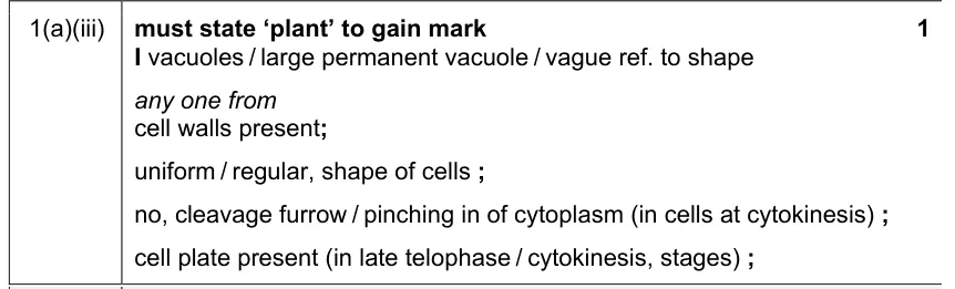
Paper 2 · May/June 2021 (22) · Q1(a)(iii) · 1 mark
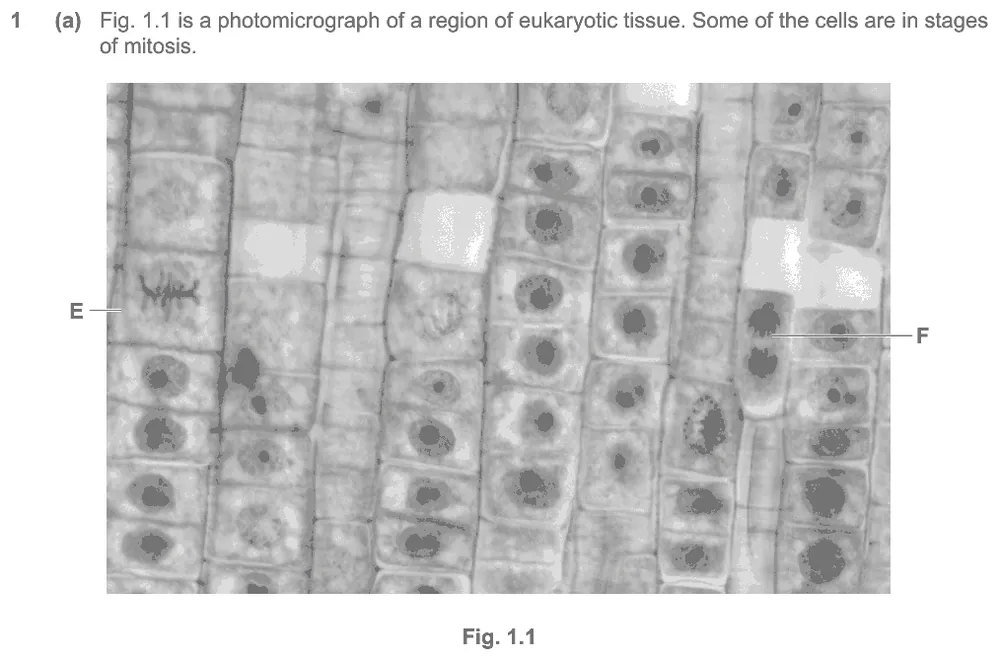
State, with a reason, whether a photomicrograph shows animal tissue or plant tissue.
Information from earlier in this question:

Show the mark scheme
Tap an image to open it full size.
From the examiners' report: Cell walls were the most common correct reason. Students who did not choose plant tissue often said that no large vacuoles could be seen, not realising that the cells of meristematic tissue do not have them.
Question 12
Paper 2 · October/November 2019 (22) · Q1(d) · 5 marks
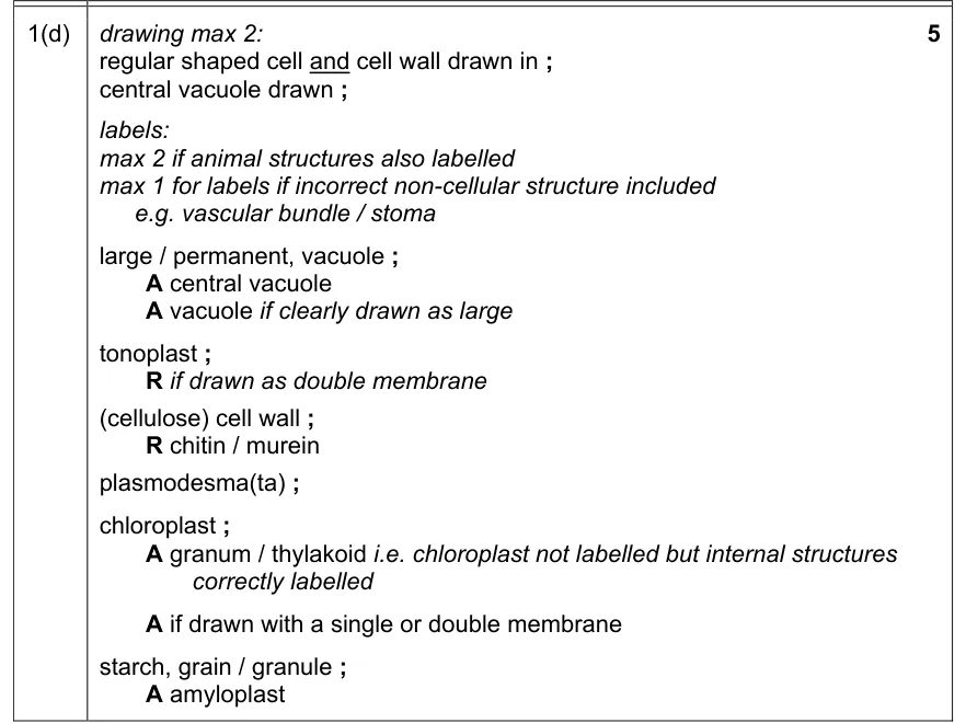
Draw a typical plant cell and label only the structures found only in plant cells.

Show the mark scheme
Tap an image to open it full size.
From the examiners' report: Quite a few answers gained only partial credit because they also labelled structures found in animal cells. In some diagrams the large permanent vacuole was too small or missing, the tonoplast was drawn as a double membrane, or there was no cell wall.
ATP from respiration (1.2.4)
Question 13
Paper 2 · February/March 2022 (22) · Q5(a)(ii) · 1 mark
Explain why blocking a small artery in the brain leads to brain damage.
Information from earlier in this question:


Show the mark scheme

Tap an image to open it full size.
From the examiners' report: Many realised that less oxygen or glucose would be delivered, so the rate of aerobic respiration would fall. Weak answers referred only to a reduced supply of blood.
Prokaryotic cells (1.2.5)
Question 14
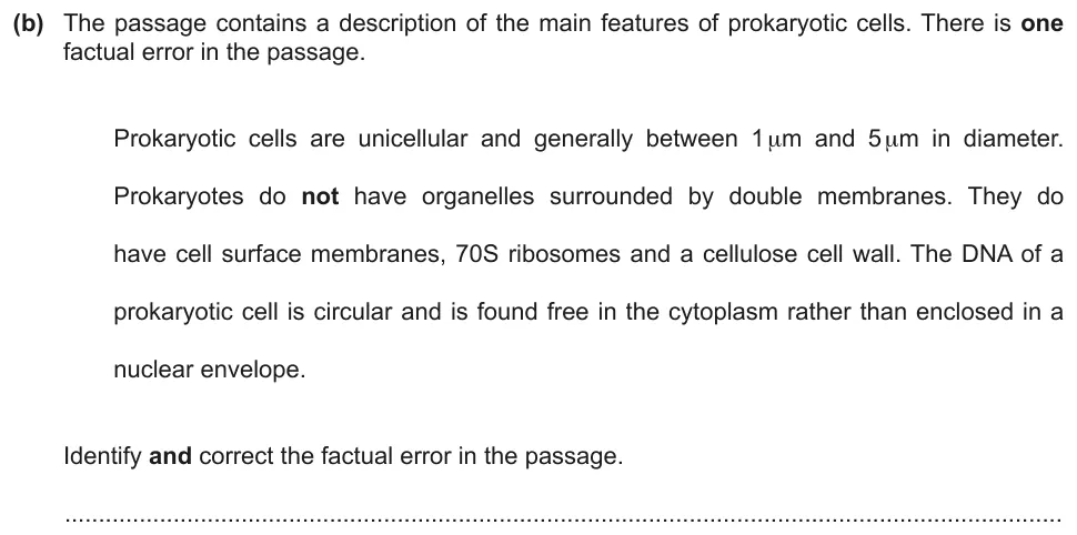
Paper 2 · February/March 2024 (22) · Q5(b) · 1 mark
Find and correct the factual error in a passage about prokaryotic cells.
Show the mark scheme

Tap an image to open it full size.
Question 15
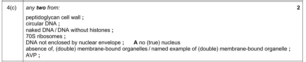
Paper 2 · February/March 2022 (22) · Q4(c) · 2 marks · Easy
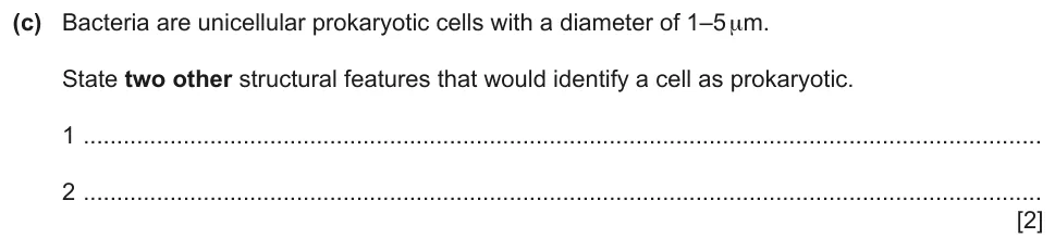
State two structural features, other than size, that identify a cell as prokaryotic.
Show the mark scheme
Tap an image to open it full size.
From the examiners' report: Most gave two correct features. Some confused prokaryotes with eukaryotes, and a small number confused them with viruses.
Question 16
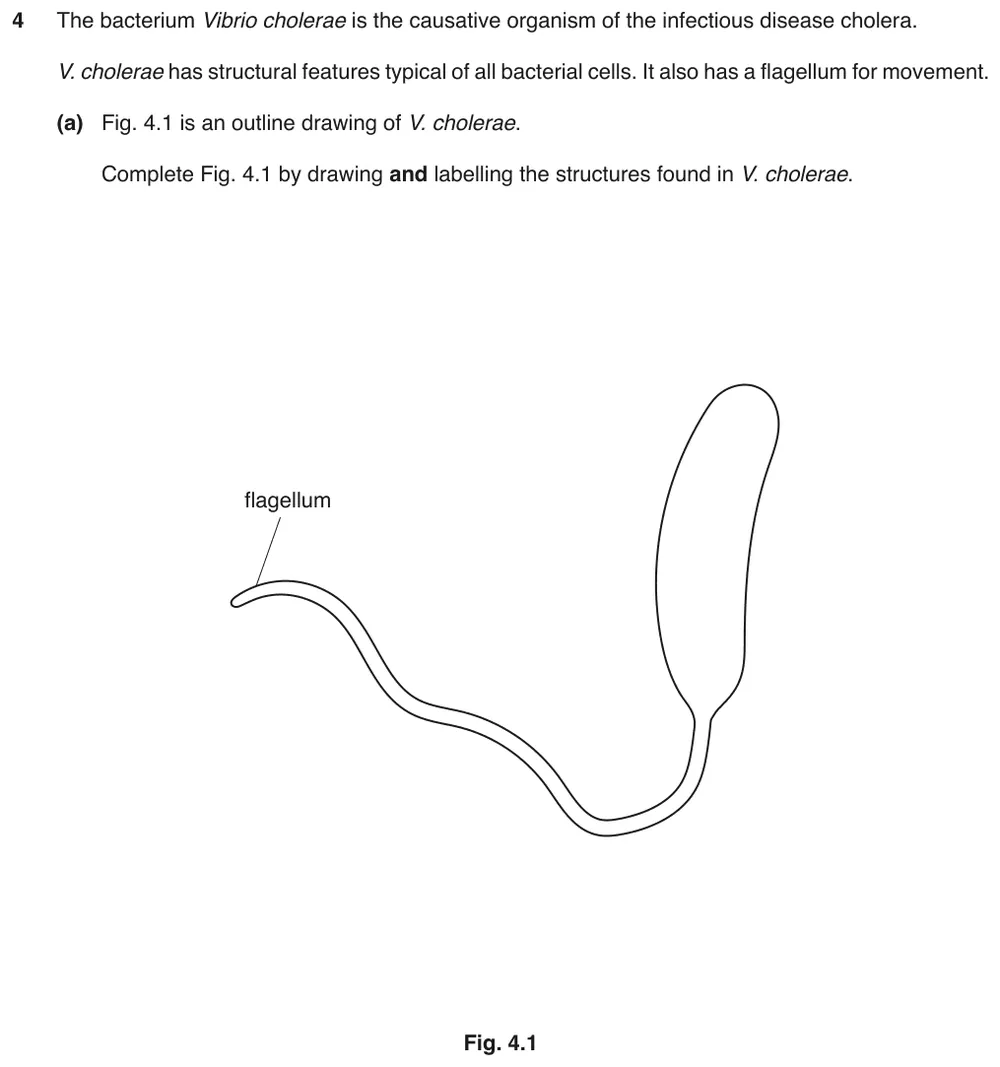
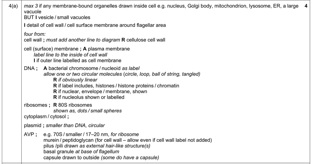
Paper 2 · May/June 2019 (22) · Q4(a) · 4 marks
Complete an outline drawing of a bacterium by drawing and labelling its structures.
Show the mark scheme
Tap an image to open it full size.
From the examiners' report: The cell wall and cell surface membrane only gained credit if another line was drawn in. Some labelled 'circular DNA' but drew unconnected lines or coils with a clear start and end, and some called the main circular DNA the plasmid. Full marks could not be given if a nucleus or mitochondrion was added.
Comparing prokaryotic and eukaryotic cells (1.2.6)
Question 17
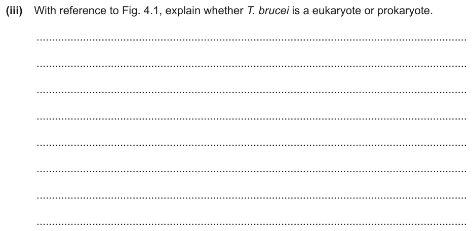
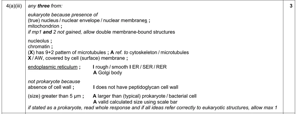
Paper 2 · May/June 2023 (22) · Q4(a)(iii) · 3 marks
Use an electron micrograph to explain whether an organism is a eukaryote or a prokaryote.
Information from earlier in this question:
Show the mark scheme
Tap an image to open it full size.
From the examiners' report: The clearest evidence is the large nucleus with its nuclear envelope. Organelles with a double membrane gained credit; organelles with a single membrane did not.
Question 18
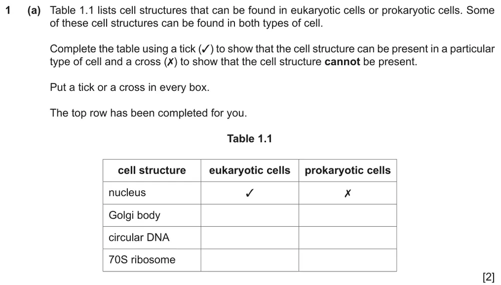
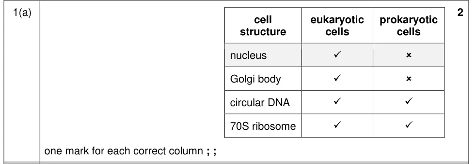
Paper 2 · February/March 2023 (22) · Q1(a) · 2 marks · Easy
Complete the table with ticks and crosses to show which structures are found in eukaryotic and prokaryotic cells.
Show the mark scheme
Tap an image to open it full size.
From the examiners' report: Most knew that prokaryotic cells have circular DNA and 70S ribosomes but no Golgi body. Fewer recognised that many eukaryotic cells also have small circular DNA and 70S ribosomes, because they have mitochondria and sometimes chloroplasts.
Question 19
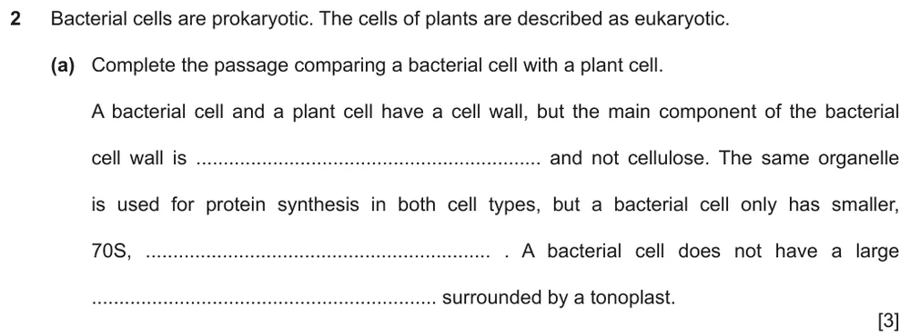
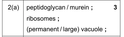
Paper 2 · May/June 2022 (23) · Q2(a) · 3 marks · Hard
Complete a passage comparing a bacterial cell with a plant cell.
Show the mark scheme
Tap an image to open it full size.
From the examiners' report: Most knew 'ribosomes' and 'vacuole'. The component of the bacterial cell wall was harder to recall: some wrote 'chitin' in place of murein or peptidoglycan.
Viruses (1.2.7)
Question 20
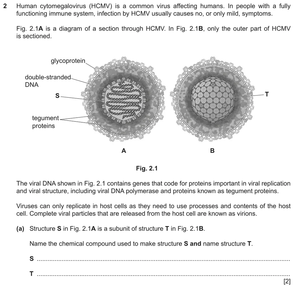
Paper 2 · October/November 2022 (22) · Q2(a) · 2 marks
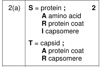
Name the chemical compound that makes up structure S and name structure T on a diagram of a virus.
Show the mark scheme
Tap an image to open it full size.
From the examiners' report: S asks for a chemical compound, so the answer is protein and not a structural term. T is the capsid or protein coat. Adding 'capsomere' to the answer for T lost the mark, and the spelling 'caspid' was not credited.
Question 21
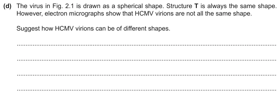
Paper 2 · October/November 2022 (22) · Q2(d) · 2 marks
Suggest how the virions of one virus can be different shapes.
Show the mark scheme

Tap an image to open it full size.
From the examiners' report: The diagram shows the capsid surrounded by an outer layer: the envelope is flexible, and this explains the change in shape. The most common wrong suggestions were mutations, or glycoproteins with different tertiary structures.
Question 22
Paper 2 · February/March 2019 (22) · Q6(d)(i) · 3 marks · Hard
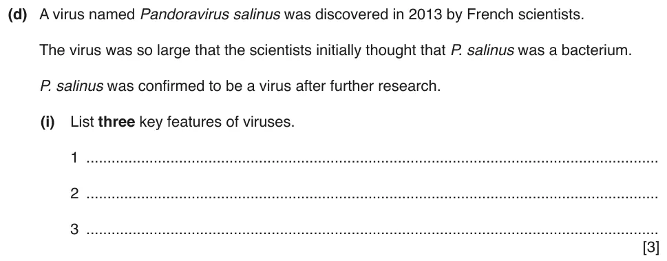
List three key features of viruses.
Show the mark scheme
Tap an image to open it full size.
From the examiners' report: Students found it hard to state three key features clearly. Some implied that viruses only exist inside host cells, and a number described the protein coat or capsid as the membrane or cell wall of the virus.
Where students lose marks (from Cambridge examiners' reports and mark schemes)
Each point below comes from the examiners' reports and mark schemes, and is followed by wording that gained marks.
Nucleolus, not nucleus. Students gave the nucleus, the ribosome or the rough endoplasmic reticulum as the structure that makes ribosomal subunits, and wrote 'centrosomes' for the centrioles.
✔ Gains credit: 'nucleolus' for 'manufactures ribosomal subunits', 'smooth endoplasmic reticulum' for 'synthesises triglycerides and other lipids', and 'pair of centrioles'.
Envelope, not nuclear membrane. Students wrote 'nuclear membrane', or described the extra detail of the whole cell.
✔ Gains credit: 'envelope' or 'two membranes', 'nuclear pores', 'outer membrane with ribosomes' and outer 'membrane continuous with RER'.
Say what centrioles do. Students wrote that centrioles are involved in mitosis, and that the Golgi body 'forms vesicles'.
✔ Gains credit: centrioles: 'formation of spindle fibres' or 'microtubule organisation'; Golgi body: 'modification' of 'proteins' or 'packaging (molecules) into vesicles'.
Label the micrograph. Students named organelles in their answer without labelling them on the micrograph, or labelled rough endoplasmic reticulum as the Golgi body.
✔ Gains credit: 'rough endoplasmic reticulum labelled' with detail such as 'for protein synthesis', and 'mitochondrion labelled' with detail such as 'produce ATP'.
Cristae, not cisternae. Students named the folds inside the mitochondrion as cisternae.
✔ Gains credit: 'mitochondrion' 'has double membrane', or 'mitochondrion has cristae' and 'peroxisome no cristae'.
Label plant-only structures. Students also labelled structures found in animal cells, drew the vacuole too small or drew the tonoplast as a double membrane.
✔ Gains credit: labels for the 'large' or 'permanent' 'vacuole', 'tonoplast', '(cellulose) cell wall', 'plasmodesma(ta)' and 'chloroplast'.
Draw circular DNA as a loop. Students labelled circular DNA but drew lines with a clear start and end, called it the plasmid, or added a nucleus or mitochondrion.
✔ Gains credit: 'DNA' drawn as a 'circle' or 'loop', 'cell wall', 'cell (surface) membrane', 'ribosomes' and 'plasmid', with no 'membrane-bound organelles drawn inside cell'.
Capsid, not caspid. Students gave a structural term for S, added 'capsomere' to their answer for T, or spelt capsid as 'caspid'.
✔ Gains credit: 'S = protein' and 'T = capsid' or 'protein coat'.
Download the questions
Download the PDF pack (22 questions, mark schemes at the end)
Keep practising
- The Microscope in Cell Studies (1.1): Cambridge (CIE) exam questions by topic
- Cambridge (CIE) A level Biology 9700 past papers, mark schemes and examiners' reports
- Cambridge (CIE) A level Biology grade thresholds
- Book a one-to-one CIE lesson or a free 15-minute discovery chat with Tom
- Register your interest in a CIE A level Biology group class
Questions, mark schemes and examiners' report extracts © Cambridge University Press & Assessment.