

The Heart (8.3): Cambridge (CIE) A Level Biology 9700 Exam Questions by Topic
The heart is sub-topic 8.3 of Topic 8, Transport in mammals, in the Cambridge International AS & A Level Biology syllabus (9700). You need to describe the external and internal structure of the mammalian heart, explain why the walls of the atria, the right ventricle and the left ventricle differ in thickness, describe the cardiac cycle by linking the blood pressure changes in systole and diastole to the opening and closing of valves, and explain the roles of the sinoatrial node, the atrioventricular node and the Purkyne tissue. This is AS content, examined on Papers 1 and 2. A level students meet it again on Paper 4.
The examiners' reports return to the same points. A thick ventricle wall generates a high pressure; answers saying that it must 'withstand high pressure' or stop the ventricle bursting gain no credit. A valve opens because of the pressures on its two sides, so compare the pressure in the ventricle with the pressure in the atrium or in the artery. The bicuspid valve is on the left and the tricuspid valve is on the right. Write about both atria and both ventricles when a question is about the whole heart. The delay at the atrioventricular node lets the atria empty and the ventricles fill before ventricular systole; it does not prevent backflow.
Studying Cambridge International A level Biology and want more help? Tom teaches CIE students one-to-one online, including students in the UAE, Saudi Arabia, Hong Kong, India and Indonesia, and every lesson is recorded. You can book a one-to-one lesson or a free 15-minute discovery chat, or register your interest in a CIE group class starting in September 2027, using the links at the bottom of this page.
What you need from IGCSE
Each question on this page assumes you're secure on these ideas. If any of them feels shaky, look it up before you start.
- The heart has four chambers, two atria and two ventricles, and a septum separates the left side from the right side.
- Arteries carry blood away from the heart and veins carry blood towards the heart.
- Mammals have a double circulation: blood passes through the heart twice in one complete circuit, once on its way to the lungs and once on its way to the rest of the body.
- Valves in the heart prevent the backflow of blood.
Key terms
These words come up in the questions and mark schemes. Examiners expect you to use them exactly.
- Systole: contraction of the heart muscle. Atrial systole is contraction of the atria; ventricular systole is contraction of the ventricles.
- Diastole: relaxation of the heart muscle.
- Atrioventricular valves: the valves between the atria and the ventricles: the bicuspid (mitral) valve on the left and the tricuspid valve on the right.
- Semilunar valves: the aortic valve and the pulmonary valve, where blood leaves the ventricles.
- Sinoatrial node (SAN): specialised muscle tissue in the wall of the right atrium that sends out waves of excitation; it is the pacemaker and initiates the heartbeat.
- Atrioventricular node (AVN): delays the impulse from the atria for a short time, then passes it on to the Purkyne tissue.
- Purkyne tissue: specialised muscle fibres that carry the impulse down the septum to the base of the ventricles, so the ventricles contract from the base upwards.
- Myogenic: describes heart muscle: the electrical activity that controls the heartbeat begins in the muscle itself.
What these questions cover (Cambridge 9700 Topic 8: Transport in mammals)
The questions test these learning outcomes from sub-topic 8.3 of the Cambridge International AS & A Level Biology syllabus (9700) for 2025 to 2027.
- 8.3.1 describe the external and internal structure of the mammalian heart
- 8.3.2 explain the differences in the thickness of the walls of the: • atria and ventricles • left ventricle and right ventricle
- 8.3.3 describe the cardiac cycle, with reference to the relationship between blood pressure changes during systole and diastole and the opening and closing of valves
- 8.3.4 explain the roles of the sinoatrial node, the atrioventricular node and the Purkyne tissue in the cardiac cycle
How to use this page
Write your answer on paper before you open the mark scheme. Allow about 1.2 minutes per mark: the 22 questions are worth 54 marks, so about 65 minutes in total. Then mark your answer strictly against the mark scheme and read the examiners' tip underneath it. If you'd rather print the questions, download the PDF pack.
Structure of the heart (8.3.1)
Question 1
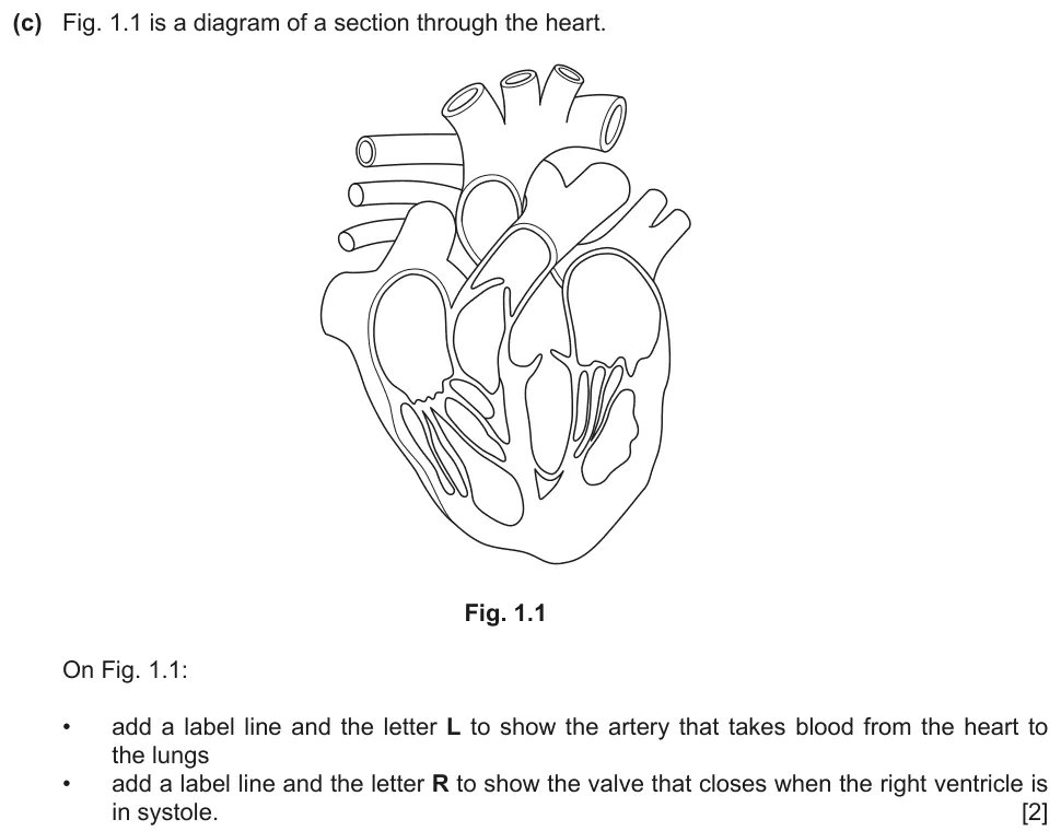
Paper 2 · October/November 2022 (22) · Q1(c) · 2 marks
Label the artery that takes blood to the lungs and the valve that closes when the right ventricle is in systole.
Show the mark scheme

Tap an image to open it full size.
From the examiners' report: Use label lines that end clearly on the structure: a letter written close to the structure gained no credit. Label lines for R often ended at the semilunar valve of the pulmonary artery. 'The right ventricle is in systole' means that the right ventricle contracts.
Question 2
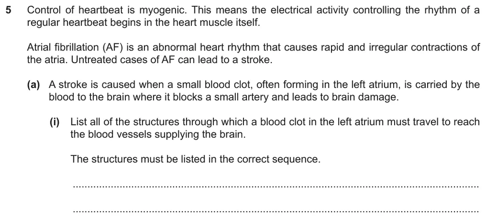
Paper 2 · February/March 2022 (22) · Q5(a)(i) · 1 mark
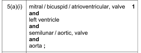
List, in the correct sequence, the structures a blood clot passes through from the left atrium to the blood vessels supplying the brain.
Show the mark scheme
Tap an image to open it full size.
From the examiners' report: Include the valves in your list. Some students wrongly took the clot through the right side of the heart and the pulmonary circulation.
Question 3
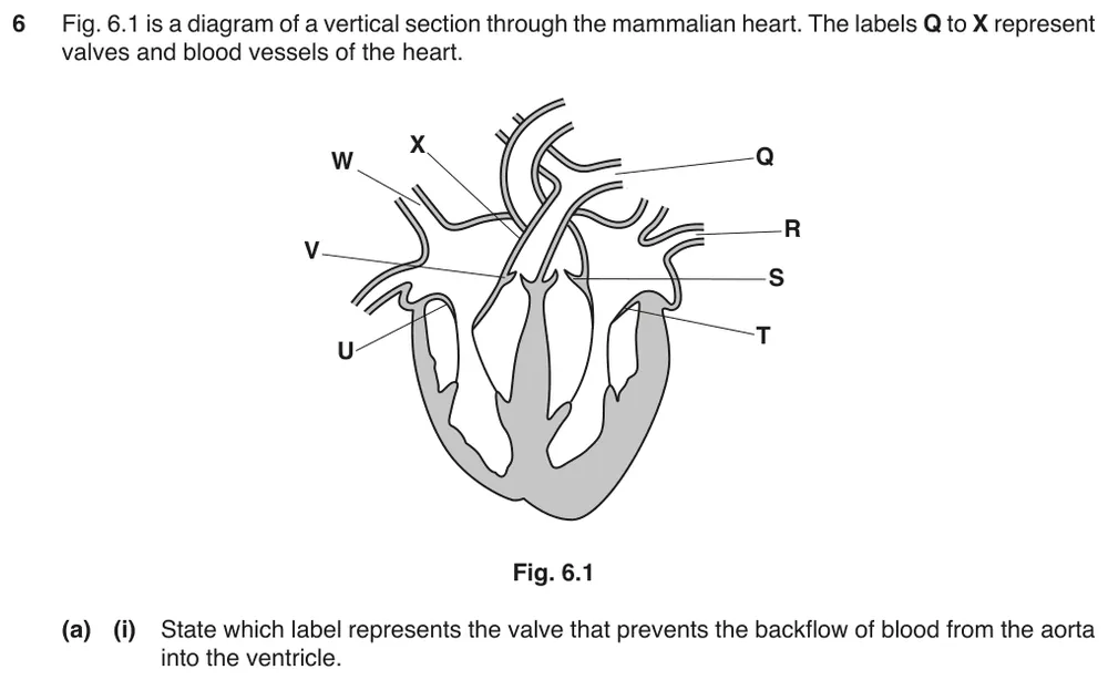
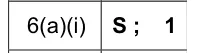
Paper 2 · October/November 2017 (22) · Q6(a)(i) · 1 mark · Easy
Identify the valve that prevents the backflow of blood from the aorta into the ventricle.
Show the mark scheme
Tap an image to open it full size.
From the examiners' report: Almost always correct. Only the weakest answers gave V, the semilunar valve between the right ventricle and the pulmonary artery.
Question 4
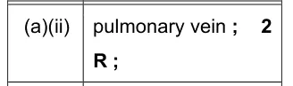
Paper 2 · October/November 2017 (22) · Q6(a)(ii) · 2 marks
Name the blood vessel that carries oxygenated blood from the lungs to the heart and give its label.

Show the mark scheme
Tap an image to open it full size.
From the examiners' report: A common wrong answer was the pulmonary artery, with the letter W.
Question 5
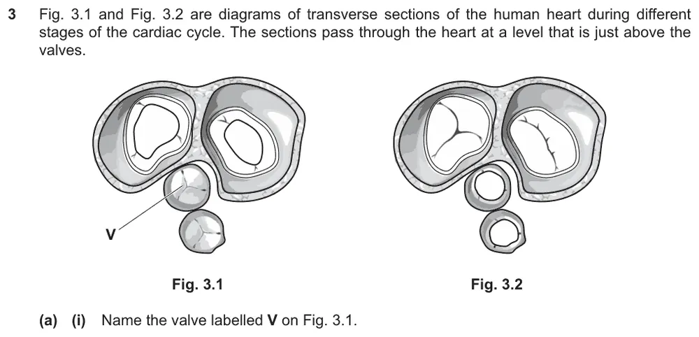
Paper 2 · February/March 2025 (22) · Q3(a)(i) · 1 mark
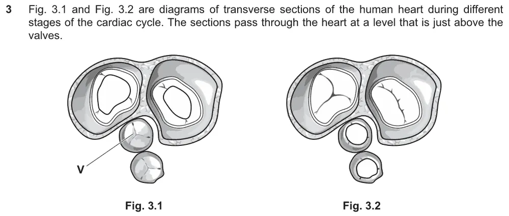
Name the valve labelled V in a transverse section of the heart.
Show the mark scheme

Tap an image to open it full size.
Thickness of the heart walls (8.3.2)
Question 6
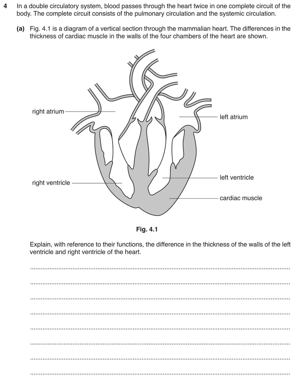
Paper 2 · February/March 2019 (22) · Q4(a) · 3 marks · Easy
Explain, from their functions, the difference in thickness between the walls of the left ventricle and the right ventricle.
Show the mark scheme

Tap an image to open it full size.
From the examiners' report: The thicker muscular wall lets the left ventricle generate a higher pressure, which is needed to overcome the greater resistance of the systemic circulation. Some students wrongly said the thicker wall is needed to withstand higher pressures.
Question 7
Paper 2 · October/November 2023 (23) · Q6(a) · 2 marks
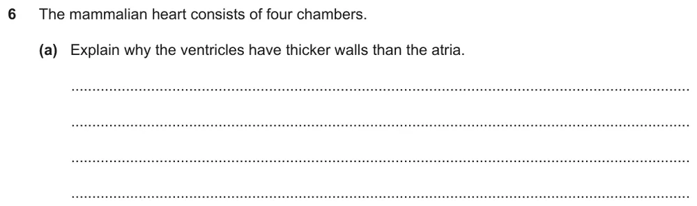
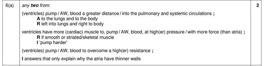
Explain why the ventricles have thicker walls than the atria.
Show the mark scheme
Tap an image to open it full size.
From the examiners' report: 'Thick walls to withstand high pressure' and 'thick walls prevent bursting' were very common and gained no credit. Say that the ventricles pump blood further, to the lungs and to the body, at a higher pressure and against a higher resistance. The muscle is cardiac muscle, not smooth muscle.
The cardiac cycle: pressure changes and valves (8.3.3)
Question 8
Paper 2 · February/March 2025 (22) · Q3(a)(ii) · 2 marks
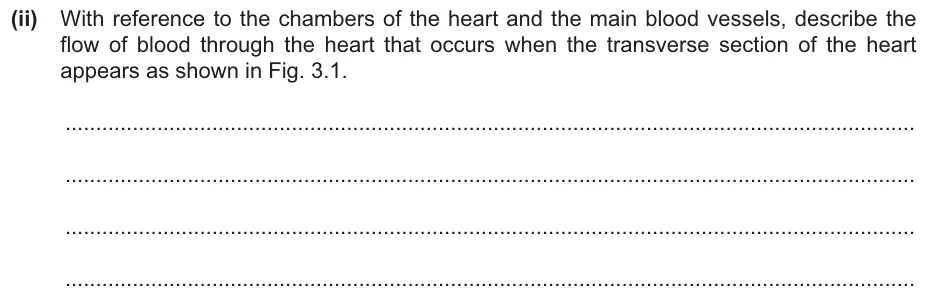
Describe the flow of blood through the heart at the stage shown in Fig. 3.1.
Information from earlier in this question:
Show the mark scheme

Tap an image to open it full size.
From the examiners' report: Describe only the blood flow at the stage shown; explanations were not needed. Write 'atria' and 'ventricles', not 'the atrium' and 'the ventricle', so that your answer covers the whole heart.
Question 9
Paper 2 · February/March 2025 (22) · Q3(b) · 2 marks
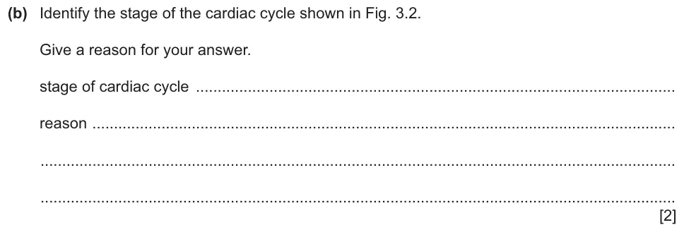
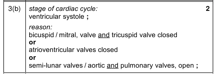
Identify the stage of the cardiac cycle shown in Fig. 3.2 and give a reason.
Show the mark scheme
Tap an image to open it full size.
Question 10
Paper 2 · May/June 2023 (22) · Q1(a) · 2 marks
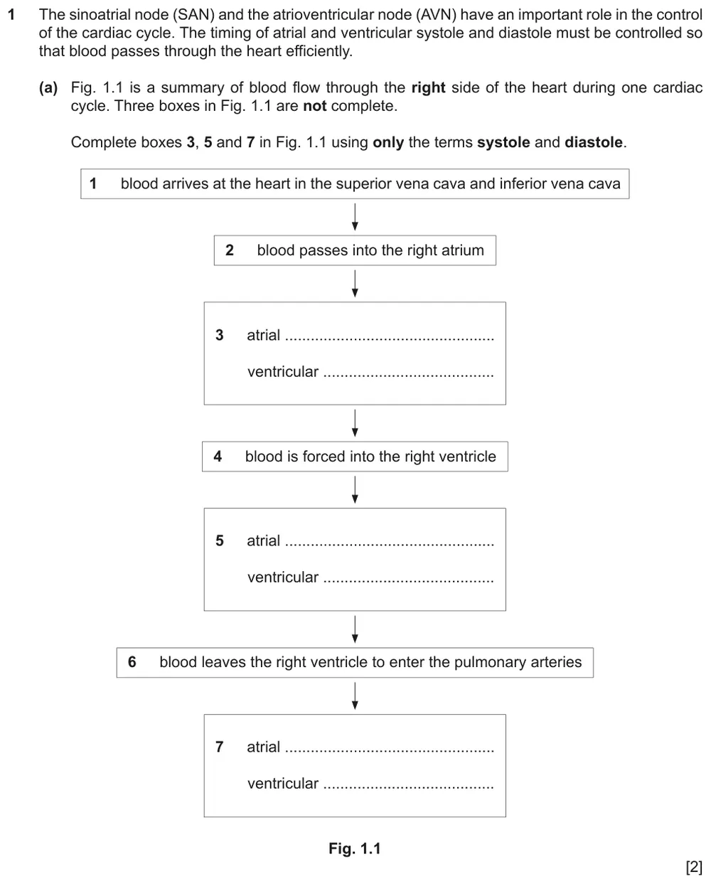
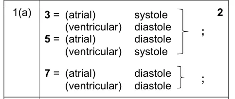
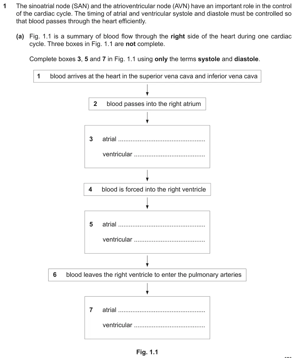
Complete the flow chart of the cardiac cycle using only the terms systole and diastole.
Show the mark scheme
Tap an image to open it full size.
From the examiners' report: Use only the terms you are told to use: some students wrote contraction and relaxation. Atrial systole and ventricular systole do not happen at the same time. Some completed each box the wrong way round.
Question 11
Paper 2 · May/June 2023 (22) · Q1(c) · 3 marks
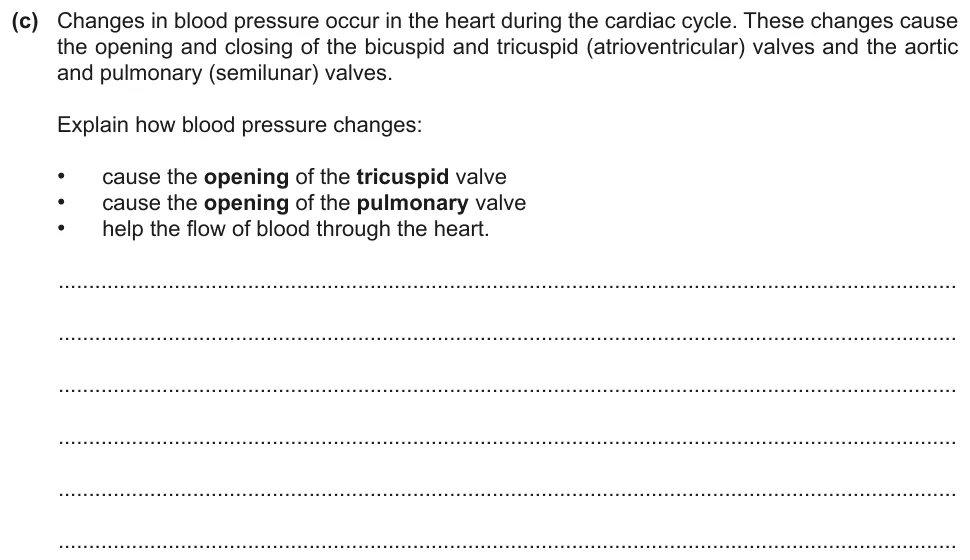
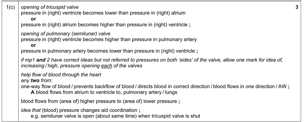
Explain how blood pressure changes open the tricuspid valve and the pulmonary valve and help the flow of blood through the heart.
Show the mark scheme
Tap an image to open it full size.
From the examiners' report: Compare the blood pressures on either side of the valve; saying that a valve opens when the pressure in the atrium increases is not a complete answer. Fewer students wrote that the pulmonary valve opens when the pressure in the ventricle exceeds the pressure in the pulmonary artery. The tricuspid and pulmonary valves are on the right side of the heart.
Question 12
Paper 2 · February/March 2020 (22) · Q3(b) · 4 marks
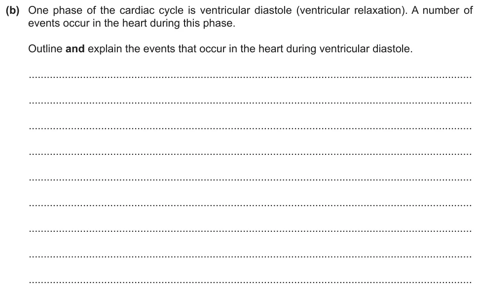
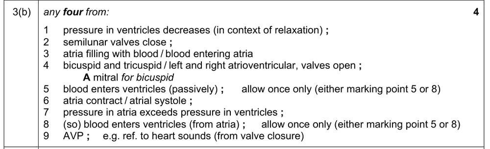
Outline and explain the events that occur in the heart during ventricular diastole.
Information from earlier in this question:

Show the mark scheme
Tap an image to open it full size.
From the examiners' report: Some answers mixed up the names of the valves and when they open or close. Others covered only one half of the heart, and a few described ventricular systole instead.
Question 13
Paper 2 · October/November 2018 (22) · Q2(d)(i) · 4 marks
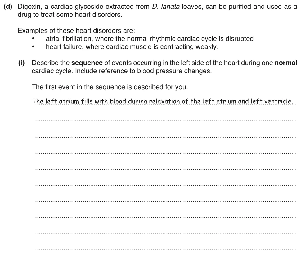
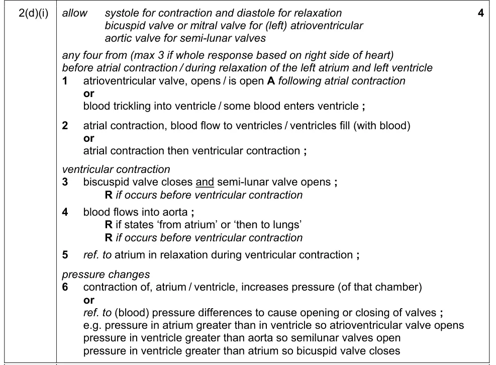
Describe the sequence of events in the left side of the heart during one cardiac cycle, including the blood pressure changes.
Show the mark scheme
Tap an image to open it full size.
From the examiners' report: Give a step-by-step account of the left side only, with the pressure differences that open and close the valves. Valves must be named correctly: the most frequent error was to call the bicuspid (left atrioventricular) valve the tricuspid valve. Some students wrote only about the pathway of blood and the valves, and did not mention atrial or ventricular contraction.
Question 14
Paper 2 · October/November 2025 (22) · Q6(a) · 5 marks
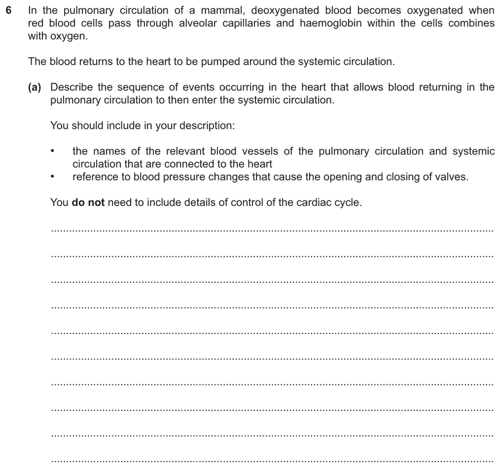
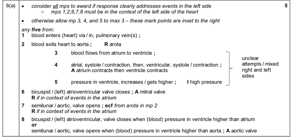
Describe how blood returning from the lungs passes through the heart and into the systemic circulation, naming the blood vessels and the pressure changes that open and close the valves.
Show the mark scheme
Tap an image to open it full size.
Pressure graphs for the cardiac cycle (8.3.2 and 8.3.3)
Question 15
Paper 2 · May/June 2020 (22) · Q2(c)(i) · 3 marks
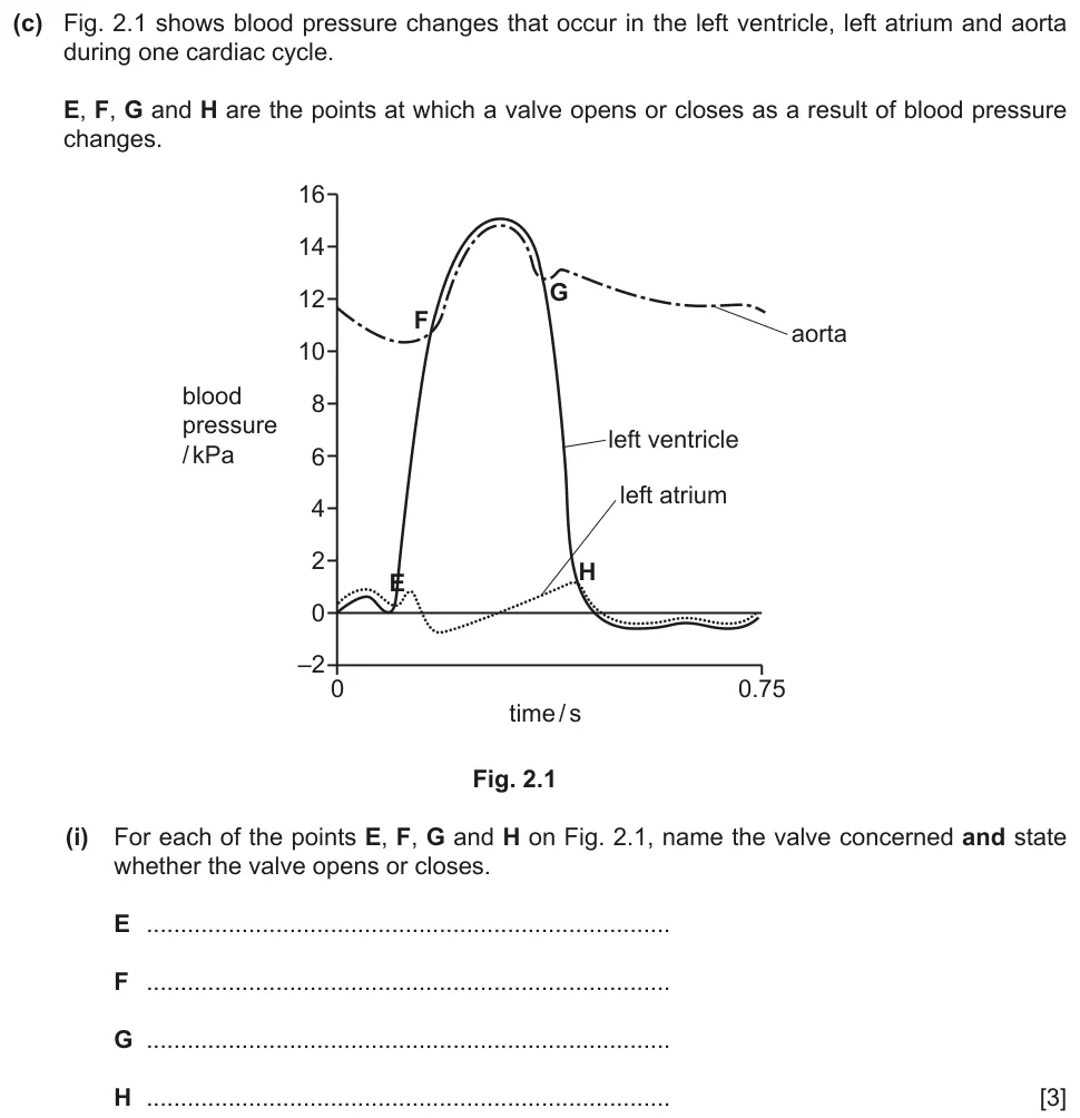
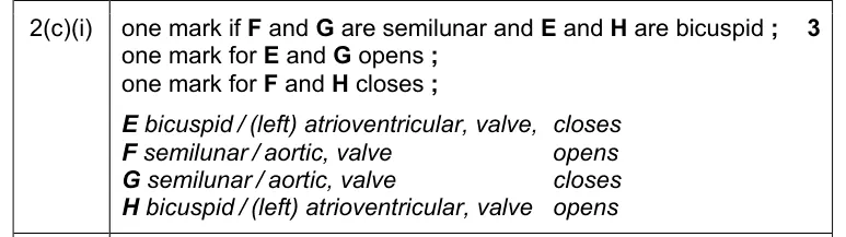
Use the pressure graph to name the valve at each of the points E, F, G and H and state whether it opens or closes.
Information from earlier in this question:

Show the mark scheme
Tap an image to open it full size.
Question 16
Paper 2 · May/June 2020 (22) · Q2(c)(ii) · 2 marks
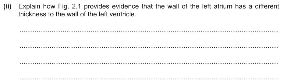
Explain how the pressure graph gives evidence that the walls of the left atrium and the left ventricle differ in thickness.
Show the mark scheme

Tap an image to open it full size.
Sinoatrial node, atrioventricular node and Purkyne tissue (8.3.4)
Question 17
Paper 2 · February/March 2025 (22) · Q3(c) · 3 marks
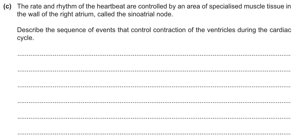
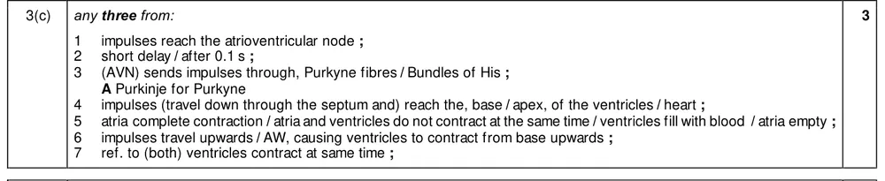
Describe the sequence of events that control the contraction of the ventricles.
Show the mark scheme
Tap an image to open it full size.
From the examiners' report: Some answers left out key events, and not all gave the events in the correct sequence.
Question 18
Paper 2 · May/June 2023 (22) · Q1(b) · 2 marks
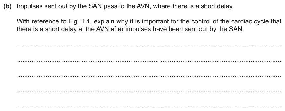
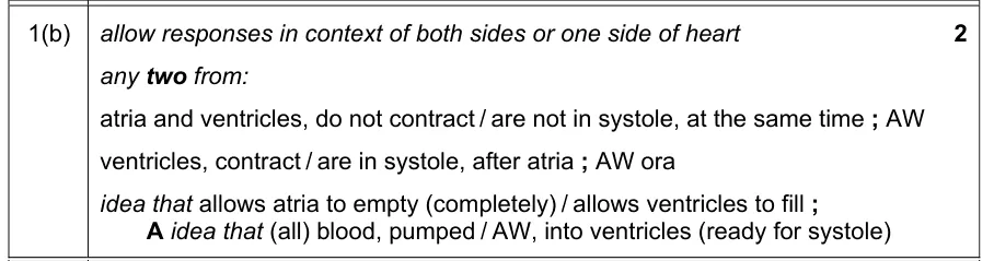
Explain why a short delay at the atrioventricular node is important for the control of the cardiac cycle.
Information from earlier in this question:
Show the mark scheme
Tap an image to open it full size.
From the examiners' report: The delay stops atrial systole and ventricular systole happening at the same time, and lets the atrium empty and the ventricle fill before ventricular systole. Do not just describe the sequence of events at the SAN and the AVN. Weak answers said the delay prevents backflow of blood or allows time for valves to open and close.
Question 19
Paper 2 · February/March 2022 (22) · Q5(b) · 3 marks · Easy
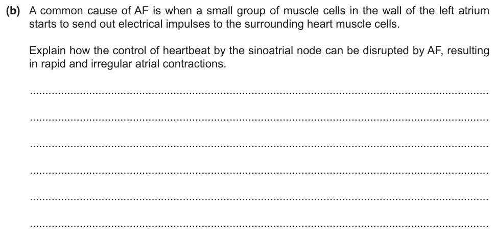
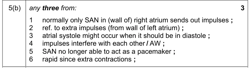
Explain how atrial fibrillation disrupts the control of the heartbeat by the sinoatrial node.
Information from earlier in this question:

Show the mark scheme
Tap an image to open it full size.
From the examiners' report: Some students only repeated the information in the question, stating that the sinoatrial node is disrupted and that this causes rapid and irregular atrial contractions. Explain how atrial fibrillation interferes with the role of the sinoatrial node as the pacemaker.
Question 20
Paper 2 · October/November 2023 (23) · Q6(b) · 4 marks
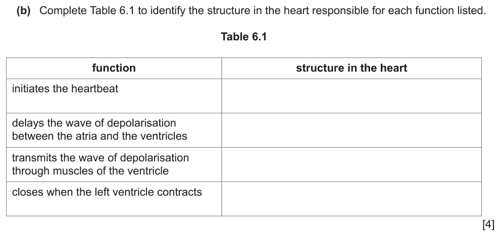
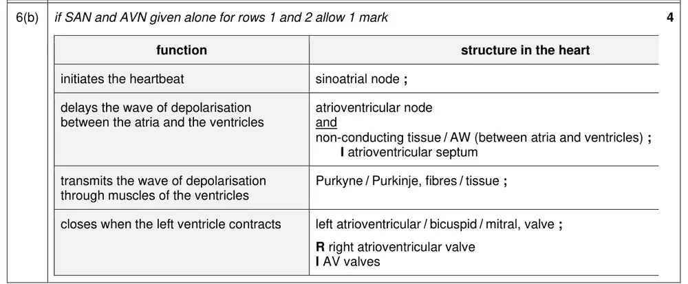
Complete the table to name the structure in the heart responsible for each function.
Show the mark scheme
Tap an image to open it full size.
From the examiners' report: Write the full names of the two nodes, not SAN and AVN, and spell 'atrioventricular' correctly. Bicuspid and mitral were accepted for the left atrioventricular valve.
Question 21
Paper 2 · October/November 2017 (22) · Q6(b) · 1 mark · Hard
State precisely where the sinoatrial node is located.

Show the mark scheme

Tap an image to open it full size.
From the examiners' report: Many knew that the sinoatrial node is in the right atrium. To be precise enough, say that it is in the wall of the right atrium.
Question 22
Paper 2 · May/June 2022 (23) · Q6(c) · 2 marks · Easy
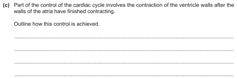
Outline how the ventricle walls are made to contract after the walls of the atria have finished contracting.
Information from earlier in this question:

Show the mark scheme

Tap an image to open it full size.
From the examiners' report: The delay at the atrioventricular node was generally well known. Stronger answers also described the band of non-conducting tissue between the atria and the ventricles. Some students misread the question and wrote about the opening and closing of valves and the direction of blood flow.
Where students lose marks (from Cambridge examiners' reports and mark schemes)
Each point below comes from the examiners' reports and mark schemes, and is followed by wording that gained marks.
Label lines must end on the structure. Students wrote the letter near the structure without a label line, or ended the line for R at the semilunar valve of the pulmonary artery.
✔ Gains credit: a 'label line pointing to' the 'tricuspid valve and labelled R', and a 'label line pointing to pulmonary artery and labelled L'.
Include the valves in the route. Students left the valves out of the route from the left atrium, or went through the right side of the heart and the pulmonary circulation.
✔ Gains credit: 'mitral / bicuspid / atrioventricular valve', 'left ventricle', 'semilunar / aortic valve' and 'aorta', in that order.
Thick walls generate pressure. Students wrote that the ventricles have thick walls 'to withstand high pressure' or to 'prevent bursting'.
✔ Gains credit: 'ventricles have more (cardiac) muscle to pump blood at high(er) pressure', 'a greater distance', 'to overcome a high(er) resistance'.
Atria and ventricles, both sides. Students wrote about the atrium and the ventricle, or explained what was happening when a description was asked for.
✔ Gains credit: blood flows 'from the atria to the ventricles' and 'into the (left and right) atria from the pulmonary vein and vena cava'.
Compare the pressures across the valve. Students said that a valve opens when the pressure increases, without comparing the pressures on its two sides.
✔ Gains credit: 'pressure in (right) ventricle becomes lower than pressure in (right) atrium' opens the tricuspid valve; 'pressure in (right) ventricle becomes higher than pressure in pulmonary artery' opens the pulmonary valve.
The bicuspid valve is on the left. Students named the left atrioventricular valve as the tricuspid valve, or based the whole answer on the right side of the heart.
✔ Gains credit: 'pressure in atrium greater than in ventricle so atrioventricular valve opens', 'bicuspid valve closes and semi-lunar valve opens', 'blood flows into aorta'.
The AVN delay lets the ventricles fill. Students said the delay prevents backflow of blood or gives the valves time to open and close, or only described the sequence of events.
✔ Gains credit: 'atria and ventricles do not contract at the same time', 'ventricles contract after atria', the delay 'allows atria to empty' and 'allows ventricles to fill'.
Full names for the two nodes. Students wrote SAN and AVN instead of the full names, and misspelt 'atrioventricular'.
✔ Gains credit: 'sinoatrial node', 'atrioventricular node', 'Purkyne fibres' and 'left atrioventricular / bicuspid / mitral valve'.
Download the questions
Download the PDF pack (22 questions, mark schemes at the end)
Keep practising
- The Circulatory System (8.1): Cambridge (CIE) exam questions by topic
- Transport of Oxygen and Carbon Dioxide (8.2): Cambridge (CIE) exam questions by topic
- Cambridge (CIE) A level Biology 9700 past papers, mark schemes and examiners' reports
- Cambridge (CIE) A level Biology grade thresholds
- Book a one-to-one CIE lesson or a free 15-minute discovery chat with Tom
- Register your interest in a CIE A level Biology group class
Questions, mark schemes and examiners' report extracts © Cambridge University Press & Assessment.