

Chromosome Behaviour in Mitosis (5.2): Cambridge (CIE) A Level Biology 9700 Exam Questions by Topic
Chromosome behaviour in mitosis is sub-topic 5.2 of Topic 5, The mitotic cell cycle, in the Cambridge International AS & A Level Biology syllabus (9700). You need to describe what the chromosomes, the nuclear envelope, the cell surface membrane and the spindle do in plant and animal cells during prophase, metaphase, anaphase and telophase. You also need to identify these stages in photomicrographs, diagrams and microscope slides. This is AS content, examined on Papers 1 and 2. A level students meet it again on Paper 4.
The examiners' reports return to the same points. Daughter chromosomes move to opposite poles, not to the 'sides' or 'ends' of the cell. Write 'nuclear envelope', not 'nuclear membrane', and say that it breaks down in prophase and re-forms around each group of chromosomes in telophase; it does not 'disappear'. A chromosome already has two sister chromatids at the start of mitosis, and each daughter chromosome after anaphase is a single chromatid. Do not confuse the centromere with a centrosome or a centriole. In photomicrographs, anaphase is often mistaken for metaphase or telophase, so look for chromosomes at the equator, separated chromatids, or a nuclear envelope re-forming. Label your drawings.
Studying Cambridge International A level Biology and want more help? Tom teaches CIE students one-to-one online, including students in the UAE, Saudi Arabia, Hong Kong, India and Indonesia, and every lesson is recorded. You can book a one-to-one lesson or a free 15-minute discovery chat, or register your interest in a CIE group class starting in September 2027, using the links at the bottom of this page.
What you need from IGCSE
Each question on this page assumes you're secure on these ideas. If any of them feels shaky, look it up before you start.
- The nucleus of a cell contains chromosomes, which are made of DNA and carry genetic information in the form of genes.
- Mitosis is nuclear division that gives genetically identical cells.
- The chromosomes are copied exactly before mitosis, and during mitosis the copies separate so that each daughter cell has the same number of chromosomes.
Key terms
These words come up in the questions and mark schemes. Examiners expect you to use them exactly.
- Prophase: the first stage of mitosis: the chromosomes condense and become visible as two sister chromatids, the nuclear envelope breaks down and the spindle begins to form.
- Metaphase: the chromosomes are arranged at the spindle equator (metaphase plate), attached to spindle fibres at their centromeres.
- Anaphase: the centromeres divide and the spindle fibres shorten, so the sister chromatids separate and move to opposite poles with their centromeres leading.
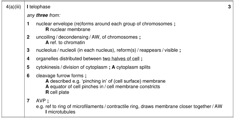
- Telophase: a nuclear envelope re-forms around each group of chromosomes, the chromosomes uncoil, the nucleoli re-form and the spindle breaks down.
- Spindle: the structure made of microtubules (spindle fibres) that attaches to the centromeres and moves the chromosomes.
- Centromere: the region that holds two sister chromatids together and attaches the chromosome to the spindle fibres.
- Nuclear envelope: the double membrane around the nucleus; it disassembles in prophase and reassembles in telophase.
- Cytokinesis: division of the cytoplasm after mitosis; in animal cells a cleavage furrow forms.
What these questions cover (Cambridge 9700 Topic 5: The mitotic cell cycle)
The questions test these learning outcomes from sub-topic 5.2 of the Cambridge International AS & A Level Biology syllabus (9700) for 2025 to 2027.
- 5.2.1 describe the behaviour of chromosomes in plant and animal cells during the mitotic cell cycle and the associated behaviour of the nuclear envelope, the cell surface membrane and the spindle
- 5.2.2 interpret photomicrographs, diagrams and microscope slides of cells in different stages of the mitotic cell cycle and identify the main stages of mitosis
How to use this page
Write your answer on paper before you open the mark scheme. Allow about 1.2 minutes per mark: the 22 questions are worth 46 marks, so about 55 minutes in total. Then mark your answer strictly against the mark scheme and read the examiners' tip underneath it. If you'd rather print the questions, download the PDF pack.
Chromosomes, the nuclear envelope and the spindle (5.2.1)
Question 1
Paper 2 · October/November 2025 (22) · Q1(e)(i) · 1 mark
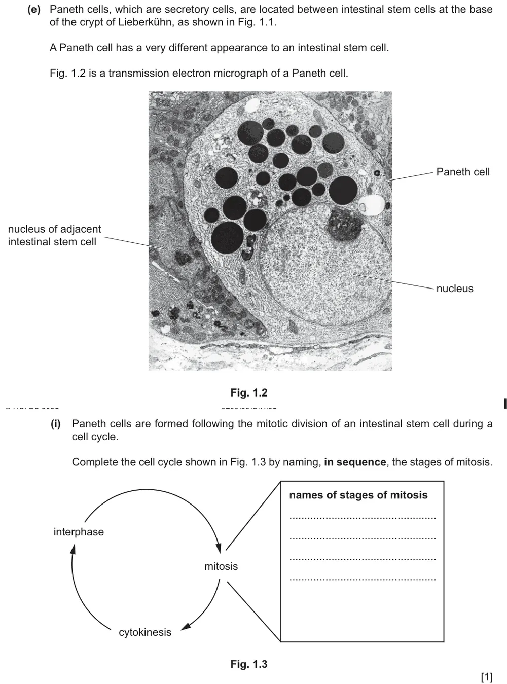
Complete the cell cycle diagram by naming the stages of mitosis in sequence.
Show the mark scheme

Tap an image to open it full size.
Question 2
Paper 2 · May/June 2024 (22) · Q5(a) · 1 mark
State what happens to chromatin so that individual chromosomes can be seen during mitosis.
Show the mark scheme

Tap an image to open it full size.
From the examiners' report: Most knew what happens to chromatin. A few contradicted themselves, for example 'it uncoils and condenses', or said that the chromatin has to be stained to make the chromosomes visible.
Question 3
Paper 2 · October/November 2023 (22) · Q1(b) · 3 marks
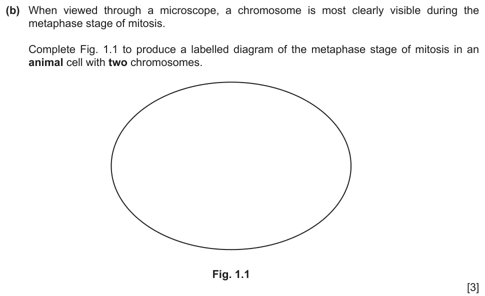
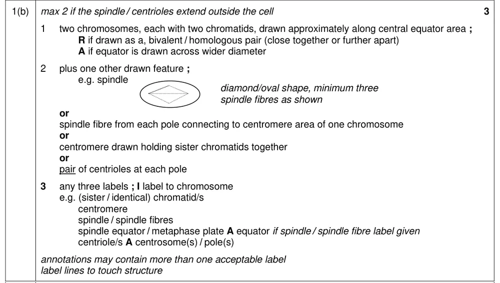
Complete a labelled diagram of metaphase of mitosis in an animal cell with two chromosomes.
Show the mark scheme
Tap an image to open it full size.
From the examiners' report: Draw the two chromosomes at the spindle equator with sister chromatids facing opposite poles, and label the diagram: labels were needed for full credit. Weaker answers often left out the spindle, or drew separated chromatids, homologous chromosomes or more than two chromosomes. A common error was to label the centrioles as centromeres.
Photomicrographs and diagrams: name the stage, then describe it (5.2.1 and 5.2.2)
Question 4
Paper 2 · February/March 2025 (22) · Q6(a)(i) · 1 mark · Easy
Put six photomicrographs of onion root tip cells into the correct sequence of the mitotic cell cycle.
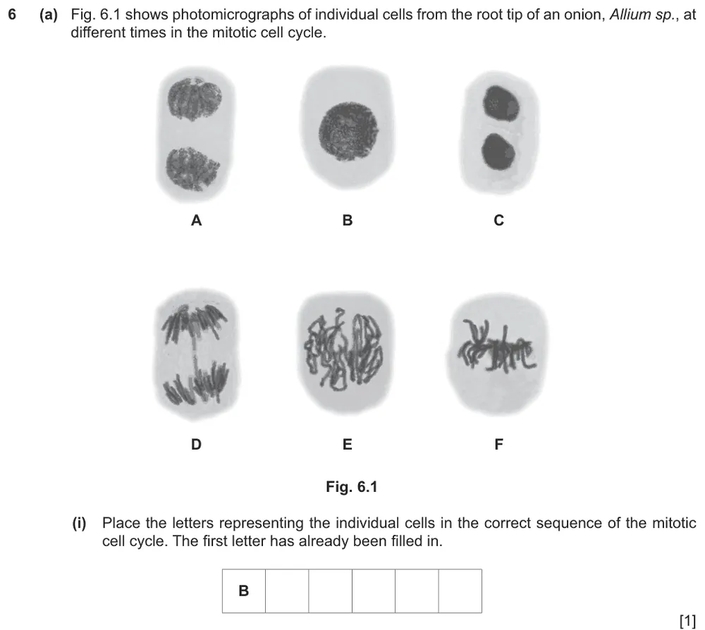
Show the mark scheme

Tap an image to open it full size.
Question 5
Paper 2 · February/March 2025 (22) · Q6(a)(ii) · 2 marks · Easy
Describe the events in the stage of mitosis shown by cell A.
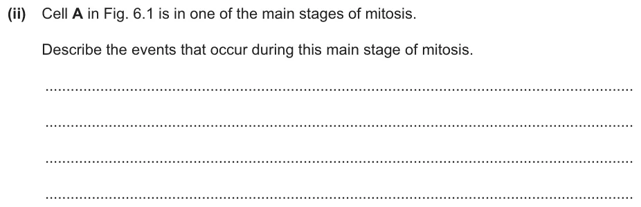
Show the mark scheme

Tap an image to open it full size.
From the examiners' report: Most answers correctly gave the chromosomes uncoiling, a nuclear envelope forming around each set of chromosomes and the spindle fibres breaking down. Some also described nucleoli appearing in each nucleus.
Question 6
Paper 2 · May/June 2023 (23) · Q1(a)(i) · 2 marks · Easy
Identify the stages of mitosis in the cells labelled B and C.
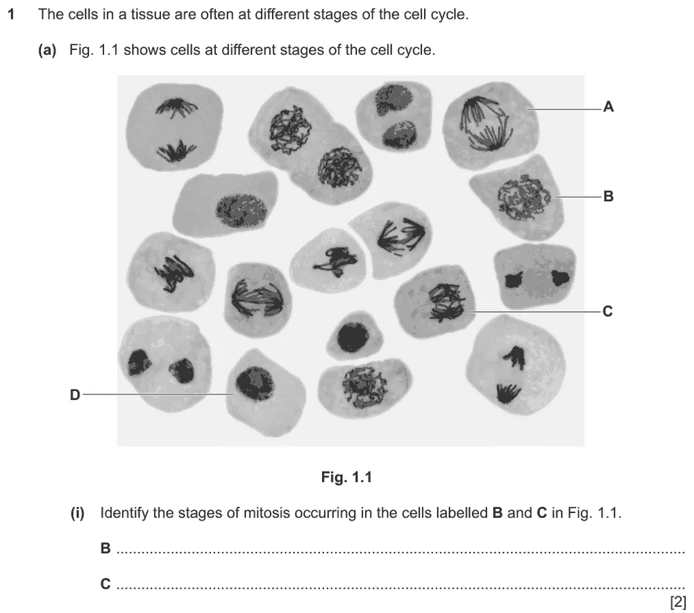
Show the mark scheme

Tap an image to open it full size.
From the examiners' report: Most identified prophase in cell B. Cell C was sometimes called anaphase rather than metaphase, perhaps because some of the darkly staining structures look slightly V-shaped; in anaphase they would not be layered on top of each other.
Question 7
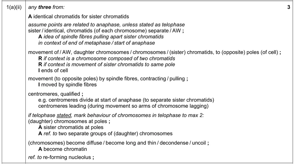
Paper 2 · May/June 2023 (23) · Q1(a)(ii) · 3 marks
Describe the behaviour of the chromosomes in the stage of mitosis shown in cell A.
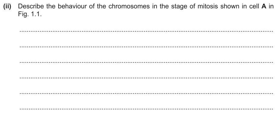
Show the mark scheme
Tap an image to open it full size.
From the examiners' report: Make it clear that the sister chromatids separate and then move to opposite poles of the cell as daughter chromosomes. 'Opposite ends of the cell' was too imprecise to be credited. Weak answers did not distinguish sister chromatids from chromosomes, or wrote about pairs of chromosomes or homologous chromosomes.
Question 8
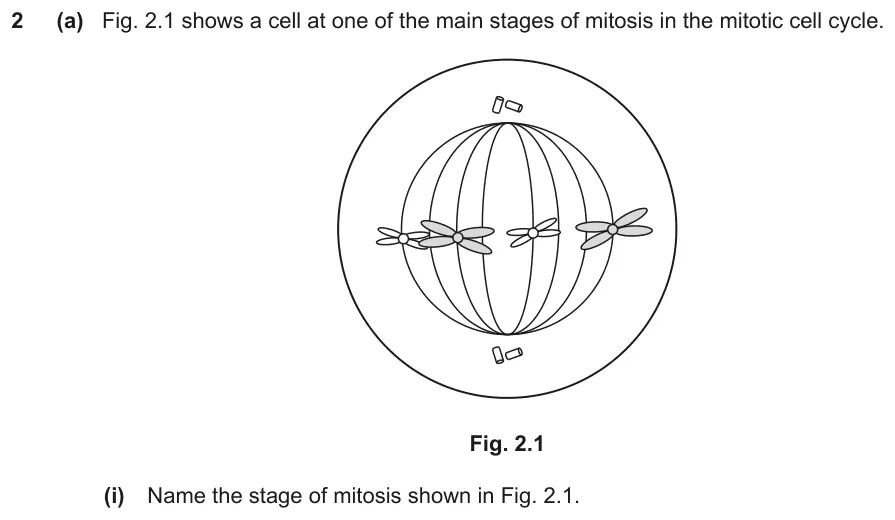
Paper 2 · February/March 2022 (22) · Q2(a)(i) · 1 mark · Easy
Name the stage of mitosis shown in the diagram.
Show the mark scheme

Tap an image to open it full size.
From the examiners' report: Most recognised the stage. The most frequent incorrect answer was anaphase.
Question 9
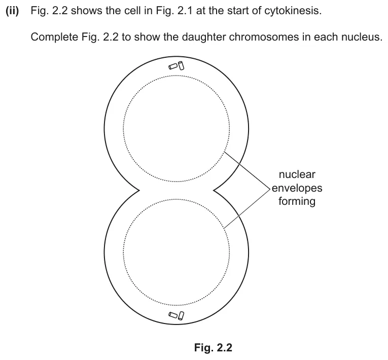
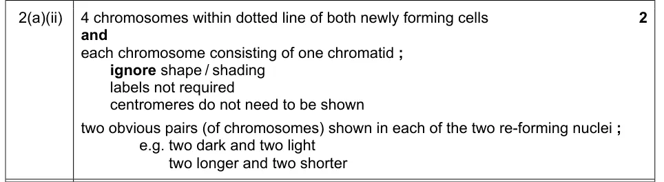
Paper 2 · February/March 2022 (22) · Q2(a)(ii) · 2 marks
Complete the diagram to show the daughter chromosomes in each nucleus at the start of cytokinesis.
Show the mark scheme
Tap an image to open it full size.
From the examiners' report: The strongest diagrams were neat and showed the relative sizes and shading of the chromosomes. Many left these details out, and some drew two chromosomes instead of four. A number wrongly drew chromosomes with two chromatids.
Question 10
Paper 2 · October/November 2022 (23) · Q4(a)(ii) · 3 marks
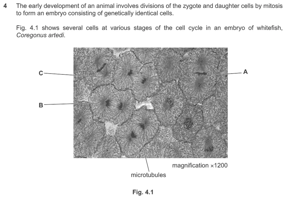
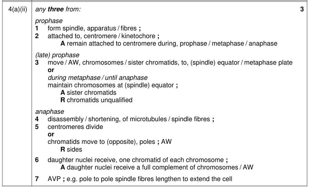
Describe the role of microtubules in mitosis.
Information from earlier in this question:
Show the mark scheme
Tap an image to open it full size.
From the examiners' report: Good answers said that microtubules form spindle fibres that attach to centromeres, arrange the chromosomes on the metaphase plate and move the chromatids to the poles in anaphase. Do not write that daughter chromosomes are pulled to the 'sides' or 'ends' of the cell, or that microtubules attach to centrosomes rather than centromeres.
Question 11
Paper 2 · October/November 2022 (23) · Q4(a)(iii) · 3 marks · Easy
State what happens in cell C until two new cells are formed.
Show the mark scheme
Tap an image to open it full size.
From the examiners' report: Naming telophase gained no credit: the changes to the nucleus and the cytoplasm had to be described. Make it clear that a nuclear envelope re-forms around each group of chromosomes. 'Nuclear membrane' was not credited, because the nucleus has a double membrane.
Question 12
Paper 2 · October/November 2022 (21) · Q6(a)(i) · 1 mark · Hard
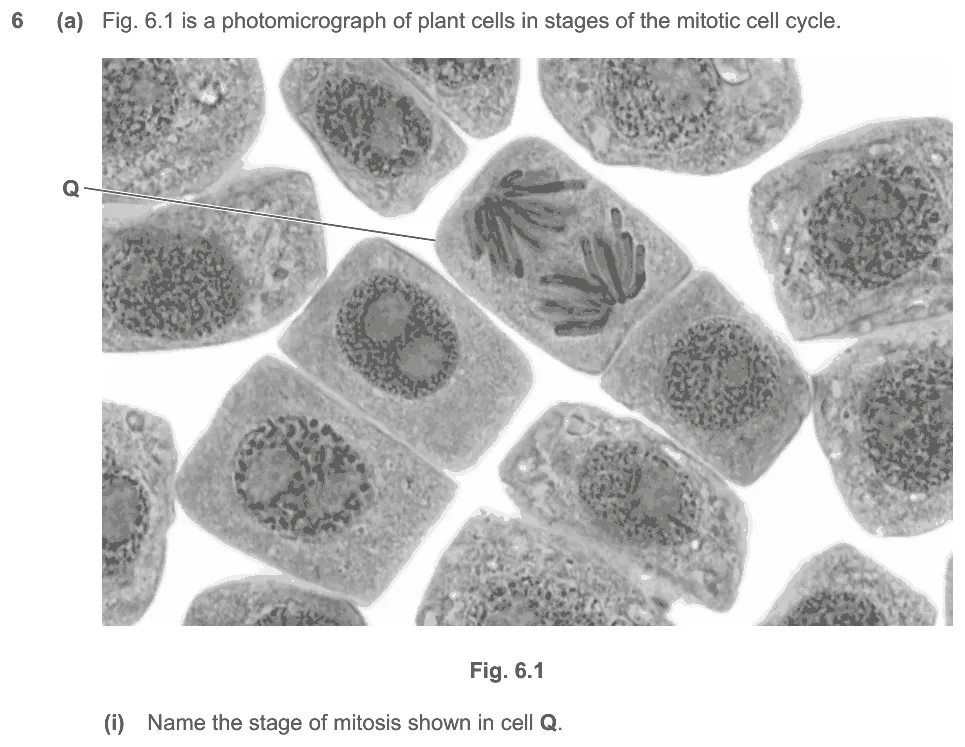
Name the stage of mitosis shown in cell Q.
Show the mark scheme

Tap an image to open it full size.
From the examiners' report: Many correctly named anaphase. The most common error was to name the stage as metaphase.
Question 13
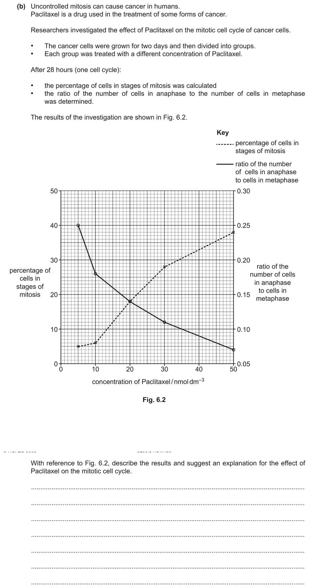
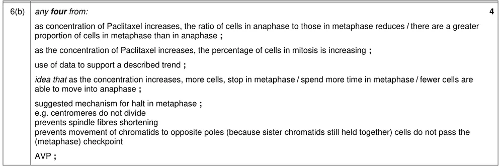
Paper 2 · October/November 2022 (21) · Q6(b) · 4 marks
Describe and explain the effect of the drug Paclitaxel on the mitotic cell cycle of cancer cells, using the graph.
Show the mark scheme
Tap an image to open it full size.
From the examiners' report: The graph has two y-axes, and some students confused the two trend lines. The strongest answers used data from the graph to support each relationship and suggested an explanation, most often that cells were stopped in metaphase, for example because the sister chromatids could not separate.
Question 14
Paper 2 · May/June 2021 (22) · Q1(a)(i) · 2 marks · Easy
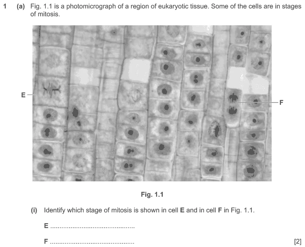
Identify the stage of mitosis shown in cell E and in cell F.
Show the mark scheme

Tap an image to open it full size.
From the examiners' report: Most were correct. Where one mark was lost, cell F was usually given as telophase rather than anaphase. There is no sign of a nuclear envelope re-forming around the daughter chromosomes, which rules out telophase.
Question 15
Paper 2 · May/June 2021 (22) · Q1(a)(ii) · 1 mark
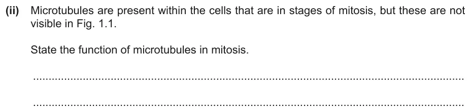
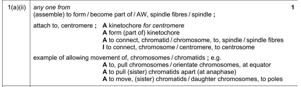
State the function of microtubules in mitosis.
Show the mark scheme
Tap an image to open it full size.
From the examiners' report: Correct answers referred to the spindle, attachment to the centromere or the movement of chromosomes. You do not need to know the terms 'centrosome' and 'kinetochore', and a number of students confused 'centrosome' with 'centromere'.
Question 16
Paper 2 · May/June 2018 (22) · Q4(a) · 3 marks · Easy
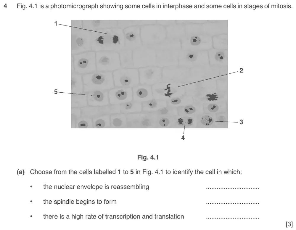
Choose the cell in which the nuclear envelope is reassembling, the cell in which the spindle begins to form and the cell with a high rate of transcription and translation.
Show the mark scheme
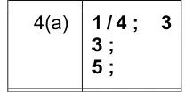
Tap an image to open it full size.
From the examiners' report: Most linked cell 5 to a high rate of transcription and translation and cell 4 to a reassembling nuclear envelope. The spindle was harder, and cell 2 was a common incorrect choice. The spindle must form before metaphase, so that the prophase chromosomes can be connected to the fibres and moved to the spindle equator.
Question 17
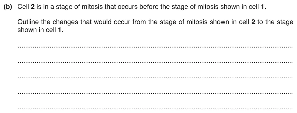
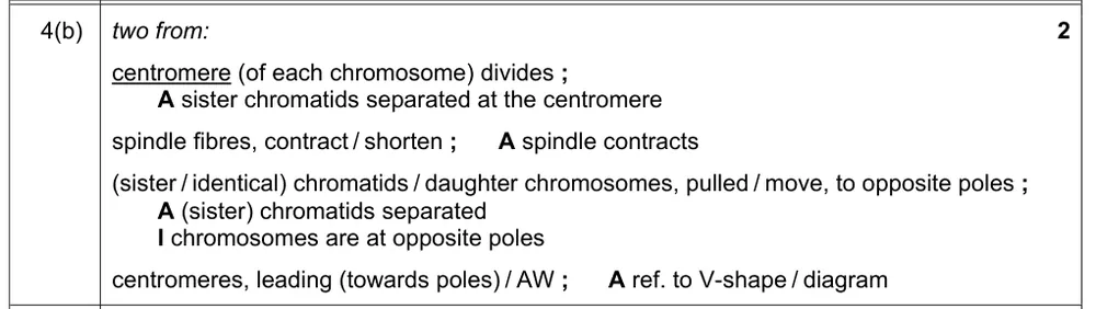
Paper 2 · May/June 2018 (22) · Q4(b) · 2 marks
Outline the changes from the stage of mitosis shown in cell 2 to the stage shown in cell 1.
Show the mark scheme
Tap an image to open it full size.
From the examiners' report: Only the events from the start of anaphase to late anaphase were needed. Say that the two sister chromatids of each chromosome are separated and moved to opposite poles by the contraction of the spindle fibres. Chromosomes do not 'form' or 'divide into' two chromatids: each already has two sister chromatids at the start of mitosis. Some confused centromeres with centrioles or centrosomes.
Question 18
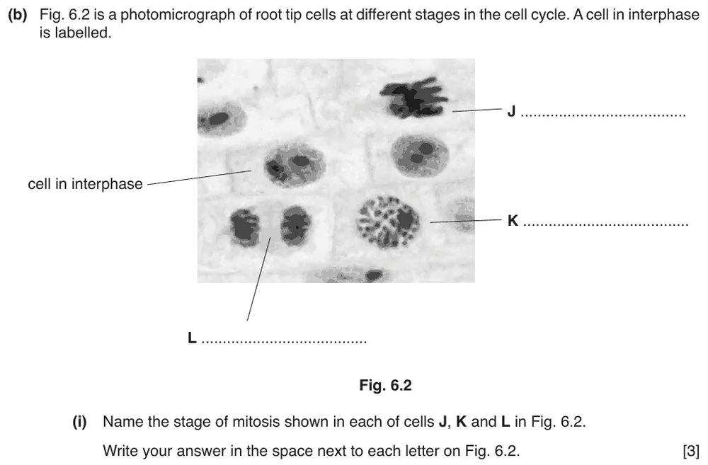
Paper 2 · February/March 2018 (22) · Q6(b)(i) · 3 marks
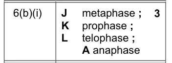
Name the stage of mitosis shown in each of cells J, K and L in the root tip photomicrograph.
Show the mark scheme
Tap an image to open it full size.
From the examiners' report: Some students were clearly well practised at identifying stages of mitosis from photomicrographs; others gained only partial credit. For cell K, prophase or early prophase gained the mark, but late prophase was rejected because chromosomes would be visible in late prophase.
Question 19
Paper 2 · October/November 2017 (22) · Q1(a) · 1 mark
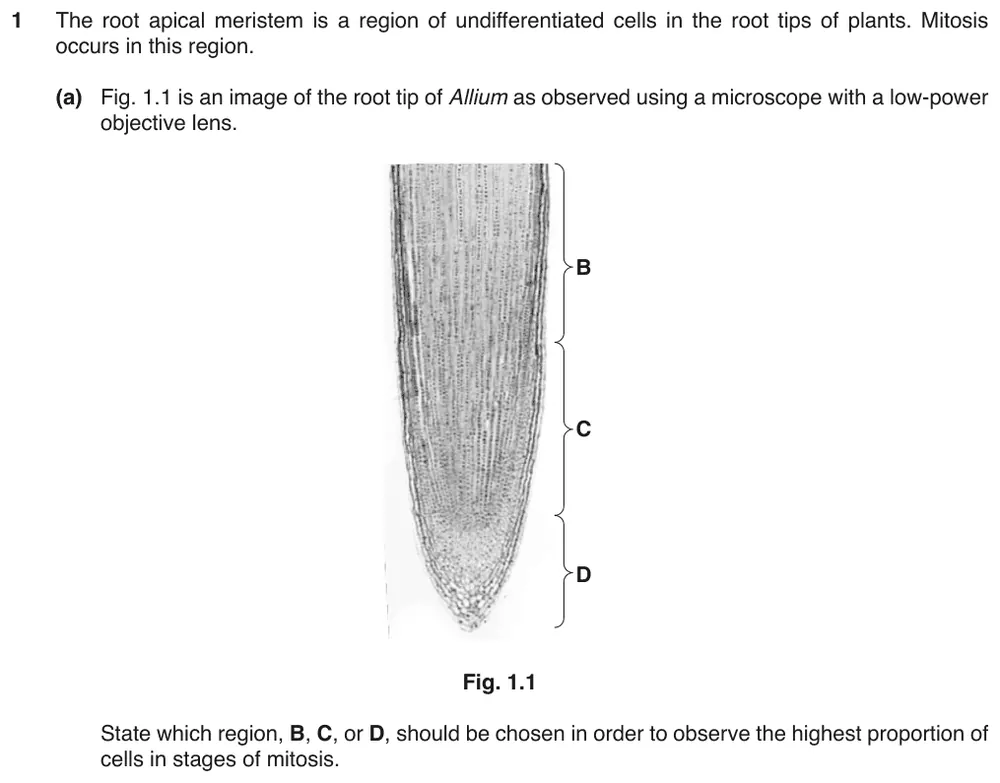
State which region of the root tip has the highest proportion of cells in stages of mitosis.
Show the mark scheme

Tap an image to open it full size.
From the examiners' report: Region C was correct, but region D was given very often. The very end of the root is the root cap, made of protective cells, so only a very small proportion of its cells are meristematic.
Question 20
Paper 2 · October/November 2017 (22) · Q1(b)(ii) · 2 marks
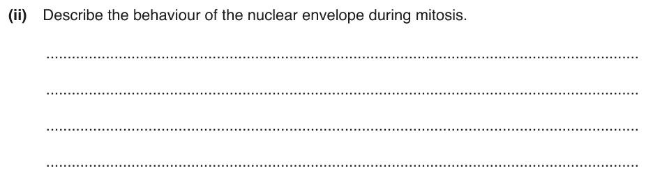
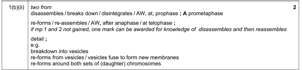
Describe the behaviour of the nuclear envelope during mitosis.
Show the mark scheme
Tap an image to open it full size.
From the examiners' report: A complete answer says that the nuclear envelope breaks down in prophase and re-forms in telophase. Write 'disassembles' or 'breaks down', not 'disappears'. It was quite common to see metaphase named instead of prophase, and cytokinesis instead of telophase.
Identifying cells in stages of mitosis (5.2.2)
Question 21
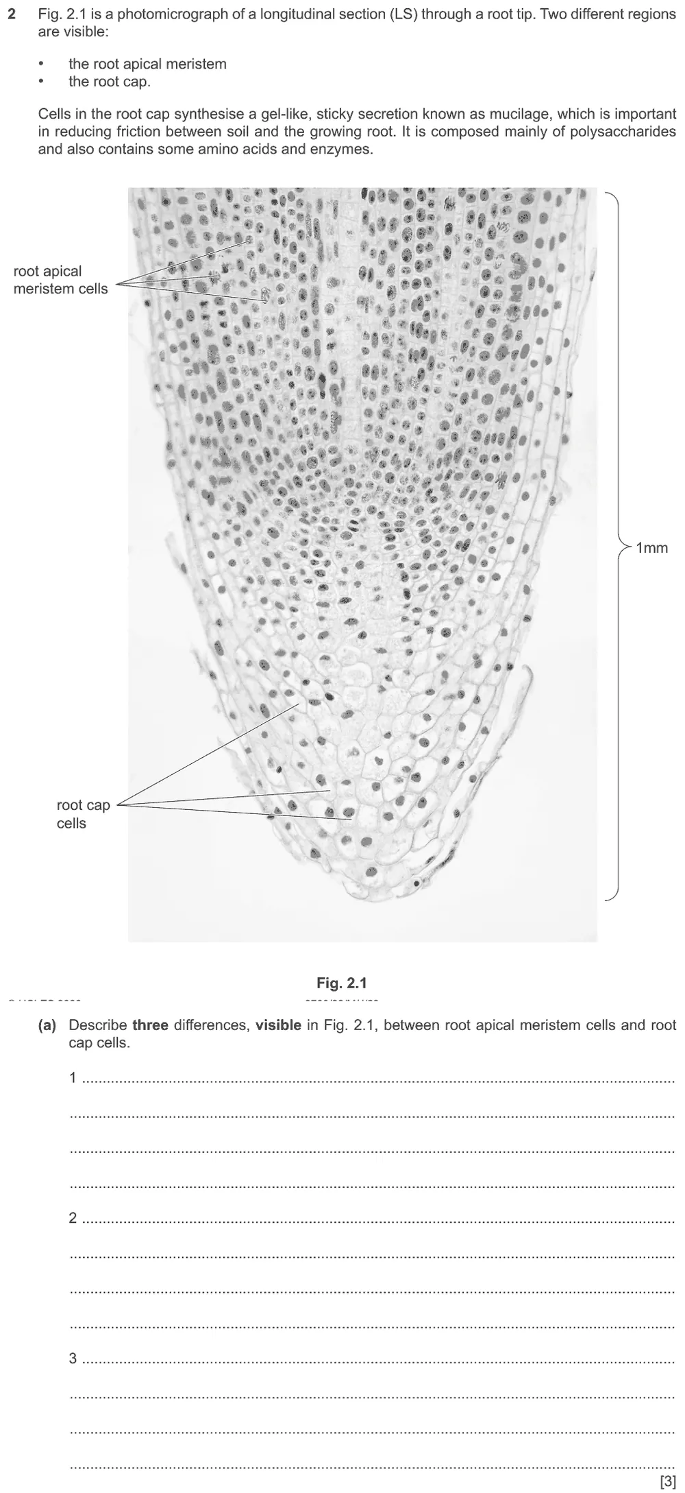
Paper 2 · May/June 2023 (22) · Q2(a) · 3 marks
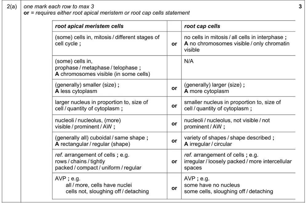
Describe three differences, visible in the photomicrograph, between root apical meristem cells and root cap cells.
Show the mark scheme
Tap an image to open it full size.
From the examiners' report: For each difference, take one feature and say what it is like in each cell type. For the nucleus, the difference is its size in proportion to the size of the cell, not its observed size. Fewer noticed that a number of meristem cells were in stages of mitosis and no root cap cells were.
Question 22
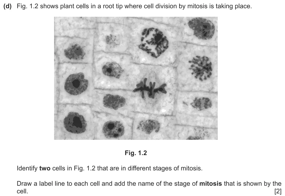
Paper 2 · October/November 2021 (22) · Q1(d) · 2 marks
Label two cells that are in different stages of mitosis and name each stage.
Show the mark scheme

Tap an image to open it full size.
From the examiners' report: Only two stages of mitosis were visible: prophase and metaphase. The metaphase cell was sometimes labelled anaphase. Some added a third label, usually interphase, which is not a stage of mitosis.
Where students lose marks (from Cambridge examiners' reports and mark schemes)
Each point below comes from the examiners' reports and mark schemes, and is followed by wording that gained marks.
Poles, not ends of the cell. Students wrote that the daughter chromosomes move to opposite ends of the cell, or did not distinguish sister chromatids from chromosomes.
✔ Gains credit: 'sister chromatids separate' and there is 'movement of daughter chromosomes to opposite poles' by 'spindle fibres contracting'.
Nuclear envelope, not nuclear membrane. Students named telophase without describing it, or wrote that a nuclear membrane forms.
✔ Gains credit: 'nuclear envelope re-forms around each group of chromosomes', 'uncoiling of chromosomes', then 'cytokinesis' as a 'cleavage furrow forms'.
Chromosomes start mitosis with two chromatids. Students wrote that chromosomes form, or divide into, two chromatids in anaphase, or confused centromeres with centrioles.
✔ Gains credit: 'centromere divides', 'spindle fibres shorten' and 'sister chromatids' are 'pulled to opposite poles' with 'centromeres leading'.
One chromatid after anaphase. Students drew daughter chromosomes with two chromatids, or drew two chromosomes instead of four.
✔ Gains credit: '4 chromosomes' in each cell, 'each chromosome consisting of one chromatid', with 'two obvious pairs' in each nucleus.
Breaks down, not disappears. Students wrote that the nuclear envelope disappears, or gave metaphase and cytokinesis as the stages.
✔ Gains credit: the nuclear envelope 'disassembles at prophase' and 're-forms at telophase', 'around both sets of daughter chromosomes'.
Anaphase is not telophase. Students called a cell with separated chromatids telophase, although no nuclear envelope was re-forming.
✔ Gains credit: 'E = metaphase' and 'F = anaphase'.
Draw the spindle and label it. Students left out the spindle, drew separated chromatids or homologous chromosomes, gave no labels, or labelled the centrioles as centromeres.
✔ Gains credit: 'two chromosomes, each with two chromatids' at the 'equator', with labels such as 'centromere', 'spindle fibres' and 'centriole'.
Read both y-axes. Students confused the two lines on a graph with two y-axes, or described the trends without data.
✔ Gains credit: 'as concentration of Paclitaxel increases, the ratio of cells in anaphase to those in metaphase reduces' and 'the percentage of cells in mitosis is increasing', because 'more cells stop in metaphase'.
Download the questions
Download the PDF pack (22 questions, mark schemes at the end)
Keep practising
- Replication and Division of Nuclei and Cells (5.1): Cambridge (CIE) exam questions by topic
- Cambridge (CIE) A level Biology 9700 past papers, mark schemes and examiners' reports
- Cambridge (CIE) A level Biology grade thresholds
- Book a one-to-one CIE lesson or a free 15-minute discovery chat with Tom
- Register your interest in a CIE A level Biology group class
Questions, mark schemes and examiners' report extracts © Cambridge University Press & Assessment.