

Receptors and control of heart rate (AQA 3.6.1.2 and 3.6.1.3) are A-level only, so they're Year 13 content. You need the structure of a Pacinian corpuscle and how stretch-mediated sodium ion channels produce a generator potential, how rods and cones explain sensitivity, colour vision and visual acuity, how the SAN, AVN and Purkyne tissue coordinate each heartbeat, and how chemoreceptors, pressure receptors and the autonomic nervous system change the heart rate.
The examiners' reports keep finding the same slips. Students write 'messages' or 'signals' instead of impulses, or 'an impulse' when the mark needs more or fewer impulses. They write 'red cones' instead of red-sensitive cones, stop before the impulses reach the optic nerve or brain, and say chemoreceptors detect oxygen. On heart rate they forget the delay at the AVN, or send impulses from the SAN when the question is about impulses to it.
Start with the Pacinian corpuscle and the retina, then the heart questions, and finish with the caffeine investigation. Write your answer on paper before you open each mark scheme. If you'd like help with this topic, my AQA masterclasses run online on Tuesdays at 5.15pm for Year 12 and on Wednesdays or Thursdays at 6.30pm for Year 13.
What you need from GCSE and Year 12
Each question on this page assumes you're secure on these GCSE and Year 12 ideas. If any of them feels shaky, revise it before you start.
- GCSE: receptors detect stimuli and send impulses along sensory neurones to the central nervous system; the retina contains light-sensitive receptor cells.
- GCSE: the heart's natural resting rate is set by a group of cells in the right atrium that act as a pacemaker.
- Year 12 (AQA 3.3.4.1): the structure of the heart and the cardiac cycle, with the atria contracting before the ventricles.
- Year 12 (AQA 3.2.3): ions such as sodium ions move through channel proteins by facilitated diffusion, down their concentration gradient.
- Year 12 (AQA 3.3.4.1): cardiac output = stroke volume × heart rate.
Key terms
These words come up in the questions and mark schemes. Examiners expect you to use them exactly.
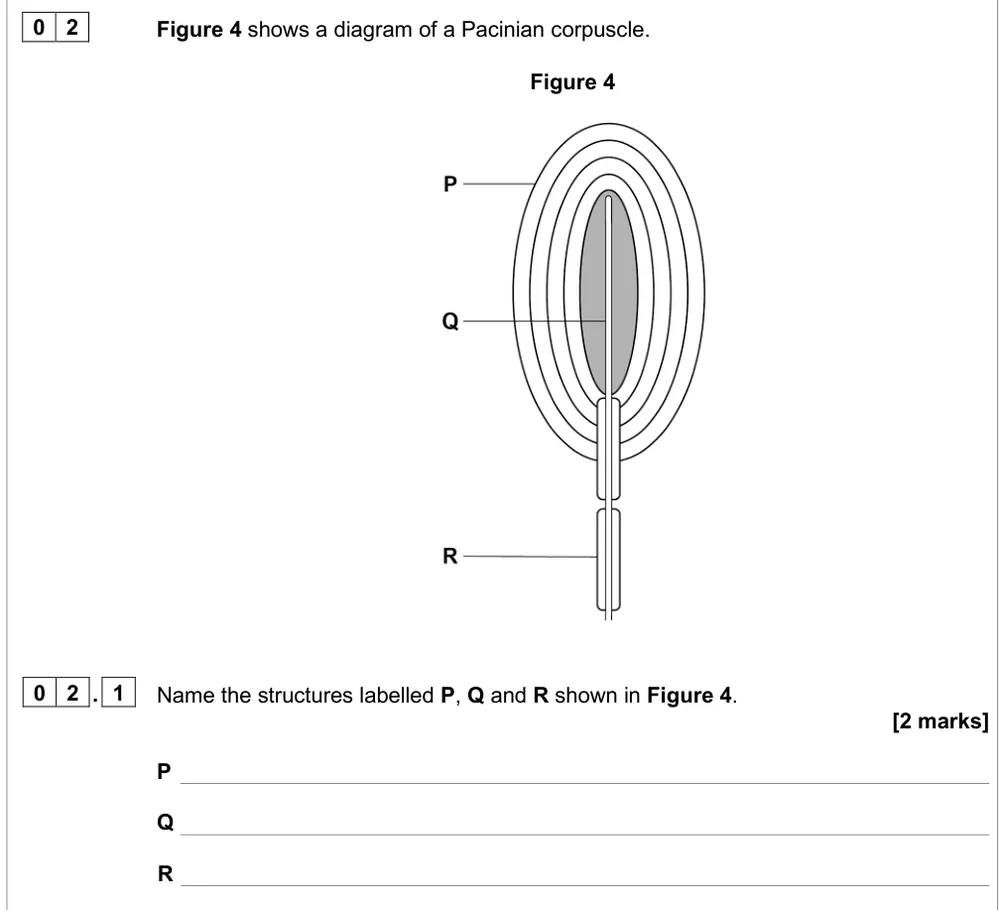
- Pacinian corpuscle: a pressure receptor: the end of a sensory neurone's axon surrounded by layers of connective tissue called lamellae, inside a capsule.
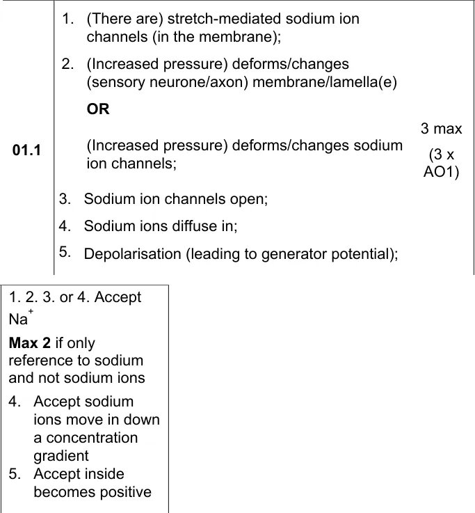
- Stretch-mediated sodium ion channels: channels in the axon membrane that open when pressure deforms the membrane, so sodium ions diffuse in.
- Generator potential: the depolarisation of a receptor's membrane when it is stimulated; if it reaches threshold it produces an action potential.
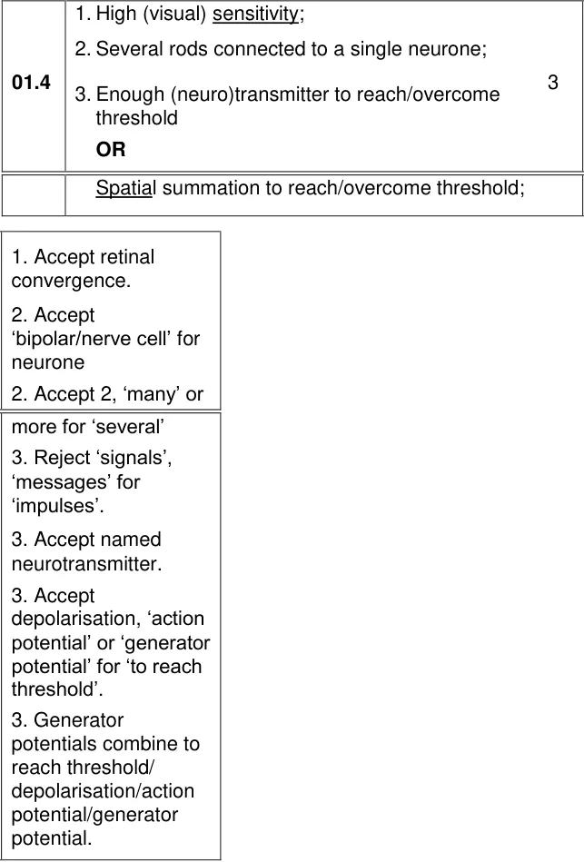
- Rods: photoreceptors with high sensitivity to light: several rods connect to one neurone, so their generator potentials combine (spatial summation) to reach threshold in dim light.
- Cones: photoreceptors with high visual acuity: each cone connects to a single neurone, so they send separate impulses to the brain.
- Red-, green- and blue-sensitive cones: three types of cone with different pigments that absorb different ranges of wavelength; examiners reject 'red cones' or 'green pigment'.
- Myogenic: the heartbeat starts in the heart muscle itself, with no impulses from nerves needed.
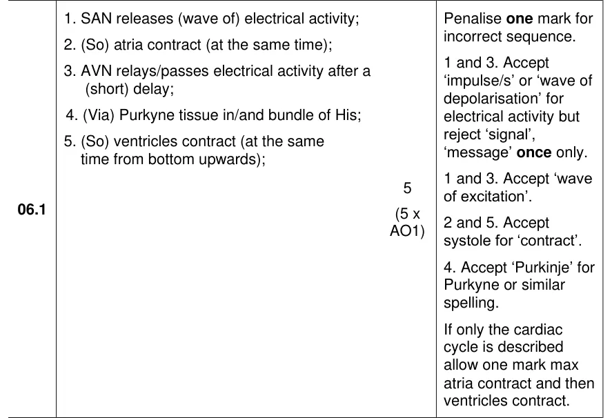
- SAN, AVN and Purkyne tissue: the SAN sends a wave of electrical activity across the atria; the AVN passes it on after a short delay, down Purkyne tissue in the bundle of His to the ventricles.
- Chemoreceptors: receptors in the aorta and carotid arteries that detect changes in carbon dioxide concentration, H⁺ or pH of the blood, not oxygen.
- Pressure receptors: receptors (baroreceptors) in the aorta and carotid arteries that detect changes in blood pressure.
- Cardiac centre: the region of the medulla that sends more impulses along sympathetic neurones to speed up the heart, or along parasympathetic (vagus) neurones to slow it, to the SAN.
What these questions cover (AQA 3.6.1.2 and 3.6.1.3)
The questions test these statements from the AQA AS and A-level Biology specification (7401/7402):
- The Pacinian corpuscle as an example of a receptor, to illustrate that: receptors respond only to specific stimuli (no written question on this point alone was available; the Pacinian corpuscle responds to pressure, and rods and cones respond to light)
- The Pacinian corpuscle as an example of a receptor, to illustrate that: stimulation of a receptor leads to the establishment of a generator potential.
- Pacinian corpuscle: The basic structure of a Pacinian corpuscle.
- Generator potential: Deformation of stretch-mediated sodium ion channels in a Pacinian corpuscle leads to the establishment of a generator potential.
- The retina: The human retina in sufficient detail to show how differences in sensitivity to light, sensitivity to colour and visual acuity are explained by differences in the optical pigments of rods and cones and the connections rods and cones make in the optic nerve. (five questions cover visual acuity, sensitivity and colour vision)
- Myogenic stimulation: Myogenic stimulation of the heart and transmission of a subsequent wave of electrical activity. The roles of the sinoatrial node (SAN), atrioventricular node (AVN) and Purkyne tissue in the bundle of His.
- Nervous control of heart rate: The roles and locations of chemoreceptors and pressure receptors and the roles of the autonomic nervous system and effectors in controlling heart rate. (the caffeine questions include the pulse rate investigation listed beside this section)
How to use this page
Write your answer on paper before you open the mark scheme. Allow about 1.3 minutes per mark: the 14 questions are worth 43 marks, so about 56 minutes in total. Then mark your answer strictly against the mark scheme and read the examiners' tip underneath it. If you'd rather print the questions, download the PDF pack.
Receptors: the Pacinian corpuscle (3.6.1.2)
Question 1
A level Paper 3 · June 2022 · Q02.1 · 2 marks
Name three labelled structures on a diagram of a Pacinian corpuscle.
Show the mark scheme
Tap an image to open it full size.
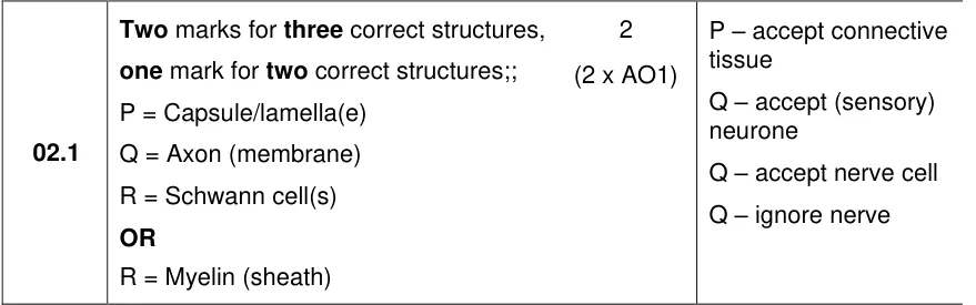
From the examiners' report: 21% got all three. P is the capsule or lamellae, not a sodium ion channel; Q needs 'axon' or 'sensory neurone', not just 'nerve'; R is a Schwann cell, not a node of Ranvier.
Question 2
A level Paper 3 · June 2023 · Q01.1 · 3 marks · Easy
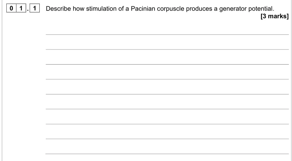
Describe how stimulation of a Pacinian corpuscle produces a generator potential.
Show the mark scheme
Tap an image to open it full size.
From the examiners' report: Nearly half got all three. Write 'stretch-mediated sodium ion channels', not a stretch-mediated membrane, say 'sodium ions' every time, and say they diffuse in rather than just 'influx'.
Rods, cones and the retina (3.6.1.2)
Question 3
A level Paper 2 · November 2020 · Q01.2 · 3 marks · Medium
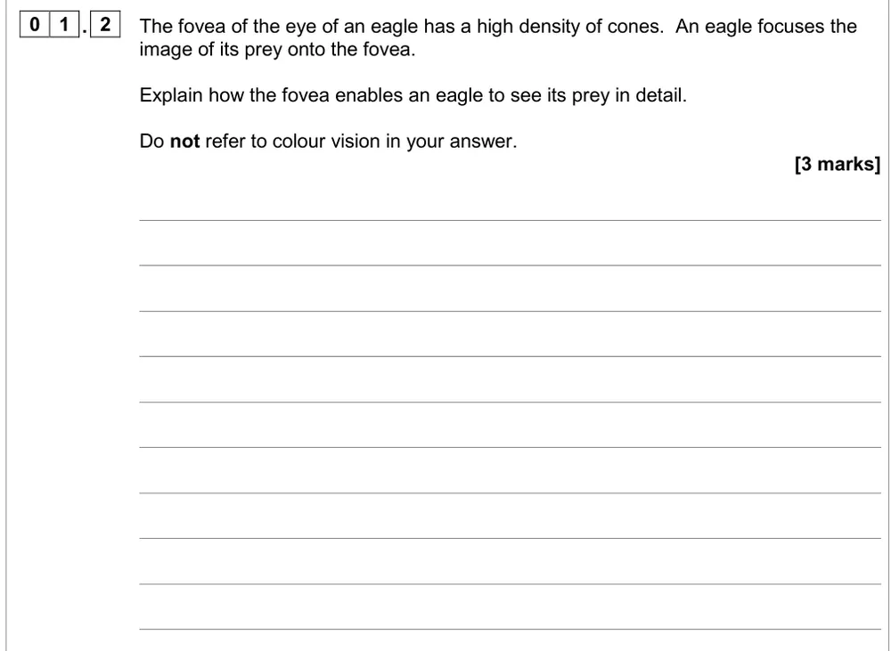
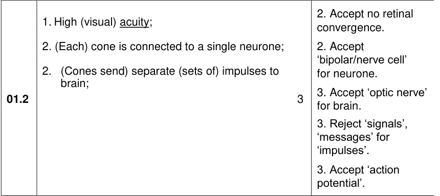
Explain how a fovea packed with cones lets an eagle see its prey in detail.
Show the mark scheme
Tap an image to open it full size.
From the examiners' report: 74% got a mark but only 14% got all three. After one cone to one neurone, say the cones send separate impulses to the brain, and leave colour vision out.
Question 4
A level Paper 2 · November 2020 · Q01.4 · 3 marks · Medium
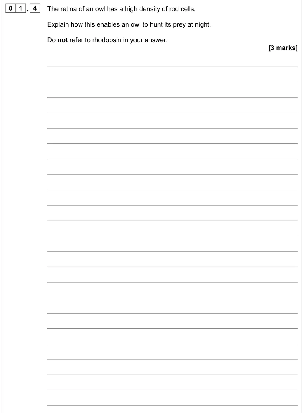
Explain how a high density of rods lets an owl hunt at night.
Show the mark scheme
Tap an image to open it full size.
From the examiners' report: Only 10% got all three. After 'several rods to one bipolar neurone', explain the threshold: their generator potentials combine by spatial summation. 'Weak impulses' and 'stimuli combining' didn't score.
Question 5
A level Paper 2 · June 2018 · Q10.1 · 3 marks · Medium
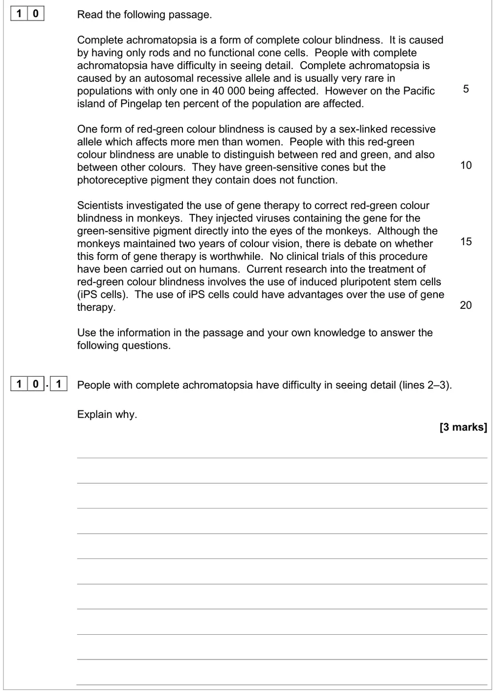
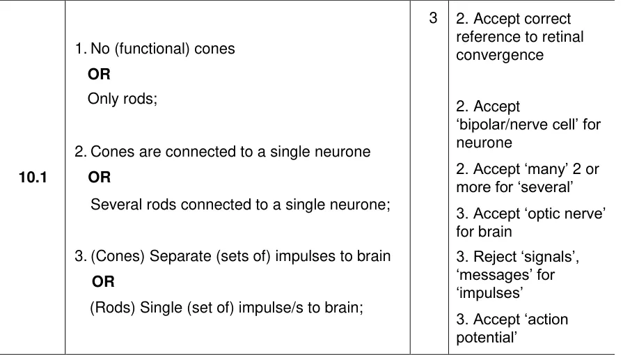
Explain why people with only rods and no working cones struggle to see detail.
Show the mark scheme
Tap an image to open it full size.
From the examiners' report: 90% got a mark but only 8% got all three. Finish the chain: several rods share one neurone, so they send a single set of impulses to the brain or optic nerve.
Question 6
A level Paper 2 · June 2018 · Q10.4 · 3 marks · Hard
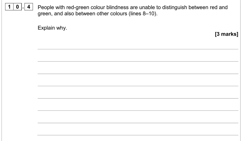
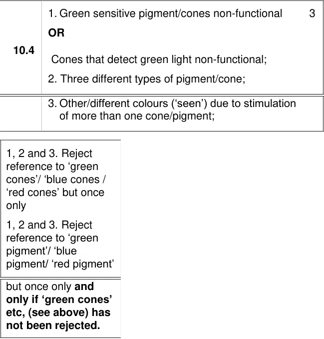
Explain why red-green colour blindness also affects other colours.
Show the mark scheme
Tap an image to open it full size.
From the examiners' report: Fewer than 1% got all three. Always write green-sensitive cones, not green cones, keep the red-sensitive cones working, and say other colours stimulate more than one type of cone at once.
Question 7
A level Paper 2 · June 2023 · Q05.1 · 3 marks · Hard
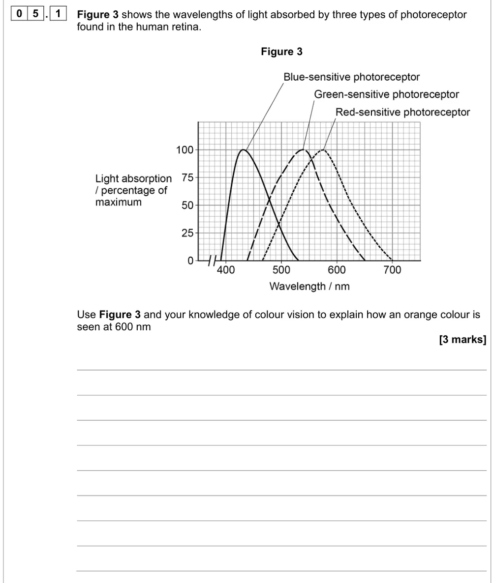
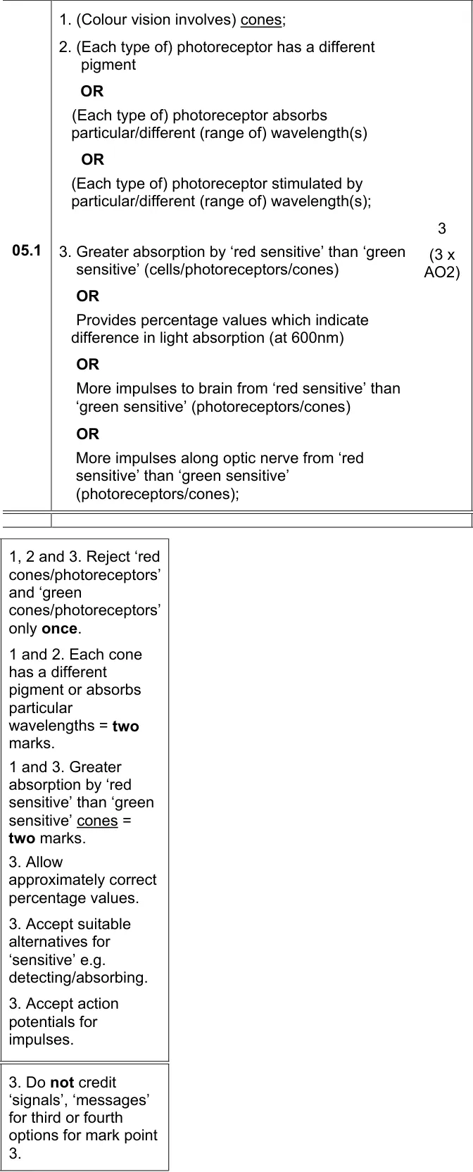
Use absorption curves to explain how orange light at 600 nm is seen.
Show the mark scheme
Tap an image to open it full size.
From the examiners' report: Only 2.6% got all three. Say cones, never rods, and write red-sensitive, not red. Talk about wavelengths absorbed, then compare how much each type absorbs at 600 nm.
Controlling the heartbeat (3.6.1.3)
Question 8
A level Paper 2 · June 2024 · Q06.1 · 5 marks · Medium
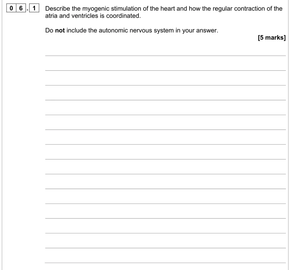
Describe myogenic stimulation and how contraction of the atria and ventricles is coordinated.
Show the mark scheme
Tap an image to open it full size.
From the examiners' report: About 28% got all five. Don't skip the atria contracting after the SAN, give the delay at the AVN, and keep SAN and AVN in the right order. Leave the nervous system out.
Question 9
A level Paper 2 · June 2017 · Q01.1 · 4 marks · Medium
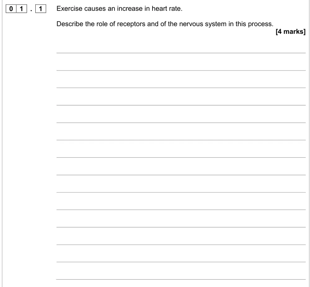
Describe the role of receptors and the nervous system in raising heart rate during exercise.
Show the mark scheme
Tap an image to open it full size.
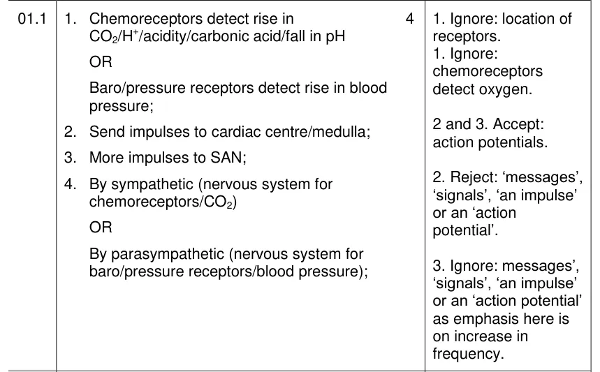
From the examiners' report: Over 25% scored zero and only 10% got all four. Use 'impulses', not messages or signals, and give the frequency: more impulses from the medulla along sympathetic neurones to the SAN.
Question 10
A level Paper 2 · November 2020 · Q10.2 · 4 marks · Medium
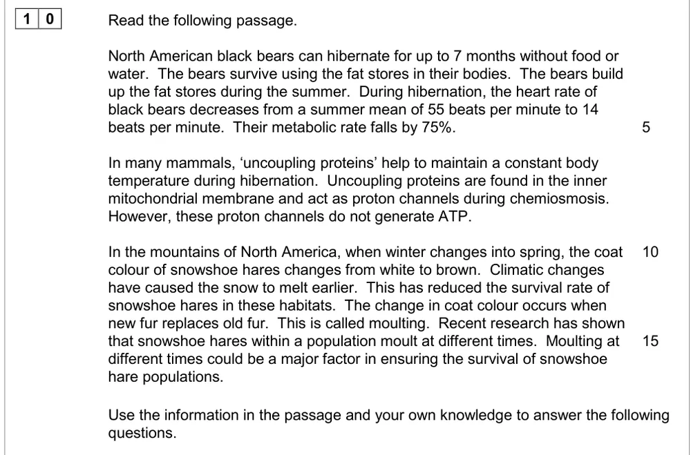
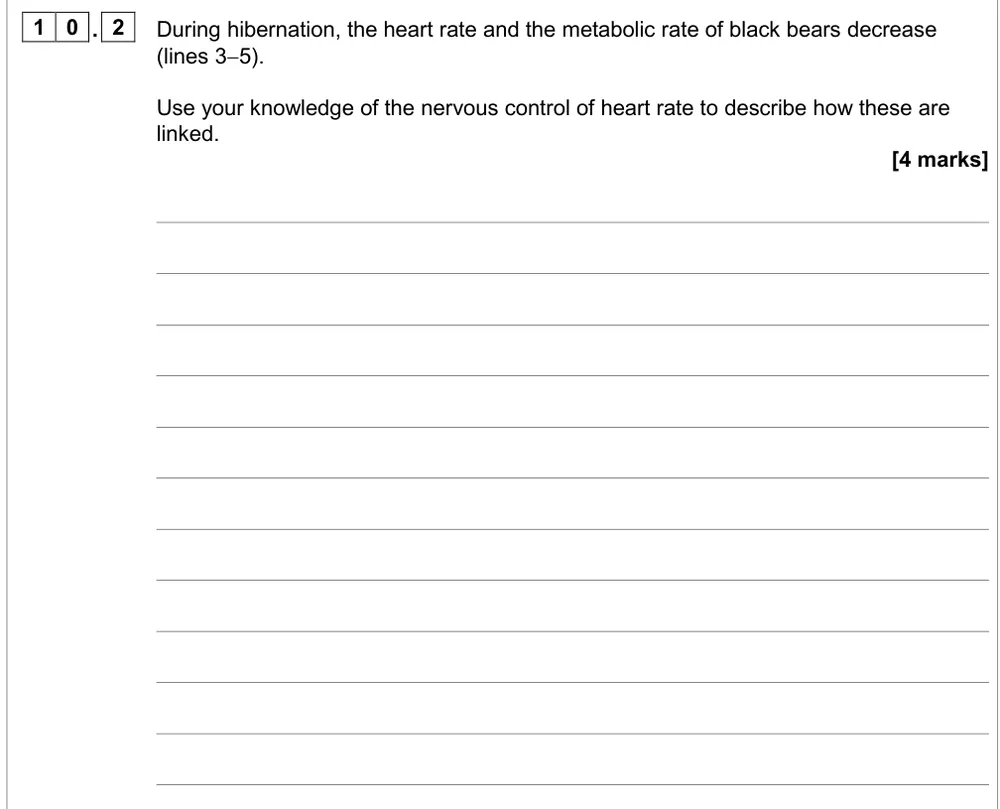
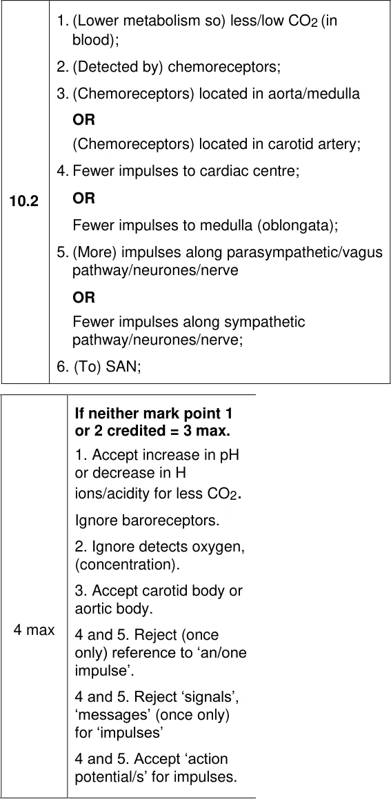
Link the lower metabolic rate and heart rate of hibernating bears through nervous control.
Information from earlier in this question:
Show the mark scheme
Tap an image to open it full size.
From the examiners' report: 36% scored zero. Start from less CO2 detected by chemoreceptors, not oxygen, then follow the impulses through the medulla and the parasympathetic nerve to the SAN.
Question 11
A level Paper 2 · June 2019 · Q10.2 · 3 marks · Hard
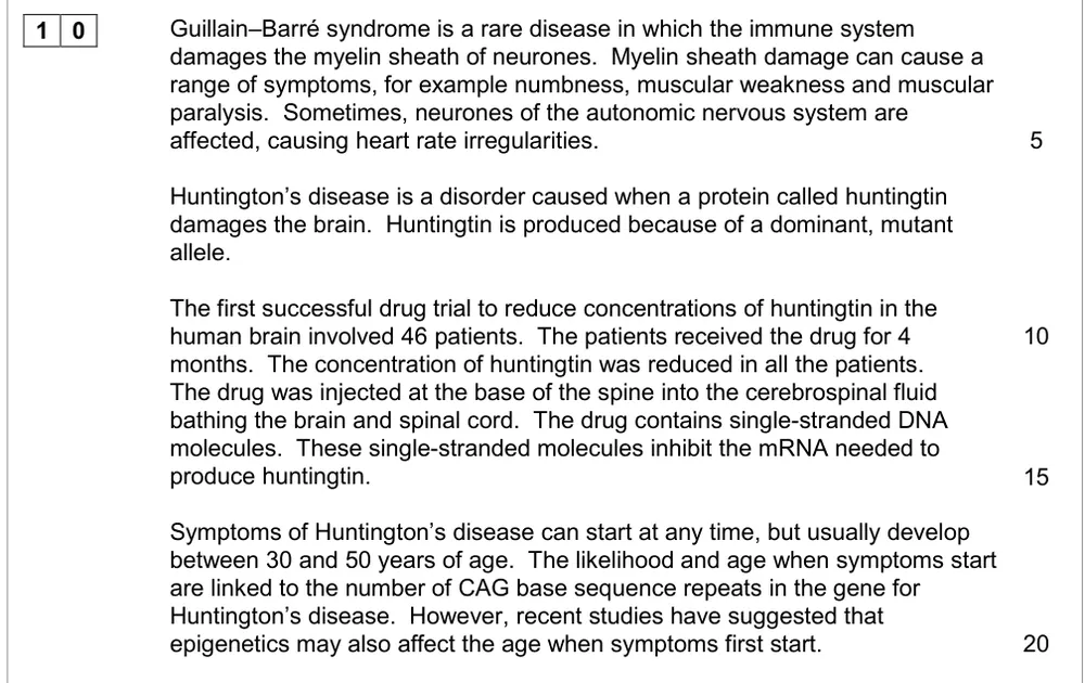
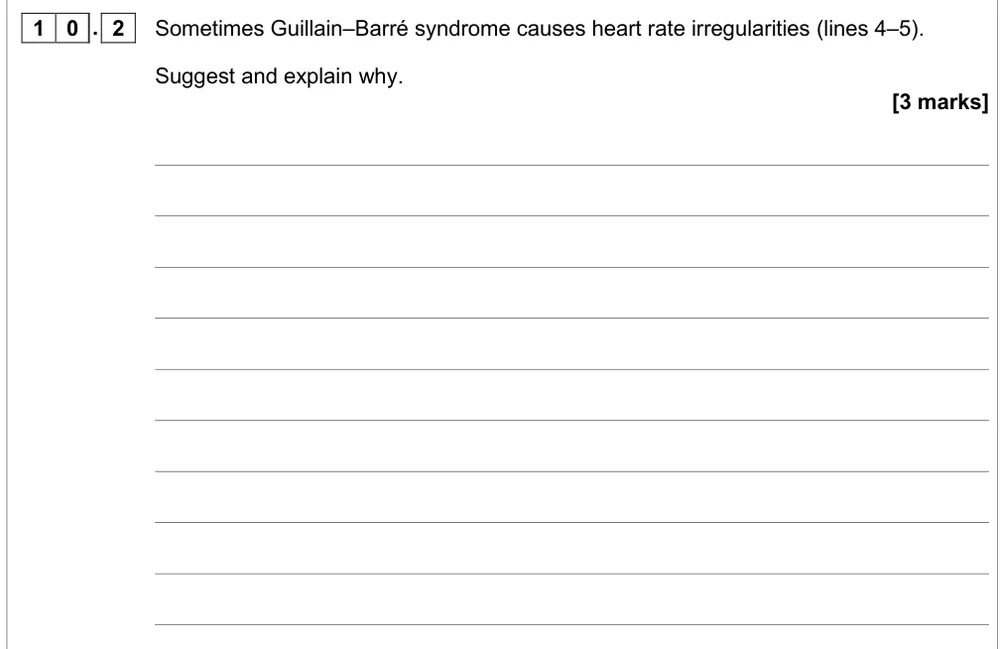
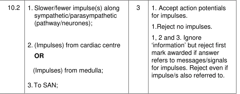
Suggest why damage to autonomic neurones causes heart rate irregularities.
Information from earlier in this question:
Show the mark scheme
Tap an image to open it full size.
From the examiners' report: Only about 4% got all three. Use the passage: the damaged neurones carry impulses from the medulla to the SAN, so explain fewer impulses to the SAN, not changes from it, and name sympathetic or parasympathetic.
Question 12
A level Paper 2 · June 2025 · Q06.3 · 3 marks · Medium
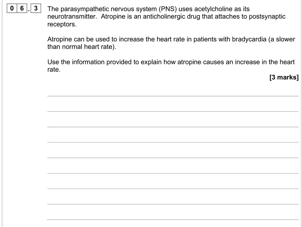
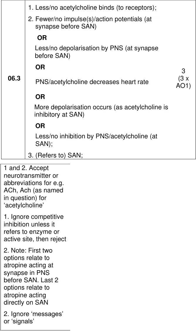
Explain how atropine, which blocks acetylcholine receptors, increases the heart rate.
Show the mark scheme
Tap an image to open it full size.
From the examiners' report: About 25% got all three. Most explained that atropine stops acetylcholine binding, but relatively few named the SAN: the parasympathetic nerve slows the SAN, so blocking it speeds the heart up.
Investigating heart rate (3.6.1.3)
Question 13
A level Paper 3 · June 2019 · Q03.4 · 2 marks · Medium
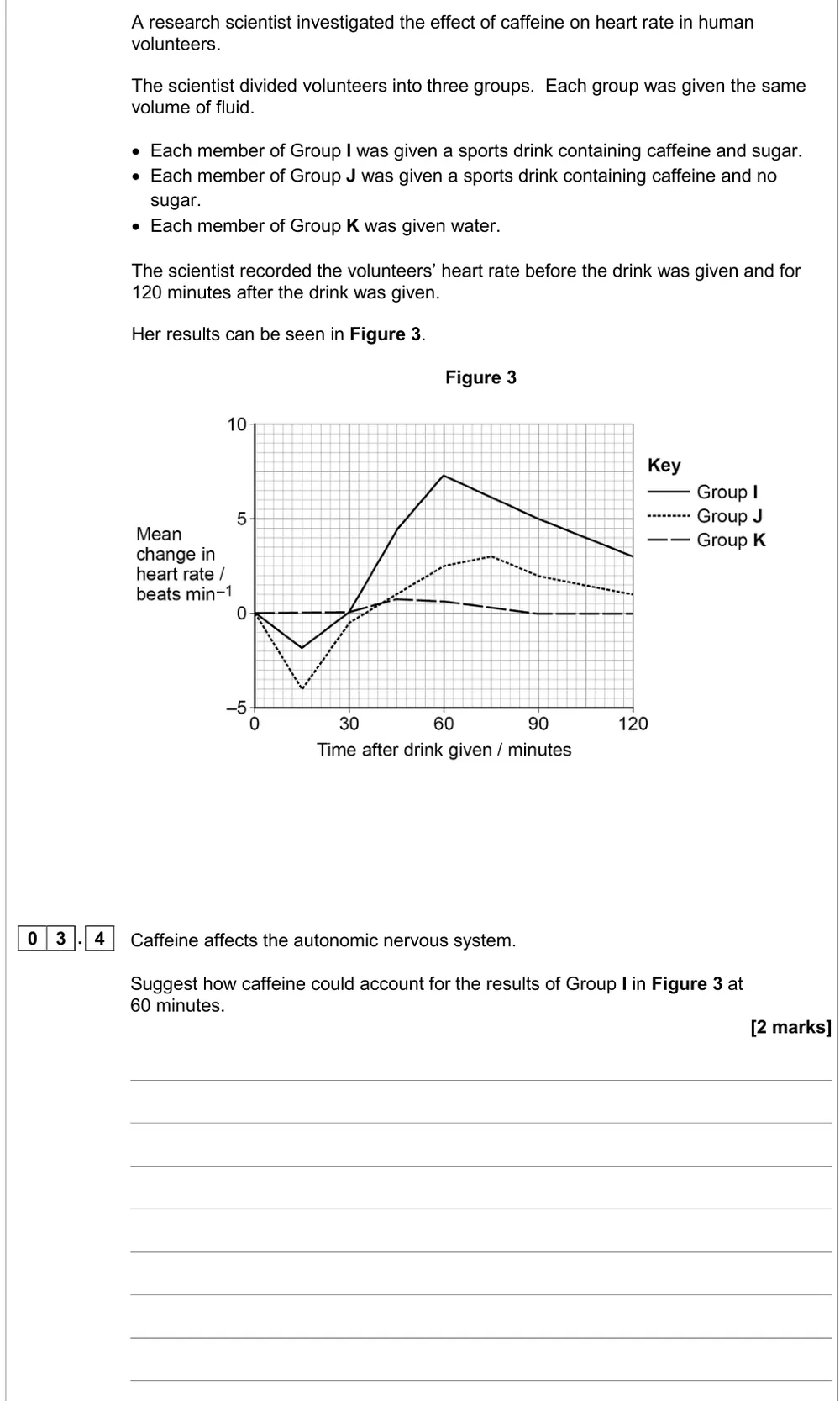
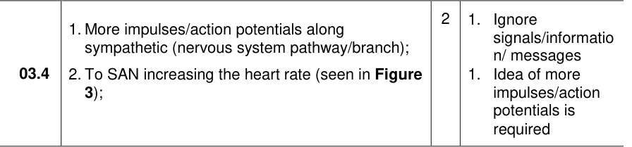
Suggest how caffeine could account for a higher heart rate at 60 minutes.
Show the mark scheme
Tap an image to open it full size.
From the examiners' report: About half got a mark. Give the idea of more impulses, not 'an impulse', name the sympathetic pathway and the SAN, and link back to the rise in heart rate. Sugar giving energy isn't the point.
Question 14
A level Paper 3 · June 2019 · Q03.6 · 2 marks · Easy
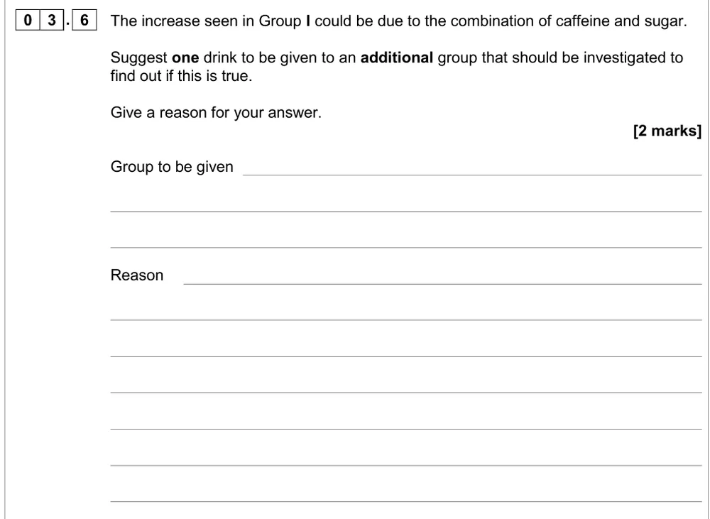
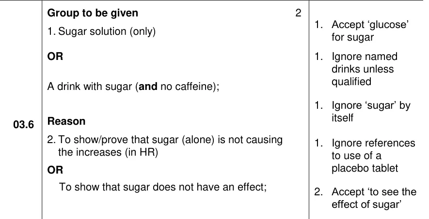
Suggest an extra drink to test whether caffeine and sugar together raise heart rate.
Show the mark scheme
Tap an image to open it full size.
From the examiners' report: Over 70% got both. 'Give sugar' on its own didn't score: say sugar with no caffeine. For the reason, 'as a control' or 'for comparison' is too vague; say it shows whether sugar alone affects heart rate.
Where students lose marks (from AQA examiners' reports and mark schemes)
Each point below is followed by wording that gained marks.
Write impulses, never messages or signals: On the exercise question over 25% scored zero and only 10% got full marks, mainly through poor terminology such as 'messages' or 'signals'. The same words cost marks on rods and cones, the hibernating bears and Guillain–Barré syndrome.
✔ Gains credit: "Send impulses to the cardiac centre or medulla"; accept "action potentials".
Heart rate changes with the number of impulses: On caffeine, those who missed the first mark often said 'an impulse' would be sent, without the idea of more impulses. Others reached the SAN but didn't link it back to the higher heart rate in Figure 3.
✔ Gains credit: "More impulses along the sympathetic (pathway); to the SAN, increasing the heart rate."
Chemoreceptors detect carbon dioxide, not oxygen: 36% scored zero on the hibernating bears. A significant minority said chemoreceptors only respond to oxygen, and some skipped the nervous system and wrote about less oxygen reaching the tissues.
✔ Gains credit: "Less CO2 in the blood; detected by chemoreceptors in the aorta or carotid artery; fewer impulses to the cardiac centre; more impulses along the parasympathetic (vagus) pathway; to the SAN."
Impulses go to the SAN, not from it: Only about 4% got all three on Guillain–Barré syndrome. Many explained changes in impulses from the SAN instead of impulses to the SAN, said no impulses would reach it, or named the autonomic nervous system without saying sympathetic or parasympathetic.
✔ Gains credit: "Slower or fewer impulses along sympathetic or parasympathetic neurones; from the cardiac centre or medulla; to the SAN."
Atria contract, then the AVN delays: On myogenic stimulation a significant minority left out atrial contraction after the SAN, and the delay at the AVN was not always given. Weaker answers confused the SAN with the AVN.
✔ Gains credit: "SAN releases a wave of electrical activity; atria contract; AVN passes on electrical activity after a short delay; via Purkyne tissue in the bundle of His; ventricles contract from the bottom upwards."
Red-sensitive cones, not red cones: Only 2.6% got all three on seeing orange at 600 nm, and fewer than 1% on red-green colour blindness. 'Red, green or blue cones' and 'green pigment' were rejected, and few said other colours stimulate more than one type of cone at once.
✔ Gains credit: "Each type of cone absorbs a different range of wavelengths; greater absorption by red-sensitive than green-sensitive cones at 600 nm."
Take acuity all the way to the brain: 74% got a mark on the eagle's fovea, usually for one cone to one neurone, but only 14% got all three because most didn't say cones send separate impulses to the brain.
✔ Gains credit: "High visual acuity; each cone connected to a single neurone; separate (sets of) impulses to the brain or optic nerve."
Sodium ions through stretch-mediated channels: On the generator potential, some wrote about a 'stretch-mediated membrane' instead of stretch-mediated sodium ion channels, and many were held to 2 marks by writing 'sodium' instead of sodium ions.
✔ Gains credit: "Pressure deforms the membrane; stretch-mediated sodium ion channels open; sodium ions diffuse in; depolarisation (generator potential)."
Download the questions
Download the PDF pack (14 questions, mark schemes at the end)
Keep practising
- Survival and response: AQA A Level Biology exam questions by topic
- AQA 3.6.1 stimuli and receptors: self-marking quiz
- The heart and circulation: AQA A Level Biology exam questions by topic
- Transport across cell membranes: AQA A Level Biology exam questions by topic
- AQA A Level Biology masterclasses
Questions, mark schemes and examiners' report extracts © AQA.