

The Gas Exchange System (9.1): Cambridge (CIE) A Level Biology 9700 Exam Questions by Topic
The gas exchange system is sub-topic 9.1 of Topic 9, Gas exchange, in the Cambridge International AS & A Level Biology syllabus (9700). You need to describe the structure of the human gas exchange system, know where cartilage, ciliated epithelium, goblet cells, squamous epithelium, smooth muscle and capillaries are found, and recognise these tissues and the airways in micrographs. You also need to describe the functions of these tissues and cells, of mucous glands and of elastic fibres, and describe gas exchange between the air in the alveoli and the blood in the capillaries. This is AS content, examined on Papers 1 and 2. A level students meet it again on Paper 4.
The examiners' reports return to the same points. Keep the tissues apart: smooth muscle contracts and relaxes, elastic fibres stretch and recoil, and cartilage supports an airway and stops it collapsing. It is the muscle that contracts, not the bronchiole. Alveoli have no smooth muscle and no ciliated epithelial cells. Mucus traps particles and pathogens, and the cilia move the mucus; it is the cilia that move, not the cells. Check whether a question asks for a cell or a tissue. When you explain a steep diffusion gradient, write about blood flowing through the alveolar capillaries, not just 'the lungs'.
Studying Cambridge International A level Biology and want more help? Tom teaches CIE students one-to-one online, including students in the UAE, Saudi Arabia, Hong Kong, India and Indonesia, and every lesson is recorded. You can book a one-to-one lesson or a free 15-minute discovery chat, or register your interest in a CIE group class starting in September 2027, using the links at the bottom of this page.
What you need from IGCSE
Each question on this page assumes you're secure on these ideas. If any of them feels shaky, look it up before you start.
- Air reaches the alveoli through the trachea, the bronchi and the bronchioles.
- A gas exchange surface has a large surface area, a thin surface, a good blood supply and good ventilation with air.
- Oxygen diffuses from the air in the alveoli into the blood, and carbon dioxide diffuses from the blood into the air in the alveoli.
- Goblet cells make mucus and ciliated cells move it, which protects the breathing system from pathogens and particles.
Key terms
These words come up in the questions and mark schemes. Examiners expect you to use them exactly.
- Cartilage: supporting tissue that keeps an airway open: C-shaped rings in the trachea and irregular plates in the bronchi. There is none in the bronchioles.
- Ciliated epithelium: the tissue lining the trachea, bronchi and bronchioles, made of ciliated epithelial cells and goblet cells.
- Goblet cell: a cell in the ciliated epithelium that secretes mucus.
- Mucous gland: a gland beneath the epithelium of the trachea and bronchi that secretes mucus.
- Squamous epithelium: the flattened cells that form the wall of an alveolus, giving a short diffusion distance.
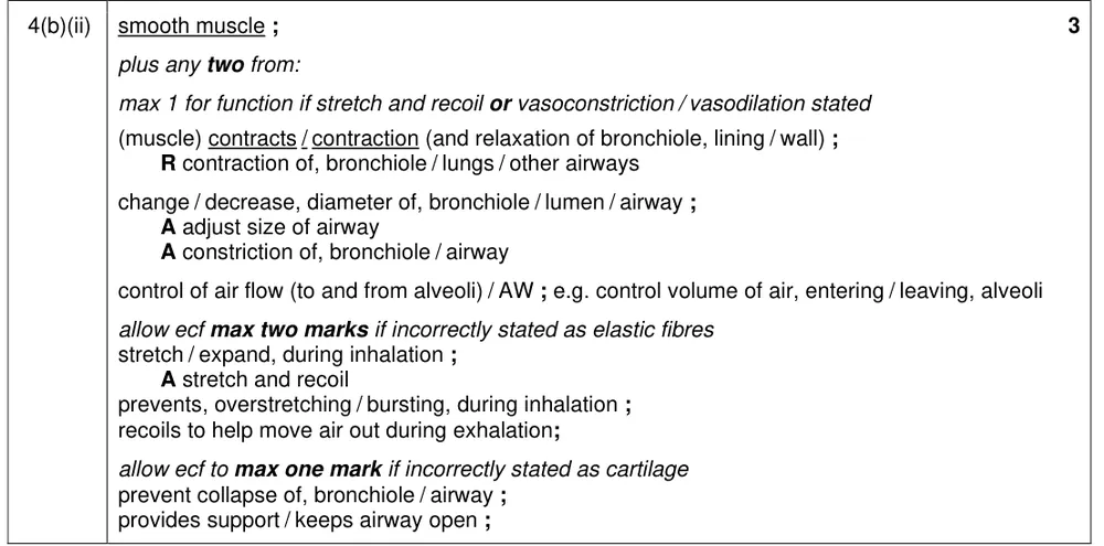
- Smooth muscle: muscle in the walls of the trachea, bronchi and bronchioles that contracts and relaxes to change the diameter of the lumen and so control the flow of air.
- Elastic fibres: fibres in the alveolar wall that stretch on inhalation and recoil on exhalation to help force air out.
What these questions cover (Cambridge 9700 Topic 9: Gas exchange)
The questions test these learning outcomes from sub-topic 9.1 of the Cambridge International AS & A Level Biology syllabus (9700) for 2025 to 2027.
- 9.1.1 describe the structure of the human gas exchange system
- 9.1.2 describe the distribution in the gas exchange system of cartilage, ciliated epithelium, goblet cells, squamous epithelium of alveoli, smooth muscle and capillaries
- 9.1.3 recognise cartilage, ciliated epithelium, goblet cells, squamous epithelium of alveoli, smooth muscle and capillaries in microscope slides, photomicrographs and electron micrographs
- 9.1.4 recognise trachea, bronchi, bronchioles and alveoli in microscope slides, photomicrographs and electron micrographs
- 9.1.5 describe the functions of ciliated epithelial cells, goblet cells and mucous glands in maintaining the health of the gas exchange system
- 9.1.6 describe the functions in the gas exchange system of cartilage, smooth muscle, elastic fibres and squamous epithelium
- 9.1.7 describe gas exchange between air in the alveoli and blood in the capillaries
How to use this page
Write your answer on paper before you open the mark scheme. Allow about 1.2 minutes per mark: the 22 questions are worth 50 marks, so about 60 minutes in total. Then mark your answer strictly against the mark scheme and read the examiners' tip underneath it. If you'd rather print the questions, download the PDF pack.
Structure of the gas exchange system (9.1.1)
Question 1
Paper 2 · May/June 2023 (22) · Q6(a) · 1 mark
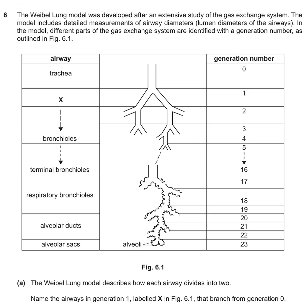
Name the airways that branch from the trachea in a model of the airways of the lung.
Show the mark scheme

Tap an image to open it full size.
From the examiners' report: The singular 'bronchus' was allowed. Write the word clearly: very poor spellings that could have been either bronchi or bronchioles were not credited.
Distribution of tissues and cells (9.1.2)
Question 2
Paper 2 · May/June 2023 (23) · Q2(c)(ii) · 3 marks
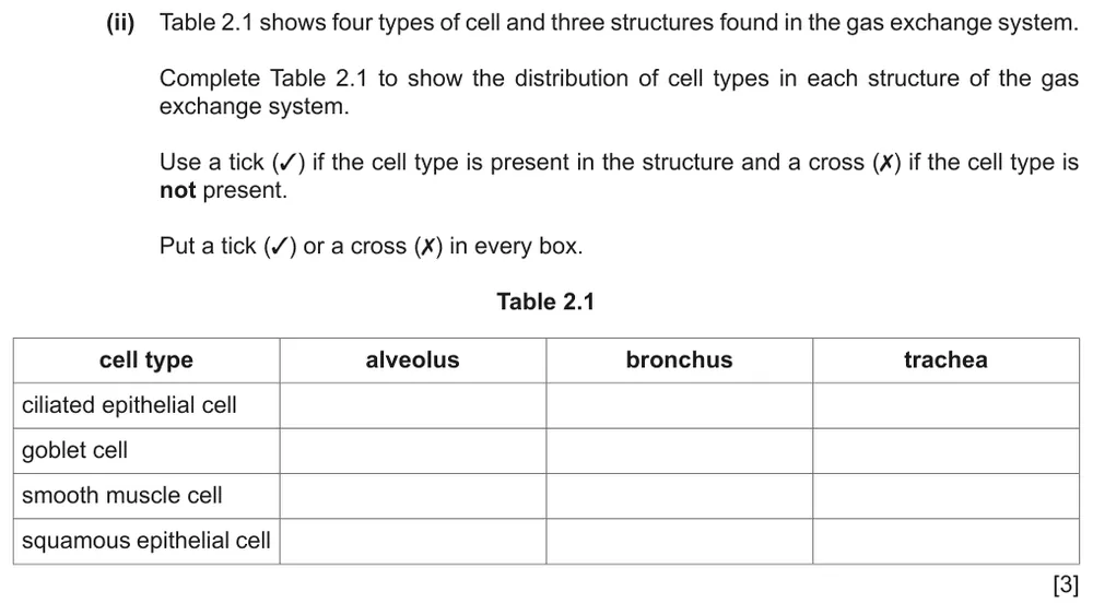
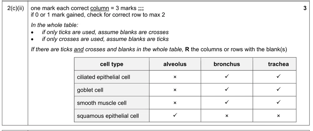
Complete the table with ticks and crosses to show which cell types are found in the alveolus, the bronchus and the trachea.
Show the mark scheme
Tap an image to open it full size.
From the examiners' report: Put a tick or a cross in every box, as instructed; a number of students used only ticks and left the other boxes blank. The alveolus column was better known than the bronchus and trachea columns. Some wrongly suggested that smooth muscle cells are present in the alveolus.
Question 3
Paper 2 · February/March 2021 (22) · Q1(c) · 1 mark
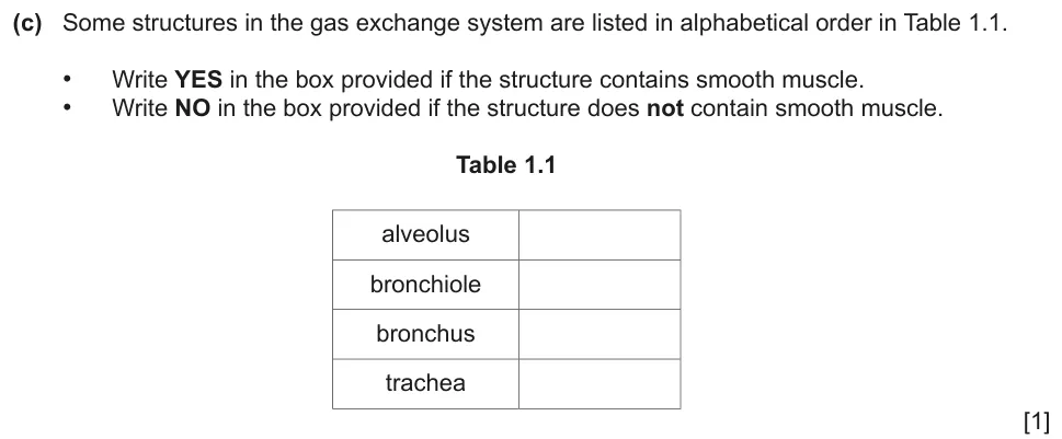
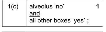
Complete the table to show which structures of the gas exchange system contain smooth muscle.
Show the mark scheme
Tap an image to open it full size.
From the examiners' report: A frequent error was to indicate that alveoli contain smooth muscle.
Question 4
Paper 2 · February/March 2020 (22) · Q4(a) · 3 marks
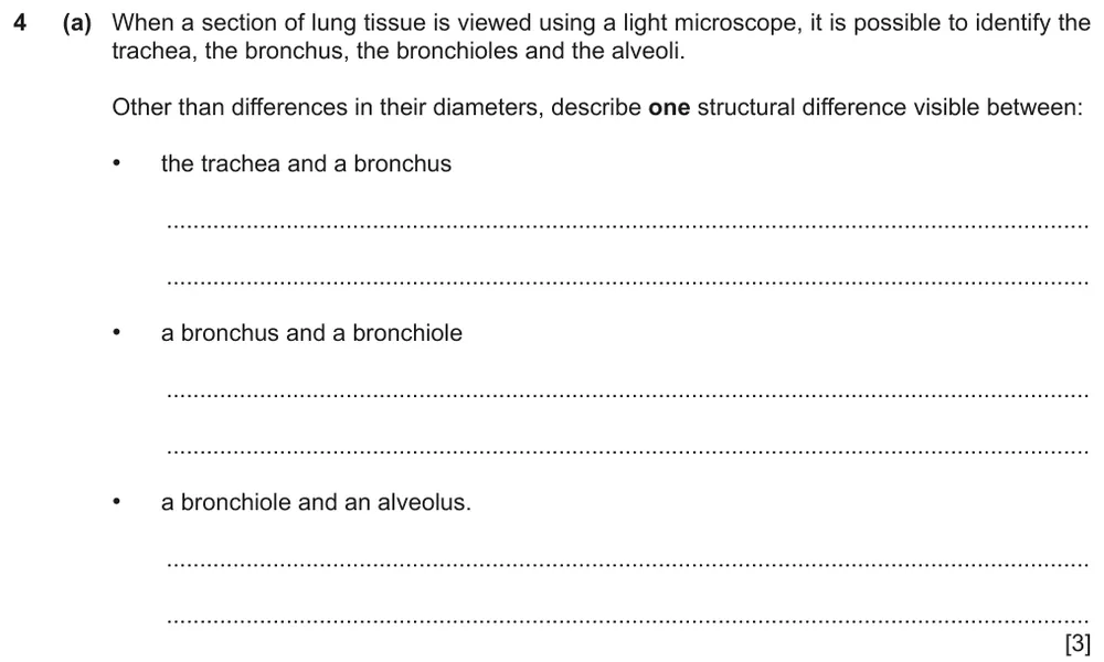
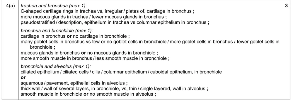
Describe one visible structural difference between the trachea and a bronchus, a bronchus and a bronchiole, and a bronchiole and an alveolus.
Show the mark scheme
Tap an image to open it full size.
From the examiners' report: Write a clear comparative sentence for each of the three differences. Weaker answers gave a relevant difference without linking it to one or other of the two structures being compared.
Recognising tissues and cells in micrographs (9.1.3)
Question 5
Paper 2 · October/November 2022 (22) · Q4(b)(i) · 3 marks · Easy
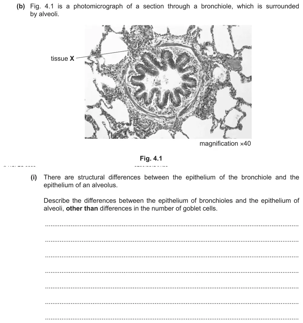
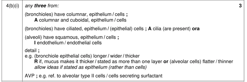
Describe the differences between the epithelium of a bronchiole and the epithelium of an alveolus.
Information from earlier in this question:

Show the mark scheme
Tap an image to open it full size.
From the examiners' report: Few knew that bronchioles have columnar, or columnar and cuboidal, epithelial cells. The epithelium is the layer facing the lumen: do not describe it as many layers thick, or say that mucus makes it thicker. Cartilage is not found in the wall of a bronchiole.
Question 6
Paper 2 · May/June 2022 (22) · Q5(a) · 1 mark
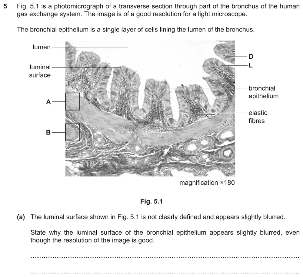
State why the luminal surface of the bronchial epithelium looks slightly blurred.
Show the mark scheme

Tap an image to open it full size.
From the examiners' report: You only needed to note that the luminal surface is where the cilia are. The image is described as having good resolution, so it is not correct to say that the cilia are too small to be seen. Some students wrongly described the cilia as microvilli.
Question 7
Paper 2 · May/June 2022 (22) · Q5(b) · 2 marks
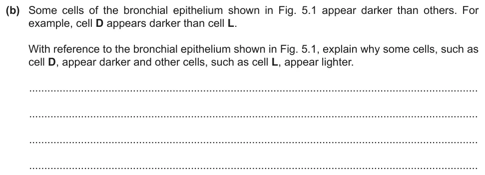
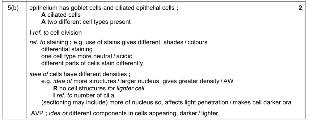
Explain why some cells of the bronchial epithelium look darker than others.
Show the mark scheme
Tap an image to open it full size.
From the examiners' report: Good answers referred to both cell D and cell L and gave two or more explanations. Only a small proportion stated that the bronchial epithelium is made of two cell types, goblet cells and ciliated epithelial cells.
Question 8
Paper 2 · May/June 2024 (21) · Q4(a)(i) · 2 marks
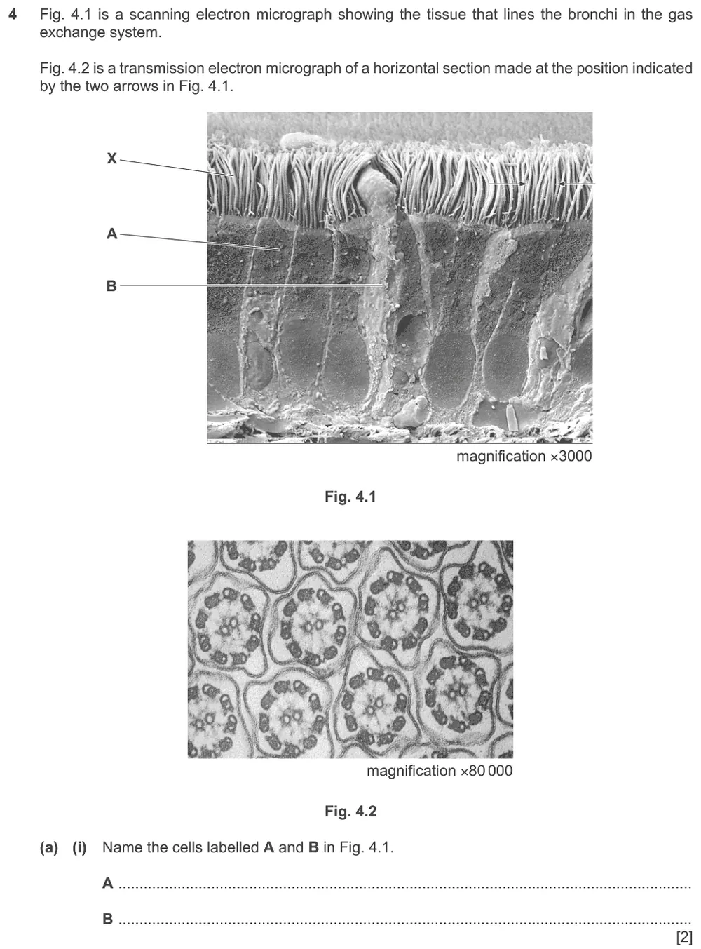
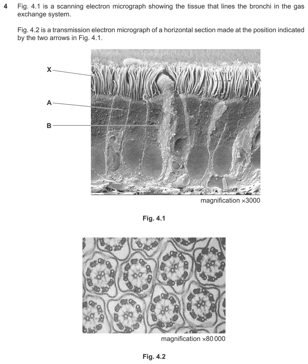
Name cells A and B in a scanning electron micrograph of the tissue lining a bronchus.
Show the mark scheme

Tap an image to open it full size.
From the examiners' report: The question asks for cells, so giving the tissue, ciliated epithelium, was a common error for A. 'Mucous gland' was often given for B, but it is not a cell type and mucous glands lie beneath the epithelium, so they would not be visible in this image.
Recognising airways in micrographs (9.1.4)
Question 9
Paper 2 · October/November 2020 (22) · Q3(a) · 3 marks
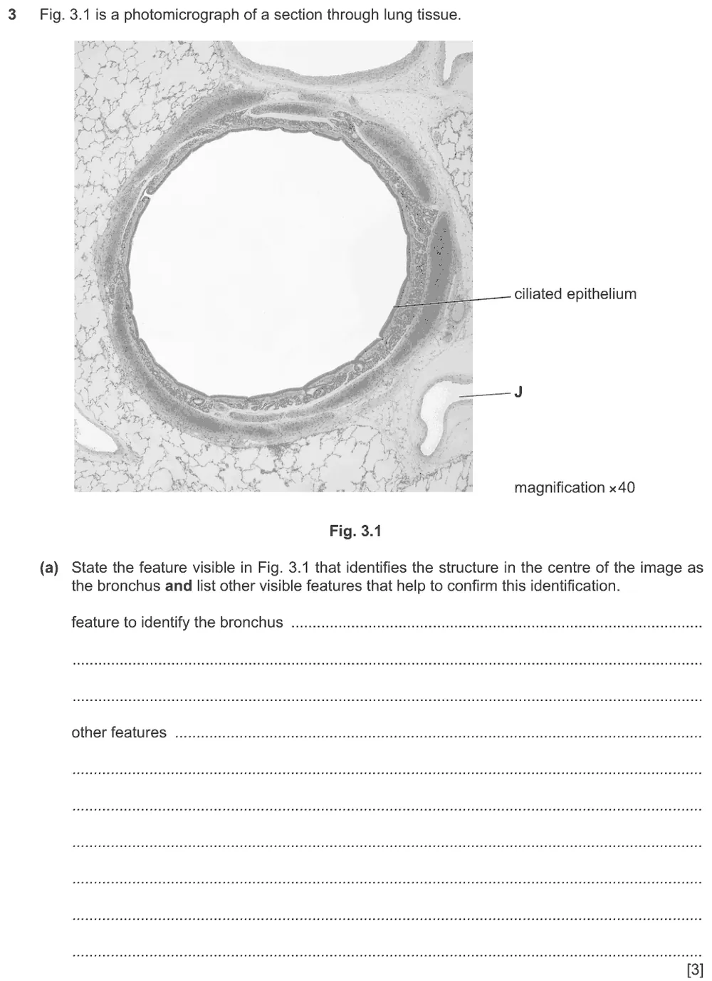
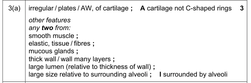
State the feature that identifies the structure as a bronchus and list other visible features that confirm this.
Show the mark scheme
Tap an image to open it full size.
From the examiners' report: Describe the cartilage as irregular or as plates, because the trachea also has cartilage, as incomplete or C-shaped rings. Smooth muscle, elastic tissue and a wide lumen were credited only as additional features. Ciliated epithelium was already labelled, so goblet cells and ciliated epithelial cells were ignored.
Question 10
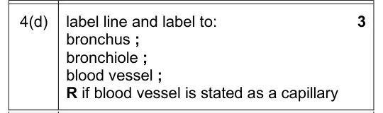
Paper 2 · May/June 2025 (22) · Q4(d) · 3 marks
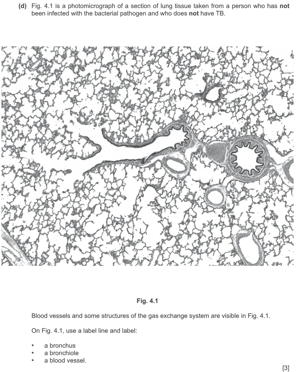
Label a bronchus, a bronchiole and a blood vessel on a photomicrograph of lung tissue.
Show the mark scheme
Tap an image to open it full size.
Ciliated epithelial cells, goblet cells and mucous glands (9.1.5)
Question 11
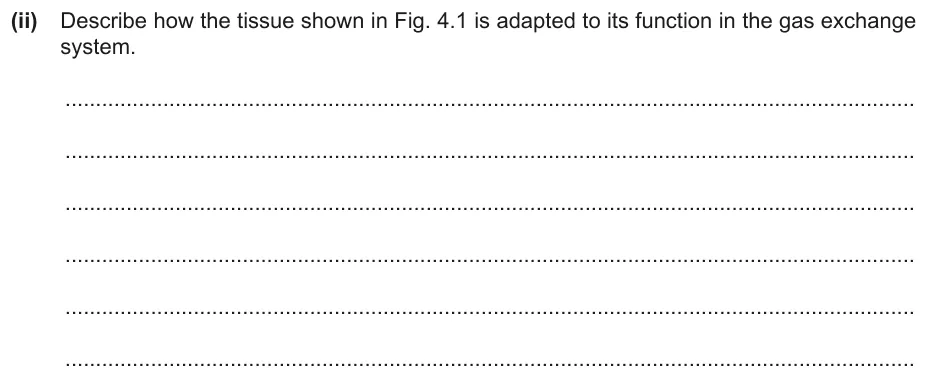
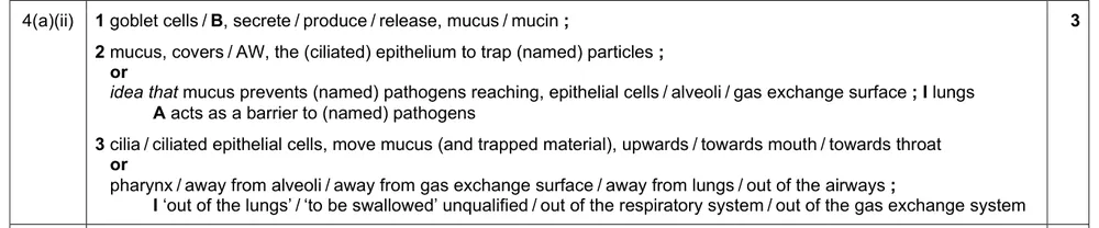
Paper 2 · May/June 2024 (21) · Q4(a)(ii) · 3 marks
Describe how the tissue lining the bronchus is adapted to its function in the gas exchange system.
Information from earlier in this question:
Show the mark scheme
Tap an image to open it full size.
From the examiners' report: Say that mucus covers the epithelium and traps particles and pathogens, and that the cilia move the mucus with the trapped material, not the pathogens on their own. This tissue is in the gas exchange system, so do not write that mucus stops pathogens reaching the gas exchange system. Cilia do not provide a large surface area for gas exchange.
Question 12
Paper 2 · October/November 2020 (22) · Q3(c) · 2 marks
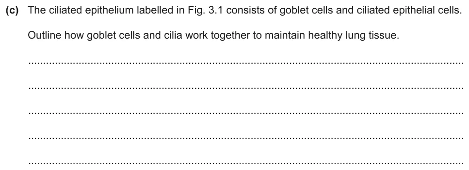
Outline how goblet cells and cilia work together to maintain healthy lung tissue.
Information from earlier in this question:
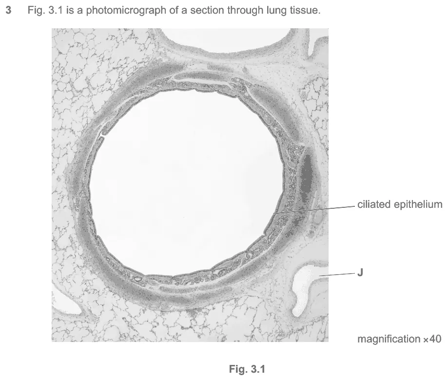
Show the mark scheme

Tap an image to open it full size.
From the examiners' report: Be precise about how the cilia move the mucus out of the airways. Do not write about hairs or finger-like structures, or describe ciliated epithelial cells moving. 'Dirt' and 'dust' are not alternative terms for pathogens, and avoid calling pathogens 'germs'.
Question 13
Paper 2 · February/March 2023 (22) · Q2(c) · 2 marks · Easy
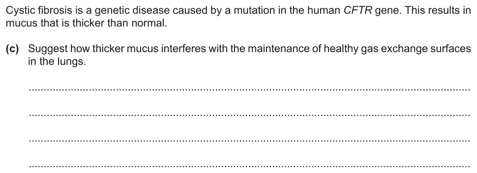
Suggest how thicker mucus interferes with keeping the gas exchange surfaces healthy.
Show the mark scheme

Tap an image to open it full size.
From the examiners' report: Most answers explained that the movement of the cilia is impaired, so mucus with trapped pathogens builds up and the risk of infection increases. The most common wrong suggestions were that thicker mucus restricts the movement of air down to the alveoli, or that the cilia are destroyed.
Question 14
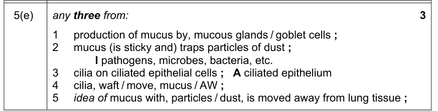
Paper 2 · February/March 2022 (22) · Q5(e) · 3 marks
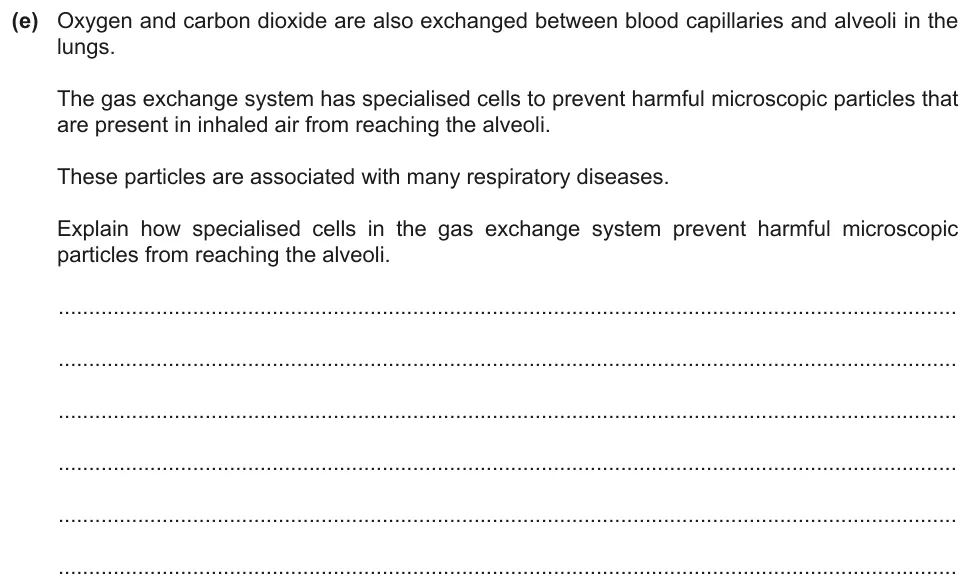
Explain how specialised cells prevent harmful microscopic particles from reaching the alveoli.
Show the mark scheme
Tap an image to open it full size.
From the examiners' report: Include the role of mucus: some answers had the particles trapped and removed by the cilia alone. It is the cilia that move, not the ciliated cells.
Functions of smooth muscle and cartilage (9.1.6)
Question 15
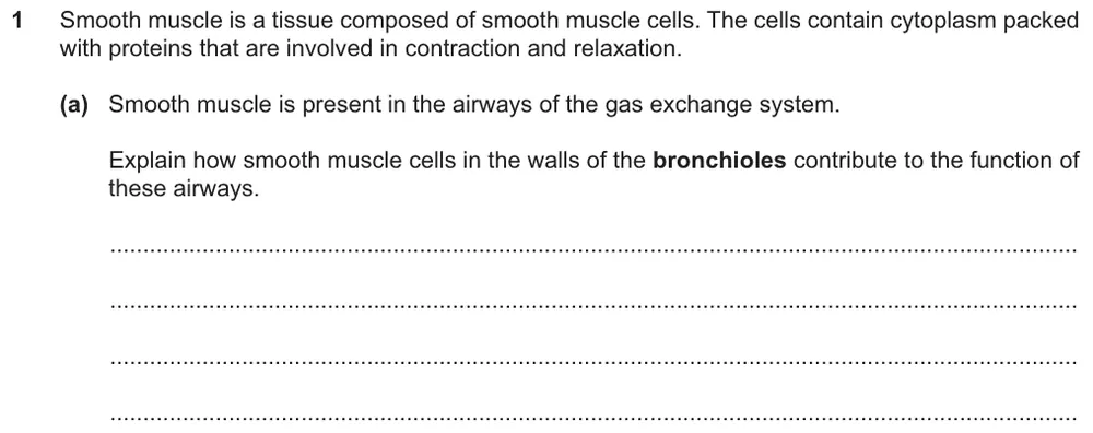
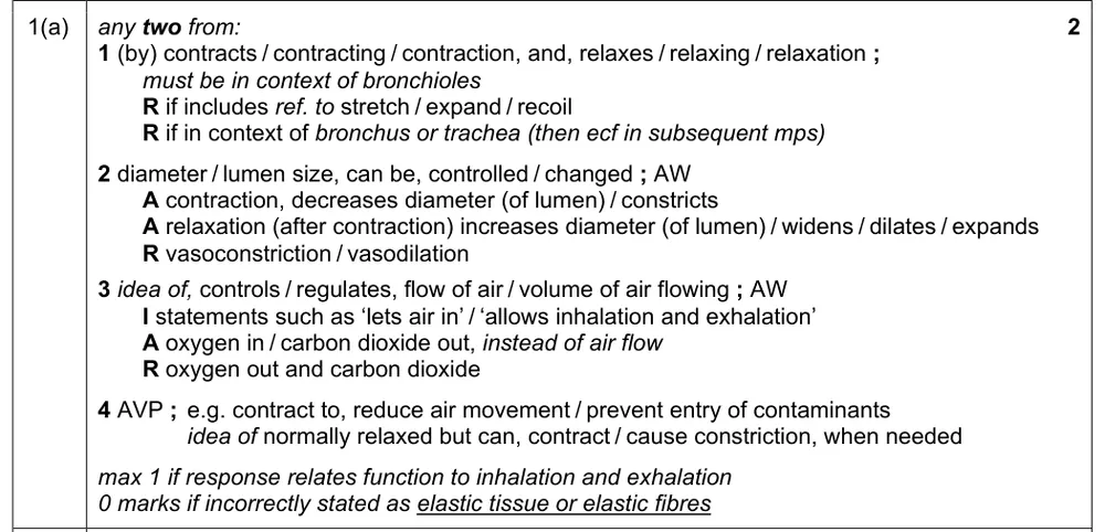
Paper 2 · May/June 2024 (22) · Q1(a) · 2 marks · Easy
Explain how smooth muscle cells in the walls of the bronchioles contribute to the function of these airways.
Show the mark scheme
Tap an image to open it full size.
From the examiners' report: Contraction narrows the lumen and relaxation widens it; some students had this the wrong way round. It is not correct to relate contraction and relaxation to breathing in and out. Vasoconstriction and vasodilation are terms for blood vessels, not airways.
Question 16
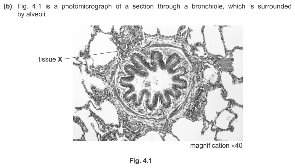
Paper 2 · October/November 2022 (22) · Q4(b)(ii) · 3 marks
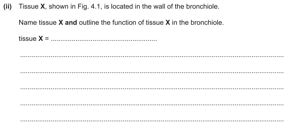
Name tissue X in the wall of the bronchiole and outline its function.
Information from earlier in this question:
Show the mark scheme
Tap an image to open it full size.
From the examiners' report: 'Muscle' was not enough: name the tissue as smooth muscle. Answers stating that the bronchiole contracted gained no credit. It was a common error to describe smooth muscle as expanding, stretching or recoiling; some gave the functions of elastic tissue or cartilage instead.
Question 17
Paper 2 · May/June 2022 (22) · Q5(c) · 3 marks · Easy
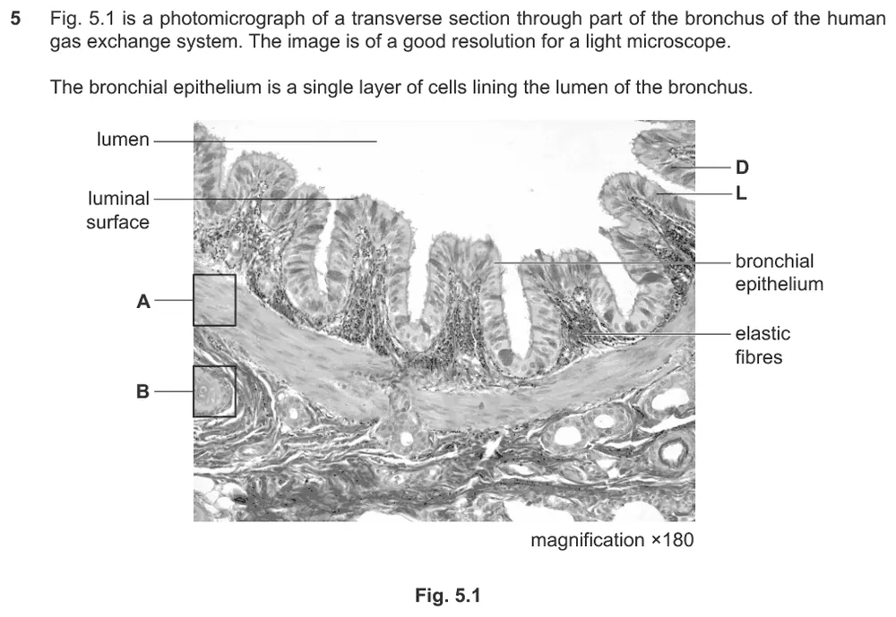
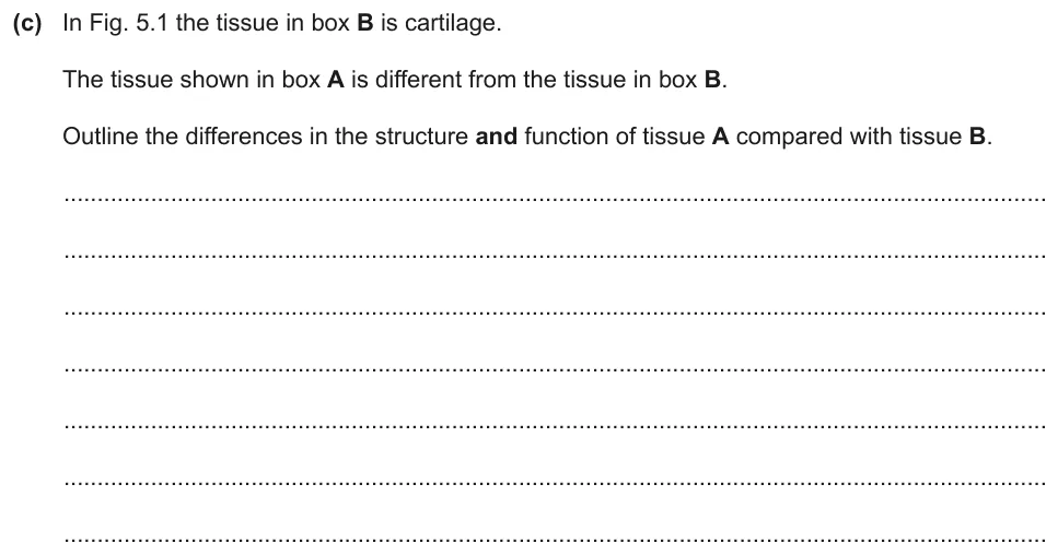
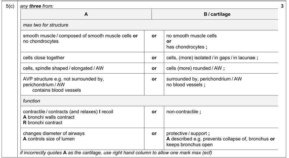
Outline the differences in structure and function between tissue A and the cartilage in box B.
Information from earlier in this question:
Show the mark scheme
Tap an image to open it full size.
From the examiners' report: The best answers named tissue A as smooth muscle, outlined its function and then compared this with the function of cartilage. Smooth muscle contracts and relaxes; stretching and recoiling is a feature of elastic tissue. Some named smooth muscle and then contradicted themselves by saying it contained elastic tissue, mucous glands or goblet cells.
Blood flow and elastic fibres in the alveoli (9.1.6 and 9.1.7)
Question 18
Paper 2 · October/November 2023 (22) · Q4(a) · 2 marks
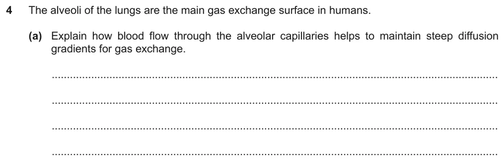
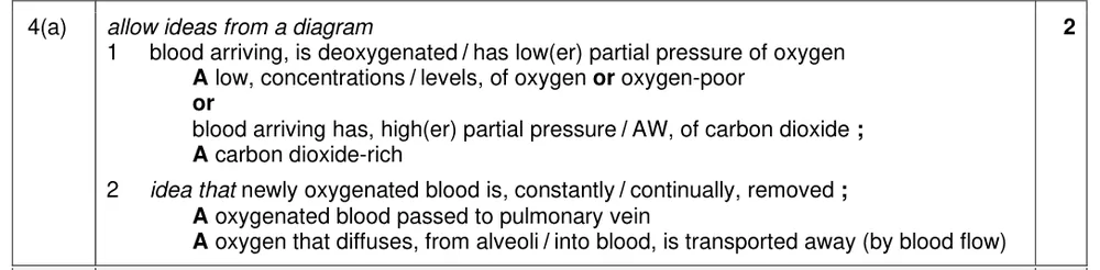
Explain how blood flow through the alveolar capillaries helps to maintain steep diffusion gradients for gas exchange.
Show the mark scheme
Tap an image to open it full size.
From the examiners' report: Focus on the flow of blood: many students only described gas exchange or the features of a good gas exchange surface. More described the blood arriving at the alveoli than noted that newly oxygenated blood is then carried away. The fullest answers covered both oxygen and carbon dioxide.
Question 19
Paper 2 · October/November 2023 (22) · Q4(b) · 2 marks
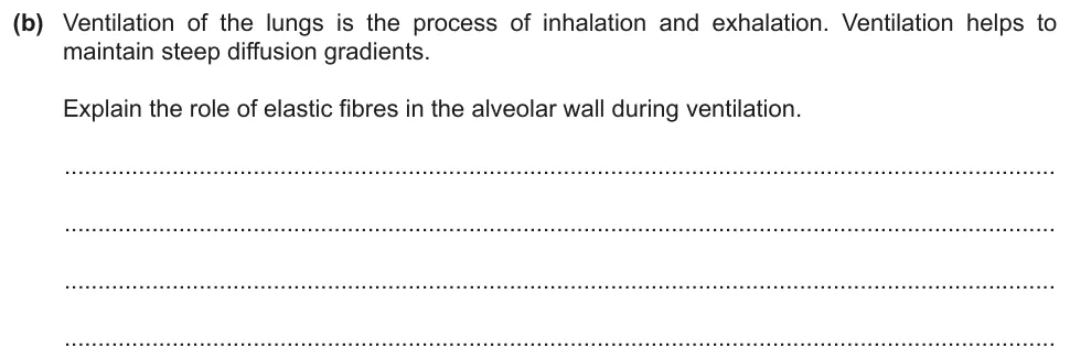
Explain the role of elastic fibres in the alveolar wall during ventilation.
Show the mark scheme

Tap an image to open it full size.
From the examiners' report: Use 'stretch' and 'recoil', not 'contract' and 'relax'. Stretch goes with inhalation and recoil with air leaving the alveoli on exhalation; a number of students had these the wrong way round.
Gas exchange between the alveoli and the blood (9.1.7)
Question 20
Paper 2 · May/June 2023 (22) · Q6(b) · 2 marks
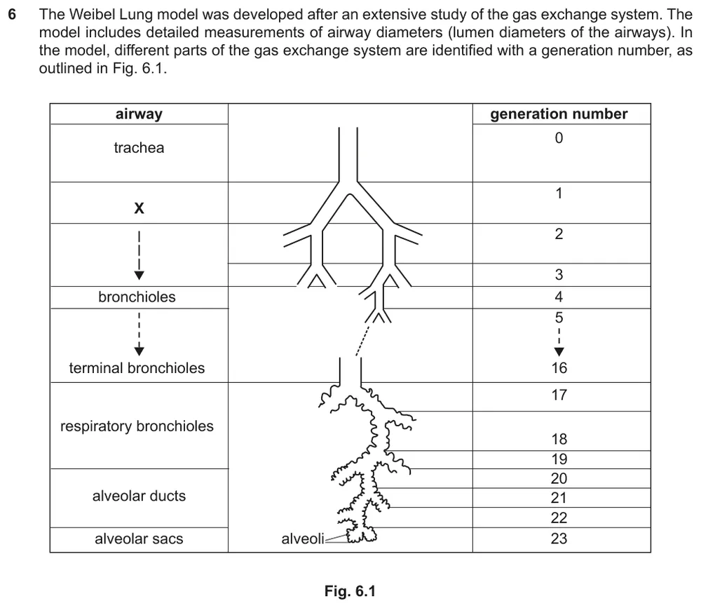
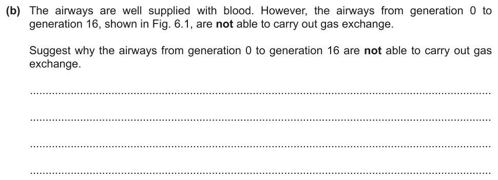
Suggest why the airways from generation 0 to generation 16 are not able to carry out gas exchange.
Information from earlier in this question:
Show the mark scheme
Tap an image to open it full size.
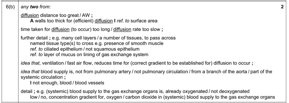
From the examiners' report: Many said that the diffusion distance is too great and supported this with the tissues in the wall of the airways, or with the absence of squamous epithelium. Stating that alveoli are not present gained no credit. The question says the airways are well supplied with blood, so 'no blood supply' is incorrect.
Question 21
Paper 2 · February/March 2023 (22) · Q5(b)(i) · 1 mark · Easy
Name a structure in the gas exchange system that has the same function as the membrane of the oxygenator.
Information from earlier in this question:


Show the mark scheme

Tap an image to open it full size.
From the examiners' report: Cell membrane, squamous epithelium and epithelium were the most common wrong answers because they are not specific to the gas exchange system.
Question 22
Paper 2 · February/March 2023 (22) · Q5(b)(ii) · 3 marks
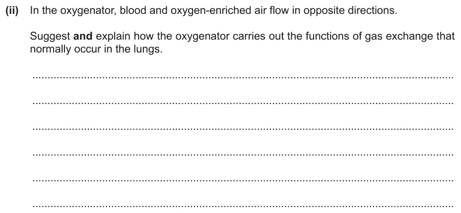
Suggest and explain how the oxygenator carries out the gas exchange that normally occurs in the lungs.
Show the mark scheme

Tap an image to open it full size.
From the examiners' report: Relate what happens in the lungs to how the oxygenator works; weaker answers often described only the lungs. Few students explained the significance of the blood and the air flowing in opposite directions.
Where students lose marks (from Cambridge examiners' reports and mark schemes)
Each point below comes from the examiners' reports and mark schemes, and is followed by wording that gained marks.
Alveoli have no smooth muscle. Students indicated that alveoli contain smooth muscle.
✔ Gains credit: 'alveolus "no" and all other boxes "yes"': the bronchiole, bronchus and trachea all contain smooth muscle.
Name the cell, not the tissue. Students gave the tissue, ciliated epithelium, when asked for a cell, or named a mucous gland, which is not a cell type.
✔ Gains credit: A is a 'ciliated epithelial (cell)' and B is a 'goblet (cell)'.
Irregular plates identify a bronchus. Students gave smooth muscle, elastic tissue or a wide lumen as the identifying feature, or wrote 'cartilage' without describing it.
✔ Gains credit: 'irregular / plates of cartilage' identifies the bronchus; 'smooth muscle', 'elastic tissue' and 'mucous glands' are other features.
Cilia move the mucus. Students wrote that cilia move or trap the pathogens, that ciliated cells move, or left mucus out altogether.
✔ Gains credit: 'goblet cells secrete mucus', mucus 'covers the (ciliated) epithelium to trap particles', and 'cilia move mucus' 'towards throat' or 'away from alveoli'.
Smooth muscle contracts; it does not recoil. Students described smooth muscle as expanding, stretching or recoiling, or wrote that the bronchiole contracts.
✔ Gains credit: 'smooth muscle', '(muscle) contracts', to 'change / decrease diameter of bronchiole / lumen' and give 'control of air flow'.
Elastic fibres stretch and recoil. Students used 'contract' and 'relax' for elastic fibres, or linked stretch and recoil to the wrong stage of breathing.
✔ Gains credit: 'allow alveoli to expand / stretch on inhalation', 'prevent alveoli from overstretching / bursting', and 'recoil to help exhalation'.
Oxygenated blood is carried away. Students described gas exchange or the features of a gas exchange surface, and did not write about blood flow.
✔ Gains credit: 'blood arriving is deoxygenated' and 'newly oxygenated blood is constantly removed'.
Airway walls are too thick for diffusion. Students wrote that the airways have no alveoli or no blood supply.
✔ Gains credit: 'diffusion distance too great', 'walls too thick for (efficient) diffusion', with 'many cell layers' and 'not squamous epithelium'.
Download the questions
Download the PDF pack (22 questions, mark schemes at the end)
Keep practising
- Cambridge (CIE) A level Biology 9700 past papers, mark schemes and examiners' reports
- Cambridge (CIE) A level Biology grade thresholds
- Book a one-to-one CIE lesson or a free 15-minute discovery chat with Tom
- Register your interest in a CIE A level Biology group class
Questions, mark schemes and examiners' report extracts © Cambridge University Press & Assessment.