

Proteins and Water (2.3 and 2.4): Cambridge (CIE) A Level Biology 9700 Exam Questions by Topic
Proteins (sub-topic 2.3) and water (sub-topic 2.4) are part of Topic 2, Biological molecules, in the Cambridge International AS & A Level Biology syllabus (9700). You need to draw the general structure of an amino acid, describe how a peptide bond forms and breaks, explain primary, secondary, tertiary and quaternary structure and the bonds that hold proteins in shape, and relate the structures of haemoglobin and collagen to their functions. You also need to explain hydrogen bonding between water molecules and relate the properties of water to its roles in living organisms. This is AS content, examined on Papers 1 and 2. A level students meet it again on Paper 4; questions marked A level on this page come from Paper 4.
The examiners' reports return to the same points. In the general structure of an amino acid the side chain is R, not H, and a peptide bond forms by condensation, not hydrolysis. Name each level of protein structure and describe the levels in order, without merging two of them in one sentence. With a ribbon diagram, say what it does not show as well as what it does. The bonds between cysteine R-groups are disulfide bonds. Keep the collagen molecule apart from the collagen fibre, and do not confuse latent heat of vaporisation with specific heat capacity.
Studying Cambridge International A level Biology and want more help? Tom teaches CIE students one-to-one online, including students in the UAE, Saudi Arabia, Hong Kong, India and Indonesia, and every lesson is recorded. You can book a one-to-one lesson or a free 15-minute discovery chat, or register your interest in a CIE group class starting in September 2027, using the links at the bottom of this page.
What you need from IGCSE
Each question on this page assumes you're secure on these ideas. If any of them feels shaky, look it up before you start.
- Proteins are large molecules made from amino acids, and contain the elements carbon, hydrogen, oxygen and nitrogen.
- Different sequences of amino acids give proteins different shapes, such as the active site of an enzyme.
- Haemoglobin in red blood cells contains iron and transports oxygen.
- Water is a solvent: substances are transported around organisms dissolved in water.
Key terms
These words come up in the questions and mark schemes. Examiners expect you to use them exactly.
- Peptide bond: the covalent bond between the carboxyl group of one amino acid and the amino group of the next; it forms by condensation and is broken by hydrolysis.
- Primary structure: the sequence of amino acids in a polypeptide.
- Secondary structure: α-helices and β-pleated sheets, held in shape by hydrogen bonds.
- Tertiary structure: the folding and coiling of a polypeptide into a three-dimensional shape, held by interactions between R-groups.
- Quaternary structure: the structure of a protein made of two or more polypeptides, such as haemoglobin.
- Disulfide bond: a covalent bond between the R-groups of two cysteine amino acids.
- Haem group: the part of each haemoglobin polypeptide that contains an iron ion, which binds one oxygen molecule.
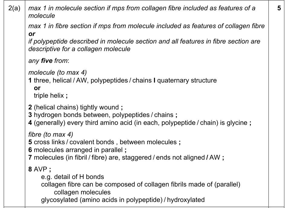
- Triple helix: three helical polypeptides wound tightly around each other to form a collagen molecule.
What these questions cover (Cambridge 9700 Topic 2: Biological molecules)
The questions test these learning outcomes from sub-topics 2.3 and 2.4 of the Cambridge International AS & A Level Biology syllabus (9700) for 2025 to 2027.
- 2.3.1 describe and draw the general structure of an amino acid and the formation and breakage of a peptide bond
- 2.3.2 explain the meaning of the terms primary structure, secondary structure, tertiary structure and quaternary structure of proteins
- 2.3.3 describe the types of interaction that hold protein molecules in shape
- 2.3.4 state that globular proteins are generally soluble and have physiological roles and fibrous proteins are generally insoluble and have structural roles
- 2.3.5 describe the structure of a molecule of haemoglobin as an example of a globular protein
- 2.3.6 relate the structure of haemoglobin to its function, including the importance of iron in the haem group
- 2.3.7 describe the structure of a molecule of collagen as an example of a fibrous protein, and the arrangement of collagen molecules to form collagen fibres
- 2.3.8 relate the structures of collagen molecules and collagen fibres to their function
- 2.4.1 explain how hydrogen bonding occurs between water molecules and relate the properties of water to its roles in living organisms
How to use this page
Write your answer on paper before you open the mark scheme. Allow about 1.2 minutes per mark: the 22 questions are worth 55 marks, so about 66 minutes in total. Then mark your answer strictly against the mark scheme and read the examiners' tip underneath it. If you'd rather print the questions, download the PDF pack.
Amino acids and peptide bonds (2.3.1)
Question 1
Paper 2 · May/June 2023 (22) · Q2(d)(ii) · 2 marks
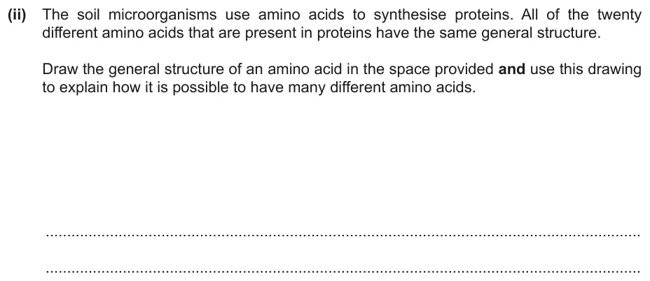
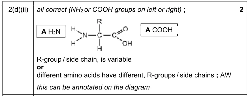
Draw the general structure of an amino acid and use it to explain how there can be many different amino acids.
Information from earlier in this question:

Show the mark scheme
Tap an image to open it full size.
From the examiners' report: On the left of the central carbon write H2N, not NH2. Some drew three hydrogen atoms on the nitrogen instead of two. Writing H in place of R gives glycine, not the general structure.
Question 2
Paper 2 · February/March 2023 (22) · Q2(a) · 1 mark · Easy
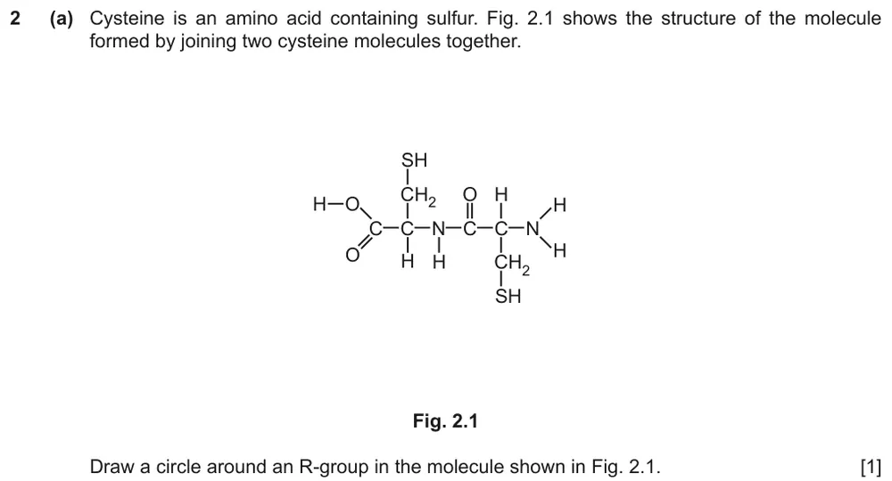
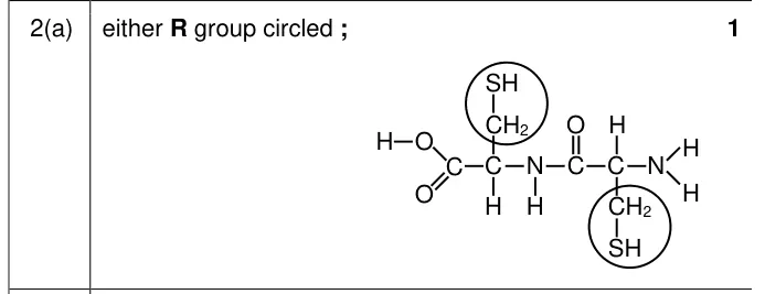
Draw a circle around an R-group in a molecule made from two cysteine molecules.
Show the mark scheme
Tap an image to open it full size.
From the examiners' report: Most circled one of the R-groups correctly, although some circled both. A common error was to circle an amino group or a carboxyl group.
Question 3
Paper 2 · October/November 2016 (22) · Q2(c) · 2 marks
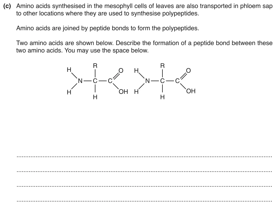
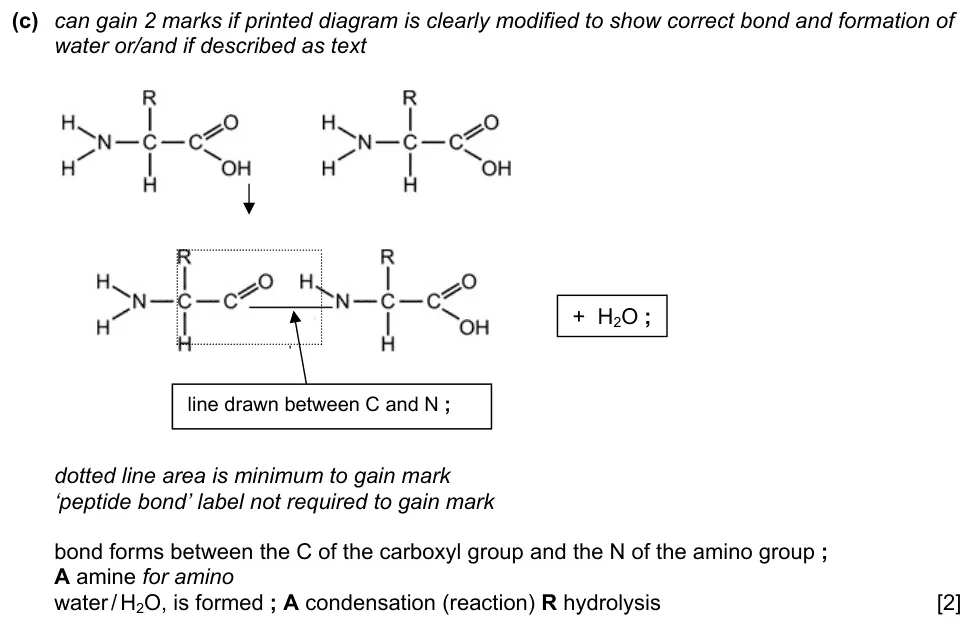
Describe the formation of a peptide bond between two amino acids.
Show the mark scheme
Tap an image to open it full size.
From the examiners' report: Many drew excellent diagrams of the dipeptide with a molecule of water given off. Calling the reaction hydrolysis contradicted the diagram and lost the mark for water. Some wrote that a glycosidic bond formed, although the question says it is a peptide bond.
Question 4
Paper 2 · February/March 2025 (22) · Q2(b)(i) · 2 marks · Easy
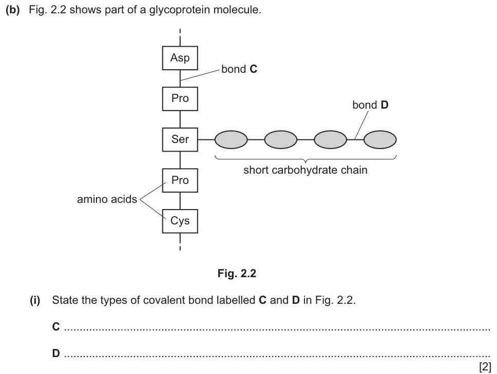
State the types of covalent bond labelled C and D in a glycoprotein molecule.
Show the mark scheme

Tap an image to open it full size.
Levels of protein structure (2.3.2)
Question 5
Paper 2 · February/March 2025 (22) · Q2(c) · 4 marks
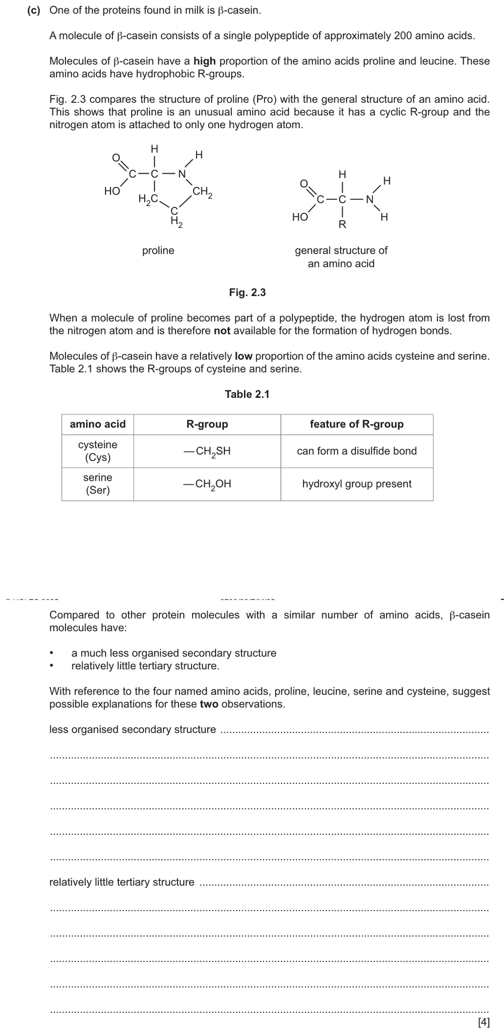
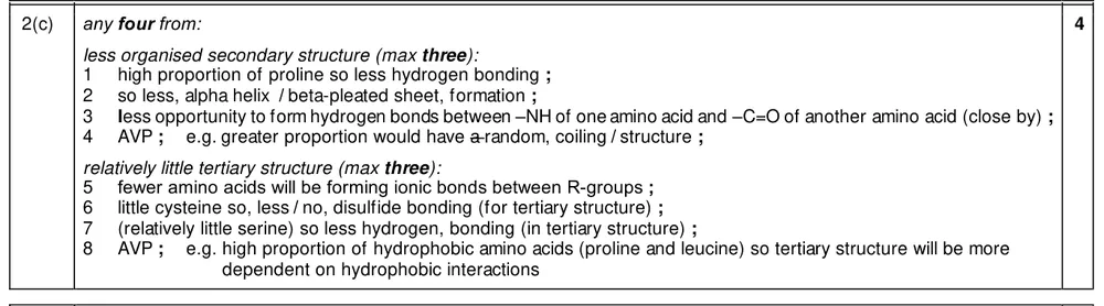
Use the information about four amino acids to explain why β-casein has little secondary and tertiary structure.
Show the mark scheme
Tap an image to open it full size.
From the examiners' report: Some linked the high proportion of proline to fewer sites for the hydrogen bonds that form and hold α-helices and β-pleated sheets. More could explain the lack of tertiary structure, usually from the low proportion of cysteine and what that means for disulfide bonds.
Question 6
Paper 2 · October/November 2023 (22) · Q2(a) · 1 mark
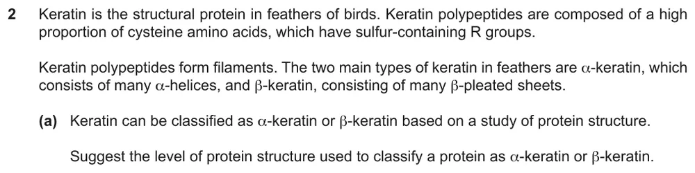
Suggest the level of protein structure used to classify keratin as α-keratin or β-keratin.
Show the mark scheme

Tap an image to open it full size.
Question 7
Paper 2 · May/June 2023 (23) · Q4(a)(i) · 3 marks · Hard
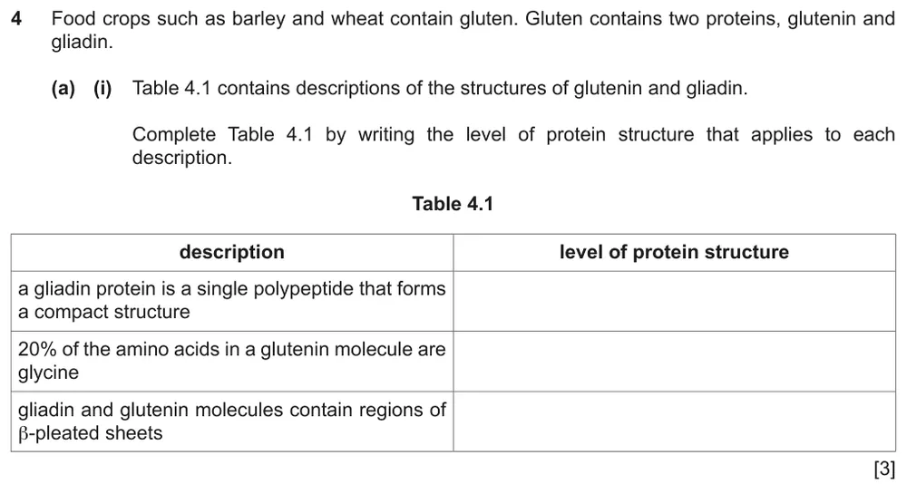
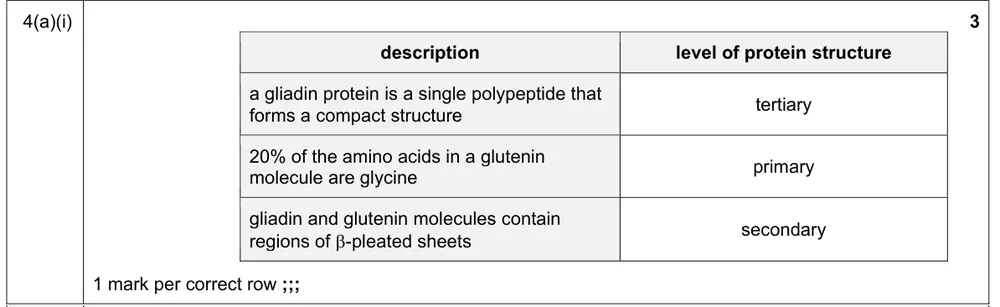
Write the level of protein structure that applies to each description of glutenin and gliadin.
Show the mark scheme
Tap an image to open it full size.
From the examiners' report: Many found this hard. β-pleated sheets point to secondary structure. A compact structure could be tertiary or quaternary, but ‘a single polypeptide’ rules out quaternary.
Question 8
Paper 2 · October/November 2021 (22) · Q3(c) · 4 marks
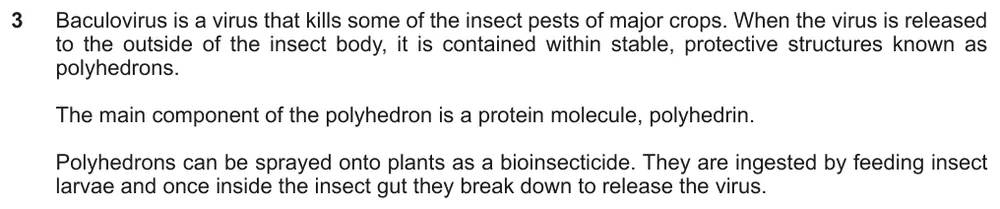
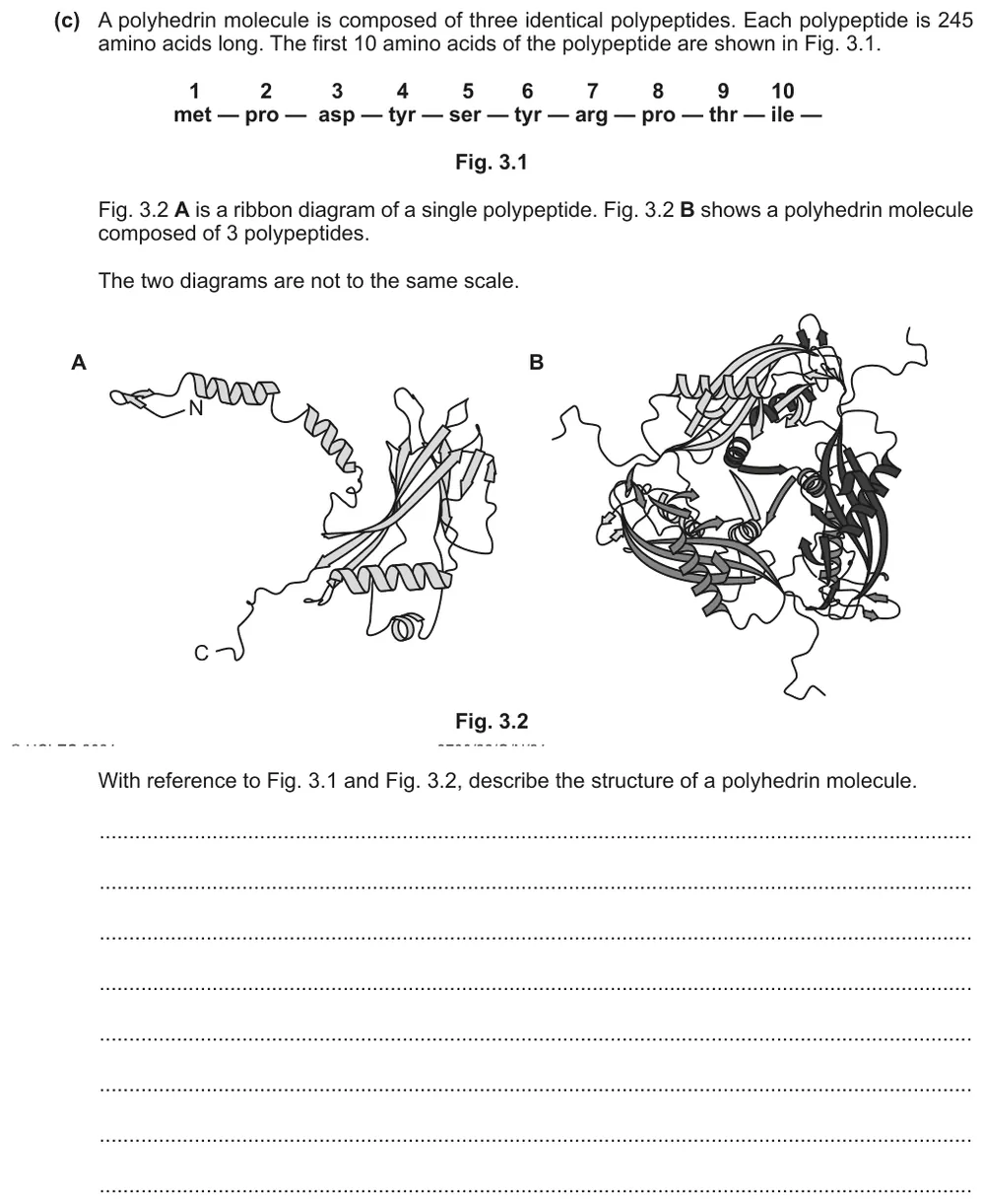
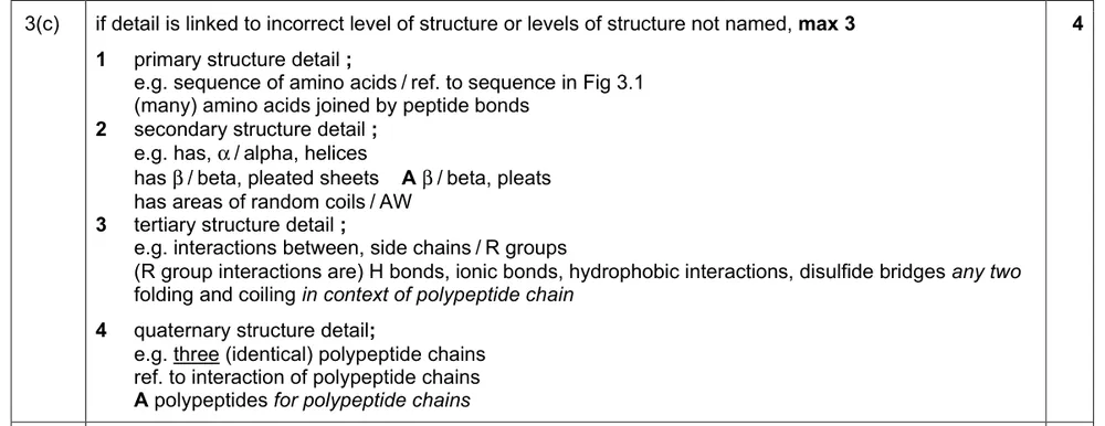
Use the amino acid sequence and ribbon diagrams to describe the structure of a polyhedrin molecule.
Information from earlier in this question:
Show the mark scheme
Tap an image to open it full size.
From the examiners' report: Use Fig. 3.1 as a reminder to describe primary structure as well as the other three levels. Work through the levels in order, using the correct terms. Answers that merged secondary with tertiary, or tertiary with quaternary, in one sentence lacked clarity.
Question 9
Paper 2 · February/March 2019 (22) · Q2(c) · 4 marks · Easy
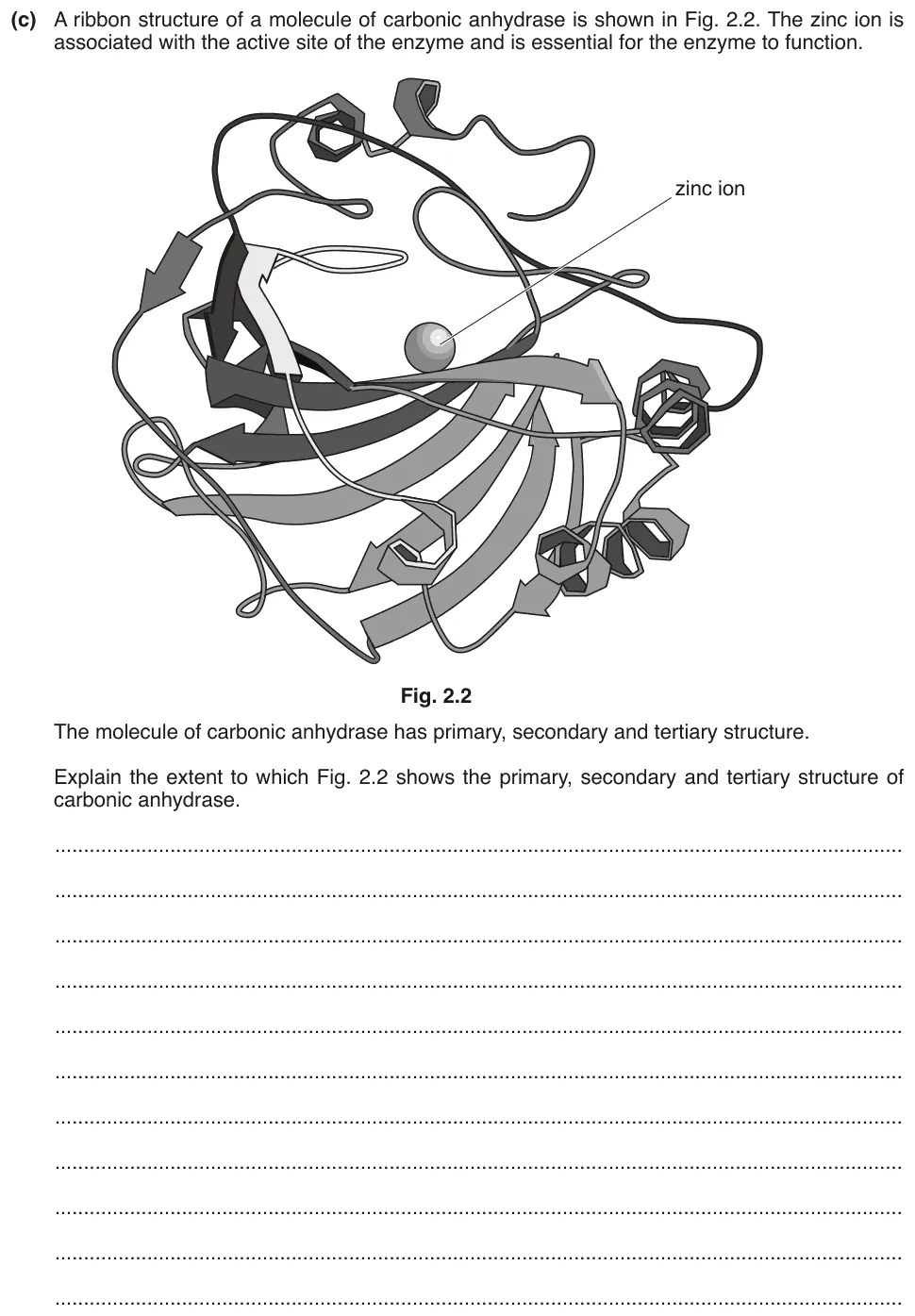
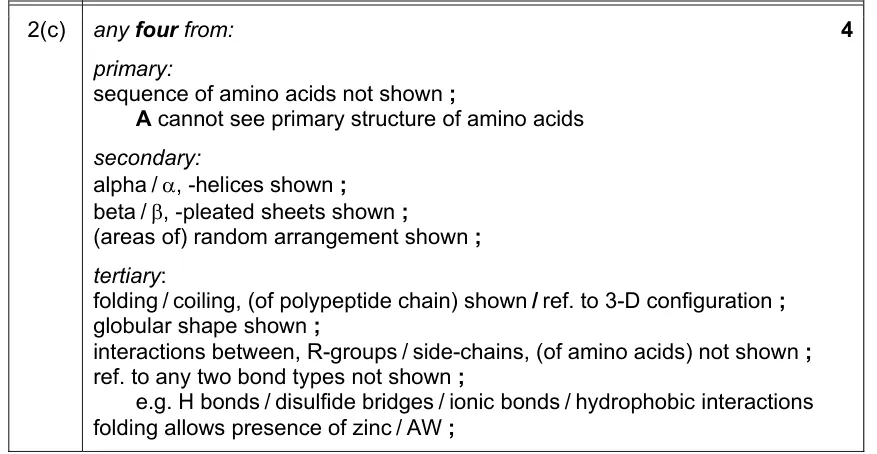
Explain how far a ribbon diagram of carbonic anhydrase shows its primary, secondary and tertiary structure.
Information from earlier in this question:

Show the mark scheme
Tap an image to open it full size.
From the examiners' report: Most recognised the α-helices and β-pleated sheets, and features of tertiary structure. Few said what the diagram does not show: there is no amino acid sequence, so no primary structure, and no detail of disulfide bonds or hydrophobic and hydrophilic interactions.
Bonds that hold proteins in shape (2.3.3)
Question 10
Paper 2 · February/March 2023 (22) · Q2(b)(i) · 1 mark · Hard
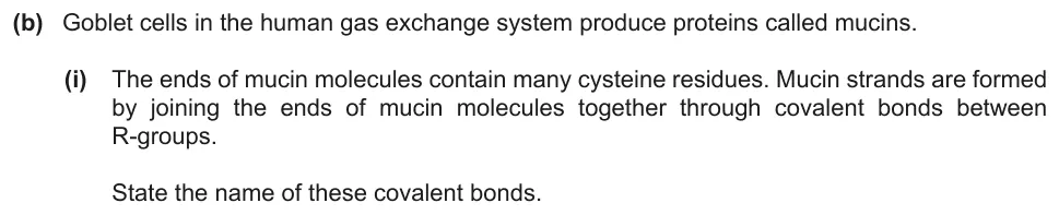
Name the covalent bonds between the R-groups of cysteine residues that join mucin molecules.
Show the mark scheme

Tap an image to open it full size.
From the examiners' report: This was not well answered. Many gave peptide, hydrogen or sulfide bonds.
Question 11
Paper 4 (A level) · February/March 2018 (42) · Q4(a) · 2 marks · Easy
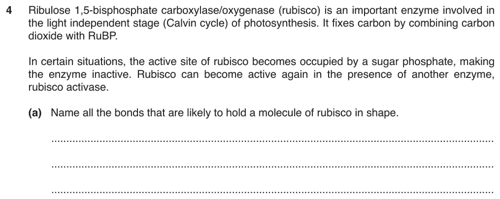
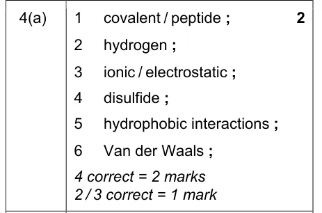
Name all the bonds that are likely to hold a molecule of rubisco in shape.
Show the mark scheme
Tap an image to open it full size.
Globular and fibrous proteins (2.3.4)
Question 12
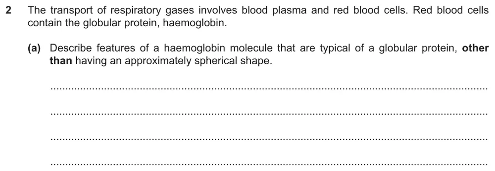
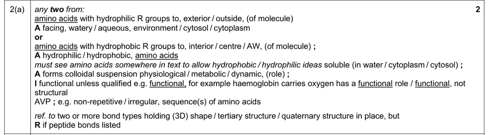
Paper 2 · May/June 2025 (22) · Q2(a) · 2 marks
Describe features of haemoglobin that are typical of a globular protein.
Show the mark scheme
Tap an image to open it full size.
Question 13
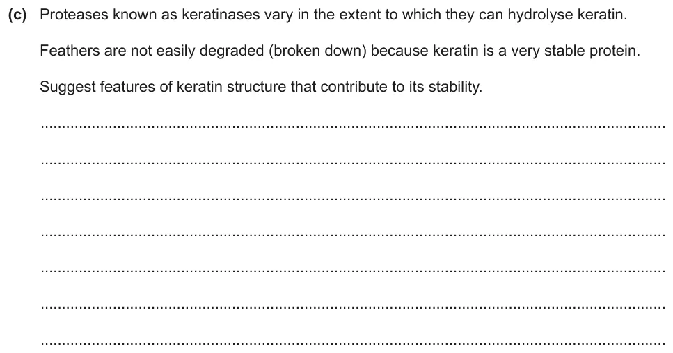
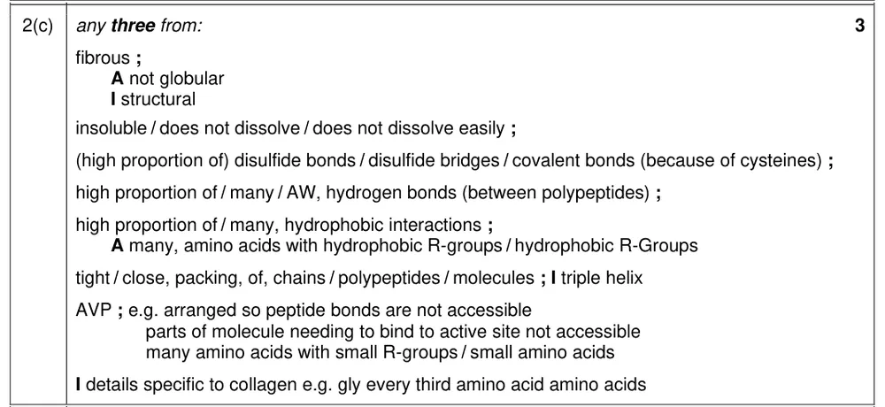
Paper 2 · October/November 2023 (22) · Q2(c) · 3 marks
Suggest features of the structure of keratin that make it a very stable protein.
Information from earlier in this question:

Show the mark scheme
Tap an image to open it full size.
From the examiners' report: Disulfide bonds were the most common correct point. For hydrogen bonds, say that there are many of them: it is wrong to call a hydrogen bond a strong bond. Describing the structure of collagen, or writing ‘collagen’ in place of ‘keratin’, gained no credit.
Structure of haemoglobin (2.3.5)
Question 14
Paper 2 · October/November 2025 (22) · Q6(b)(i) · 2 marks
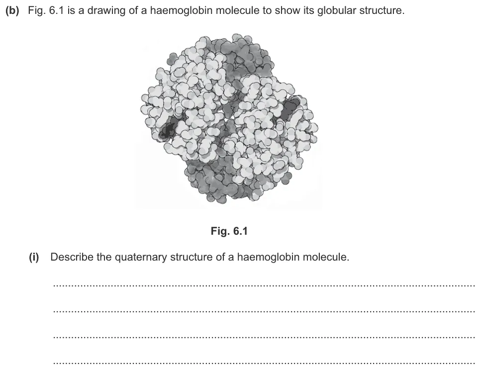
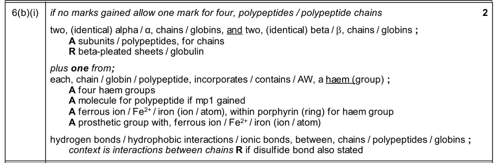
Describe the quaternary structure of a haemoglobin molecule.
Show the mark scheme
Tap an image to open it full size.
Haemoglobin and its function (2.3.6)
Question 15
Paper 2 · October/November 2025 (22) · Q6(b)(ii) · 2 marks
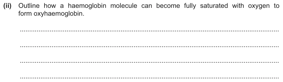
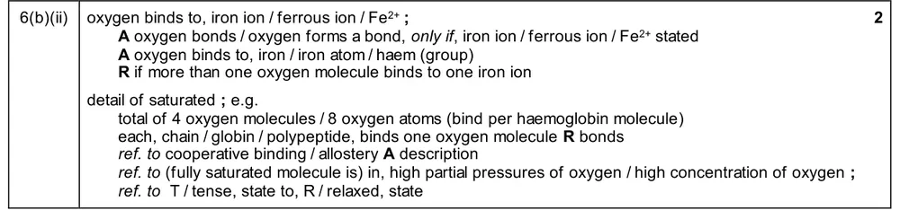
Outline how a haemoglobin molecule becomes fully saturated with oxygen.
Show the mark scheme
Tap an image to open it full size.
Question 16
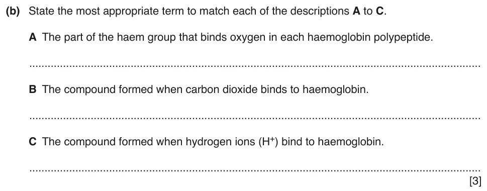
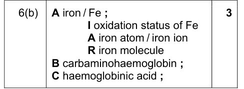
Paper 2 · October/November 2018 (22) · Q6(b) · 3 marks
Give the terms for the part of haem that binds oxygen and for two compounds formed by haemoglobin.
Show the mark scheme
Tap an image to open it full size.
From the examiners' report: A was the best known; ‘prosthetic group’ and ‘iron molecule’ were wrong. For B, carboxyhaemoglobin was often given in place of carbaminohaemoglobin. For C, the abbreviation on its own was not enough: write haemoglobinic acid.
Structure of collagen (2.3.7)
Question 17
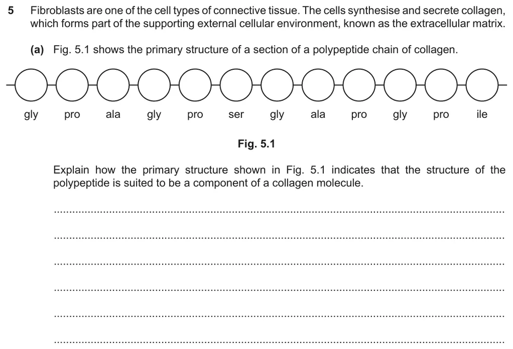
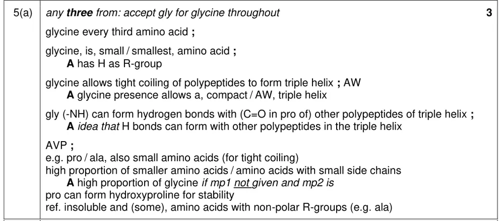
Paper 2 · October/November 2022 (22) · Q5(a) · 3 marks
Explain how the primary structure shown suits the polypeptide to being part of a collagen molecule.
Show the mark scheme
Tap an image to open it full size.
From the examiners' report: Most saw that glycine is every third amino acid and is the smallest amino acid. Full credit needed a link to the collagen molecule: three polypeptides coiled tightly into a triple helix. Strong covalent bonds and a parallel, staggered arrangement belong to the fibril or fibre, not to the molecule.
Question 18
Paper 2 · May/June 2024 (23) · Q2(a) · 5 marks
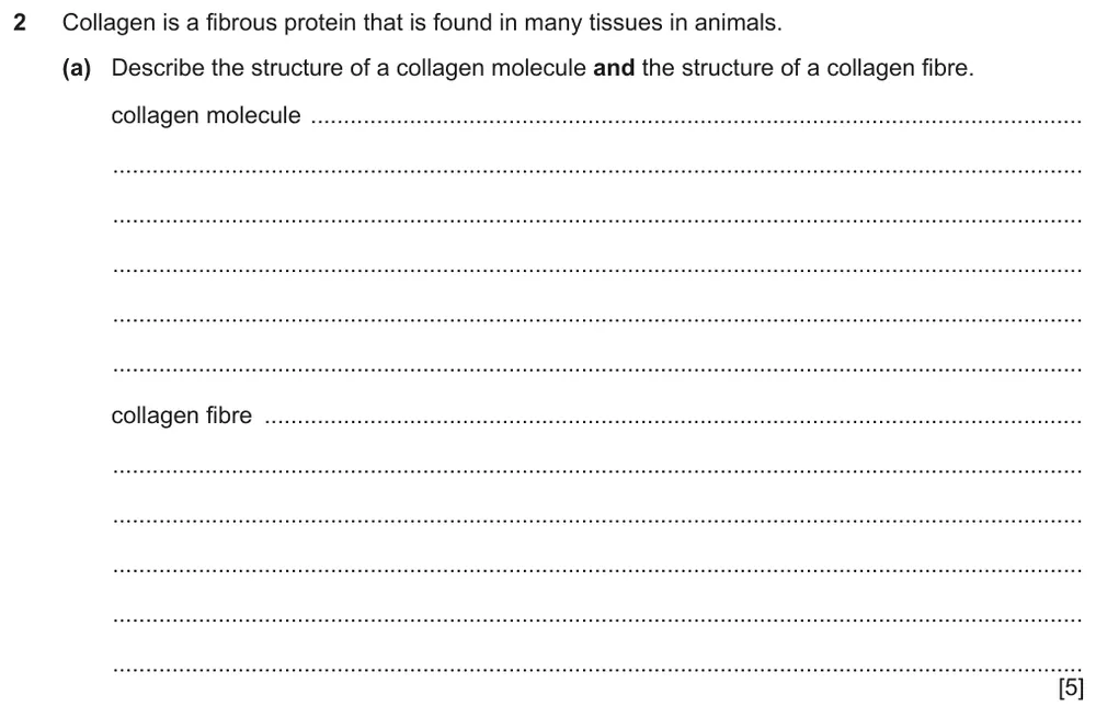
Describe the structure of a collagen molecule and the structure of a collagen fibre.
Show the mark scheme
Tap an image to open it full size.
From the examiners' report: The fibre was described less well than the molecule. A collagen polypeptide is not an alpha helix, and glycine does not form covalent bonds with the other polypeptides. Keep each fact in the right section: correct points written under the wrong heading could not gain credit.
Collagen structure and function (2.3.8)
Question 19
Paper 2 · February/March 2023 (22) · Q6(a) · 3 marks · Hard
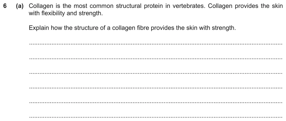
Explain how the structure of a collagen fibre provides the skin with strength.
Show the mark scheme

Tap an image to open it full size.
From the examiners' report: Most described the collagen molecule, not the fibre, which did not answer the question. Few gave features of fibres, such as the cross-links between molecules and the staggered arrangement.
Water and hydrogen bonding (2.4.1)
Question 20
Paper 2 · October/November 2024 (22) · Q1(b) · 2 marks
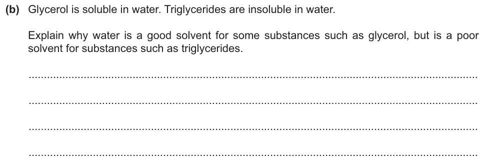
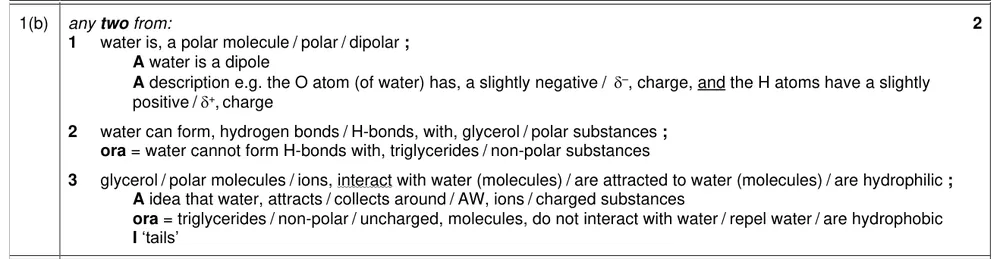
Explain why water is a good solvent for glycerol but a poor solvent for triglycerides.
Show the mark scheme
Tap an image to open it full size.
Question 21
Paper 2 · February/March 2023 (22) · Q3(b)(i) · 2 marks
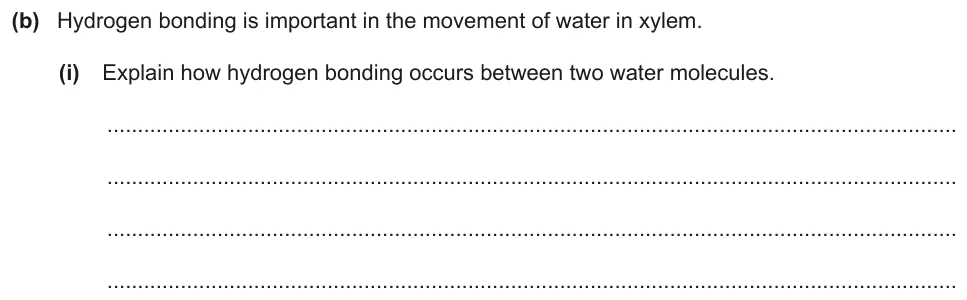
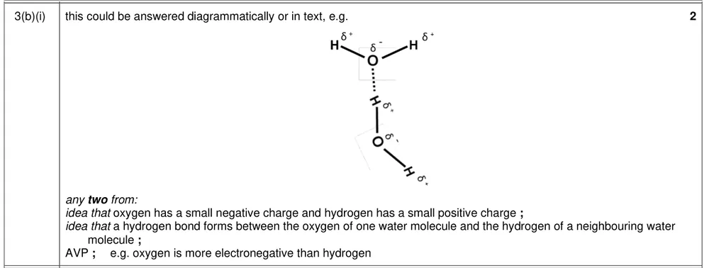
Explain how hydrogen bonding occurs between two water molecules.
Show the mark scheme
Tap an image to open it full size.
From the examiners' report: This was well understood. Do not write about hydrogen ions, oxygen ions or OH– ions, and do not call a hydrogen bond a covalent bond.
Question 22
Paper 2 · February/March 2023 (22) · Q3(b)(iii) · 2 marks
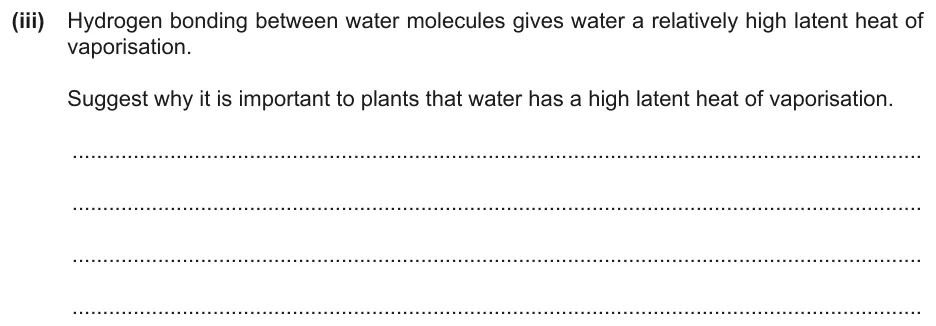
Suggest why a high latent heat of vaporisation of water is important to plants.
Show the mark scheme

Tap an image to open it full size.
From the examiners' report: Some confused a high latent heat of vaporisation with a high specific heat capacity. Others wrote about properties of water that the question did not ask for.
Where students lose marks (from Cambridge examiners' reports and mark schemes)
Each point below comes from the examiners' reports and mark schemes, and is followed by wording that gained marks.
R, not H, in the general structure. Students wrote H where the R group should be, which gives glycine, or joined NH2 to the central carbon the wrong way round.
✔ Gains credit: a structure that is 'all correct', with 'H2N' accepted, and 'different amino acids have different R-groups' or 'side chains'.
A peptide bond forms by condensation. Students drew the dipeptide and water correctly and then called the reaction hydrolysis, or wrote that a glycosidic bond forms.
✔ Gains credit: the 'bond forms between the C of the carboxyl group and the N of the amino group' and 'water is formed'; 'condensation' is accepted.
One polypeptide cannot be quaternary. Students could not match the descriptions to the levels of protein structure, and missed that a single polypeptide rules out quaternary structure.
✔ Gains credit: a single polypeptide that forms a compact structure is 'tertiary'; the proportion of an amino acid is 'primary'; regions of β-pleated sheets are 'secondary'.
Say what the diagram leaves out. Students recognised α-helices and β-pleated sheets but did not comment on what the ribbon diagram does not show.
✔ Gains credit: 'sequence of amino acids not shown', 'alpha-helices shown', 'beta-pleated sheets shown', and 'interactions between R-groups not shown'.
Disulfide, not sulfide. Students named the covalent bonds between cysteine R-groups as peptide, hydrogen or sulfide bonds.
✔ Gains credit: 'disulfide (bonds)'.
Many hydrogen bonds, not strong ones. Students stated that hydrogen bonds are strong bonds, or described the structure of collagen when the question was about keratin.
✔ Gains credit: 'disulfide bonds', 'many hydrogen bonds (between polypeptides)', 'many hydrophobic interactions', 'fibrous' and 'insoluble'.
Collagen molecule or collagen fibre? Students described the collagen molecule when the question asked about the fibre, or put correct facts in the wrong section.
✔ Gains credit: for the fibre: 'covalent bonds between collagen molecules', molecules 'staggered' 'so no weak areas'; for the molecule: 'three helical polypeptides', 'tightly wound'.
Latent heat is not specific heat capacity. Students confused a high latent heat of vaporisation with a high specific heat capacity, or wrote about other properties of water.
✔ Gains credit: 'large cooling effect' or 'removal of heat energy by evaporation', which 'reduces enzyme denaturation'.
Download the questions
Download the PDF pack (22 questions, mark schemes at the end)
Keep practising
- Testing for Biological Molecules, Carbohydrates and Lipids (2.1 and 2.2): Cambridge (CIE) exam questions by topic
- Cambridge (CIE) A level Biology 9700 past papers, mark schemes and examiners' reports
- Cambridge (CIE) A level Biology grade thresholds
- Book a one-to-one CIE lesson or a free 15-minute discovery chat with Tom
- Register your interest in a CIE A level Biology group class
Questions, mark schemes and examiners' report extracts © Cambridge University Press & Assessment.