

Homeostasis in Mammals (14.1): Cambridge (CIE) A Level Biology 9700 Exam Questions by Topic
Homeostasis in mammals is sub-topic 14.1 of Topic 14, Homeostasis, in the Cambridge International AS & A Level Biology syllabus (9700). You need to explain homeostasis and negative feedback, describe the structure of the kidney and the nephron, and explain how urine is formed by ultrafiltration in the Bowman’s capsule and selective reabsorption in the proximal convoluted tubule. You also need the roles of the hypothalamus, posterior pituitary gland, ADH and aquaporins in osmoregulation, cell signalling by glucagon, the control of blood glucose concentration by insulin and glucagon, and how test strips and biosensors measure glucose. This is A level content, examined on Paper 4.
The examiners' reports ask for precision. The mechanism that keeps a factor at its set point is negative feedback; 'homeostasis' was not accepted when a question asked for the mechanism. In ultrafiltration, a high hydrostatic pressure forces fluid through the basement membrane, so do not write about diffusion. In selective reabsorption, it is the water potential of the cells that decreases, not that of the blood. Write that ADH is secreted, and that less ADH is released when blood water potential rises, not none. When a question says explain, do not just describe the graph.
Studying Cambridge International A level Biology and want more help? Tom teaches CIE students one-to-one online, including students in the UAE, Saudi Arabia, Hong Kong, India and Indonesia, and every lesson is recorded. You can book a one-to-one lesson or a free 15-minute discovery chat, or register your interest in a CIE group class starting in September 2027, using the links at the bottom of this page.
What you need from AS Level
Each question on this page assumes you're secure on these ideas. If any of them feels shaky, look it up before you start.
- Active transport uses ATP and carrier proteins to move ions against a concentration gradient; facilitated diffusion and cotransport use membrane proteins too.
- Water moves by osmosis from a higher (less negative) water potential to a lower (more negative) water potential.
- In cell signalling, a ligand binds to a complementary receptor on the cell surface membrane of its target cell.
- Glycogen is a branched polysaccharide of α-glucose, stored in liver and muscle cells.
Key terms
These words come up in the questions and mark schemes. Examiners expect you to use them exactly.
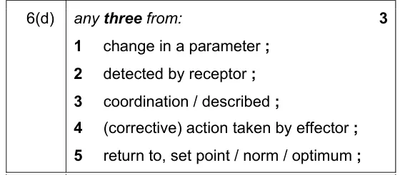
- Negative feedback: a control mechanism in which a change in a factor away from its set point is detected by receptors and effectors act to return it to the set point.
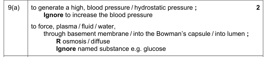
- Ultrafiltration: filtration under high hydrostatic pressure: fluid is forced out of the blood in the glomerulus, through the basement membrane, into the Bowman’s capsule.
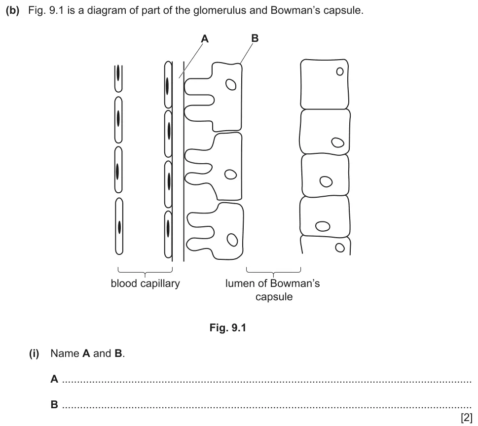
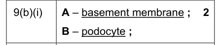
- Basement membrane: the layer between the capillary endothelium and the podocytes that acts as the filter; red blood cells and large proteins cannot pass through it.
- Podocyte: a cell of the inner wall of the Bowman’s capsule, with gaps or slits between its projections that let the filtrate pass into the capsule.
- Selective reabsorption: the movement of useful substances such as glucose, amino acids, ions and water from the filtrate in the proximal convoluted tubule back into the blood.
- Osmoreceptors: receptor cells in the hypothalamus that detect changes in the water potential of the blood.
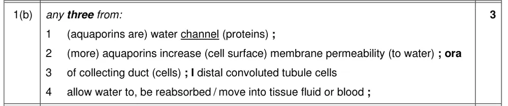
- Aquaporin: a water channel protein; ADH causes more aquaporins to be added to the cell surface membranes of collecting duct cells, increasing their permeability to water.
- Glycogenolysis: the breakdown of glycogen to glucose in liver cells, stimulated by glucagon.
What these questions cover (Cambridge 9700 Topic 14: Homeostasis)
The questions test these learning outcomes from sub-topic 14.1 of the Cambridge International AS & A Level Biology syllabus (9700) for 2025 to 2027.
- 14.1.1 explain what is meant by homeostasis and the importance of homeostasis in mammals
- 14.1.2 explain the principles of homeostasis in terms of internal and external stimuli, receptors, coordination systems (nervous system and endocrine system), effectors (muscles and glands) and negative feedback
- 14.1.3 state that urea is produced in the liver from the deamination of excess amino acids
- 14.1.4 describe the structure of the human kidney
- 14.1.5 the parts of a nephron and its associated blood vessels and structures
- 14.1.6 describe and explain the formation of urine in the nephron
- 14.1.7 relate the detailed structure of the Bowman’s capsule and proximal convoluted tubule to their functions in the formation of urine
- 14.1.8 describe the roles of the hypothalamus, posterior pituitary gland, antidiuretic hormone (ADH), aquaporins and collecting ducts in osmoregulation
- 14.1.9 describe the principles of cell signalling using the example of the control of blood glucose concentration by glucagon
- 14.1.10 explain how negative feedback control mechanisms regulate blood glucose concentration, with reference to the effects of insulin on muscle cells and liver cells and the effect of glucagon on liver cells
- 14.1.11 explain the principles of operation of test strips and biosensors for measuring the concentration of glucose in blood and urine, with reference to glucose oxidase and peroxidase enzymes
How to use this page
Write your answer on paper before you open the mark scheme. Allow about 1.2 minutes per mark: the 22 questions are worth 79 marks, so about 95 minutes in total. Then mark your answer strictly against the mark scheme and read the examiners' tip underneath it. If you'd rather print the questions, download the PDF pack.
Homeostasis (14.1.1)
Question 1
Paper 4 · October/November 2022 (41) · Q10 · 5 marks
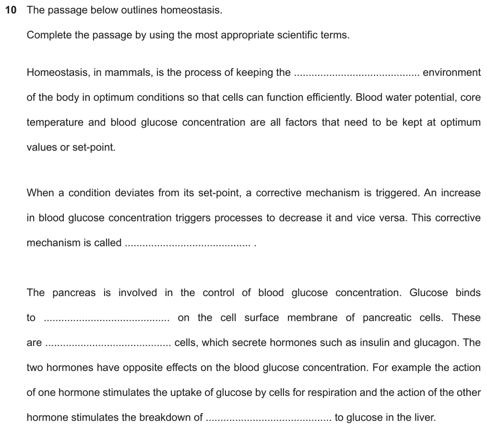
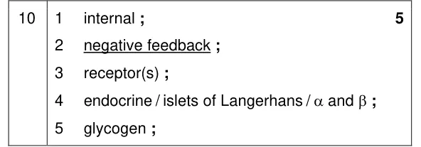
Complete a passage about homeostasis using the most appropriate scientific terms.
Show the mark scheme
Tap an image to open it full size.
From the examiners' report: 'Internal' and 'receptor' were the easiest answers, and negative feedback was often correct. The last two spaces were harder: some students wrote 'secretory' instead of endocrine, and 'glucagon' instead of glycogen.
Negative feedback (14.1.2)
Question 2
Paper 4 · May/June 2024 (42) · Q10(a) · 1 mark · Hard
Name the mechanism that maintains blood glucose concentration around a set point.

Show the mark scheme

Tap an image to open it full size.
From the examiners' report: Many students wrote homeostasis. The mechanism is negative feedback.
Question 3
Paper 4 · May/June 2021 (42) · Q6(d) · 3 marks · Easy
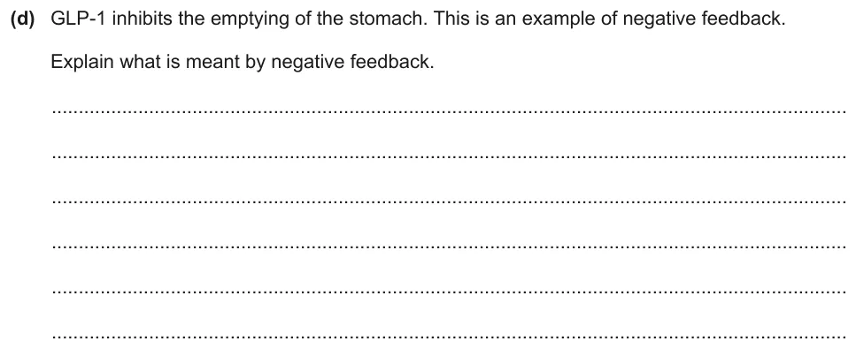
Explain what is meant by negative feedback.
Show the mark scheme
Tap an image to open it full size.
From the examiners' report: Some students knew that negative feedback returns a parameter to its set point or normal value, but did not then outline the sequence of events.
Urea production (14.1.3)
Question 4
Paper 4 · October/November 2024 (41) · Q6(a)(i) · 2 marks
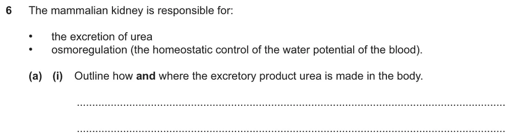
Outline how and where urea is made in the body.
Show the mark scheme

Tap an image to open it full size.
Structure of the kidney (14.1.4)
Question 5
Paper 4 · February/March 2023 (42) · Q1(a) · 4 marks · Easy
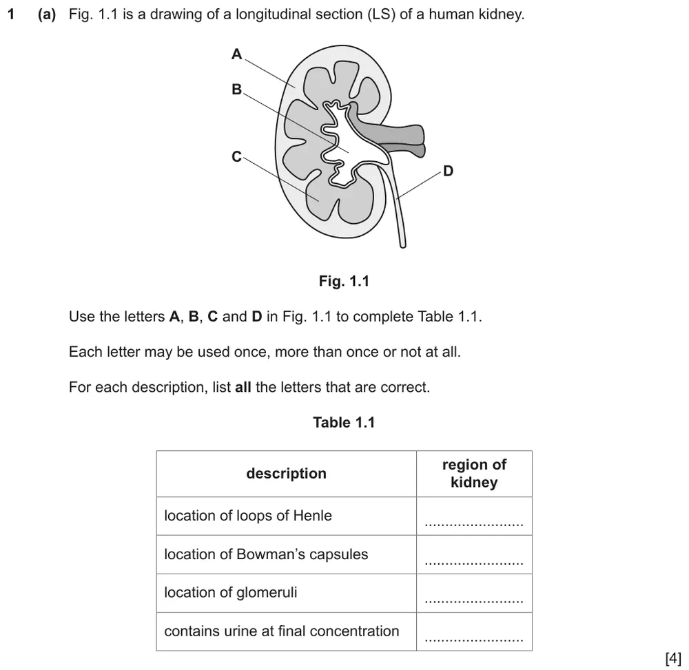
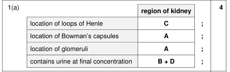
Match regions of a kidney in longitudinal section to parts of the nephron and to where urine is at its final concentration.
Show the mark scheme
Tap an image to open it full size.
From the examiners' report: Most located the loops of Henle, Bowman’s capsules and glomeruli. Few recognised that both region B, the pelvis, and region D, the ureter, contain urine at its final concentration.
Identifying parts of the nephron (14.1.5)
Question 6
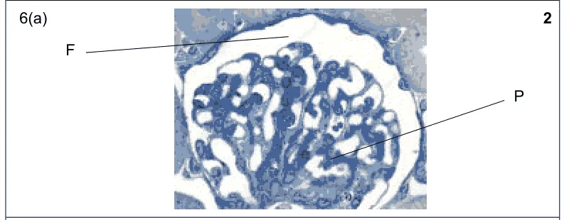
Paper 4 · May/June 2023 (42) · Q6(a) · 2 marks
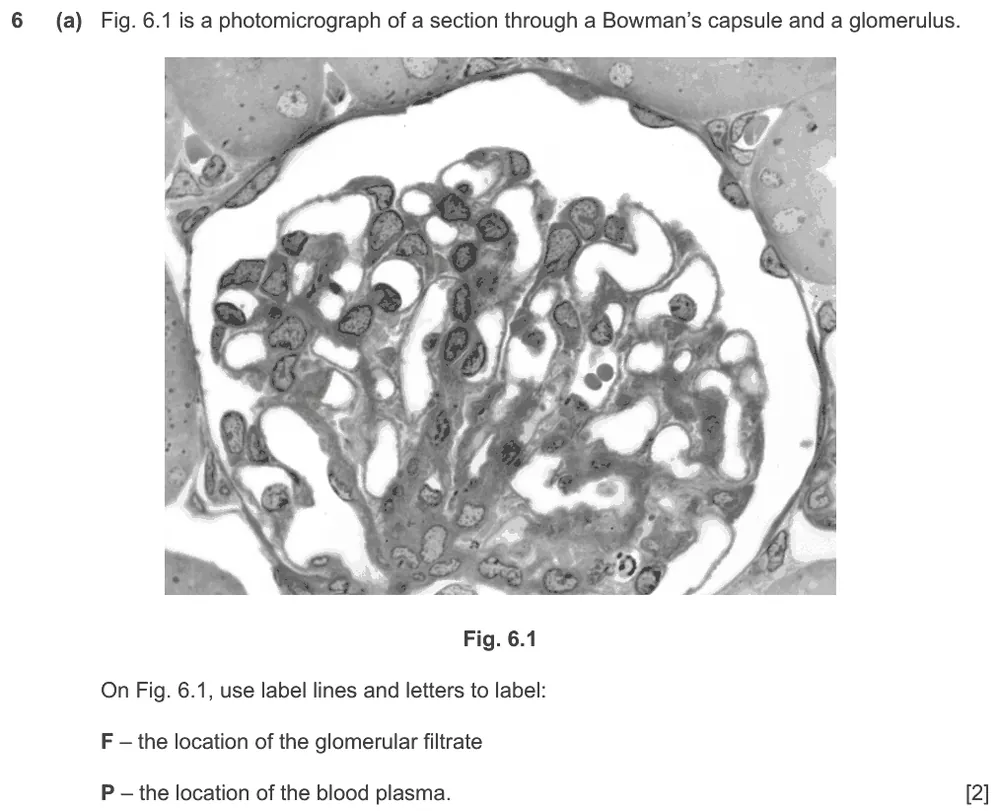
Label the glomerular filtrate and the blood plasma on a photomicrograph of a Bowman’s capsule and glomerulus.
Show the mark scheme
Tap an image to open it full size.
From the examiners' report: Many could not label where the glomerular filtrate and the blood plasma are found. A few wrote the letters F and P on the image instead of using label lines as instructed.
Question 7
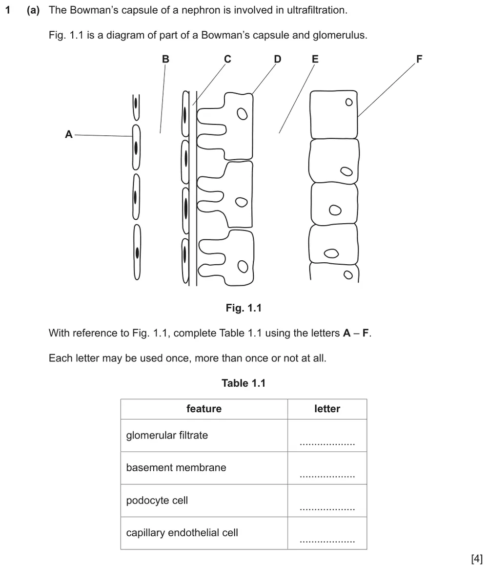
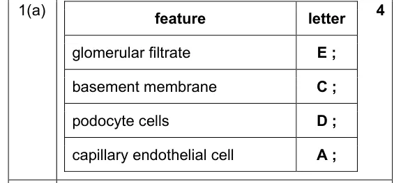
Paper 4 · October/November 2021 (42) · Q1(a) · 4 marks · Hard
Match letters on a diagram of a Bowman’s capsule and glomerulus to four features.
Show the mark scheme
Tap an image to open it full size.
From the examiners' report: Weaker answers found it hard to identify the basement membrane or where the glomerular filtrate would be.
Ultrafiltration in the Bowman’s capsule (14.1.6 and 14.1.7)
Question 8
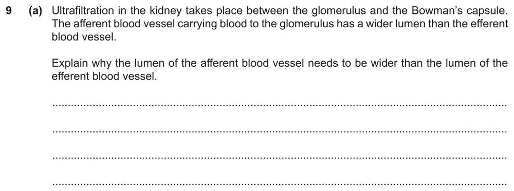
Paper 4 · May/June 2022 (42) · Q9(a) · 2 marks
Explain why the lumen of the afferent blood vessel needs to be wider than the lumen of the efferent blood vessel.
Show the mark scheme
Tap an image to open it full size.
From the examiners' report: Most found this very hard. Write that it generates a high blood pressure or hydrostatic pressure: 'higher' or 'increased' pressure, and 'pressure' on its own, did not gain credit. Then say that fluid is forced through the basement membrane or into the Bowman’s capsule, and avoid words for passive movement such as diffusion.
Question 9
Paper 4 · May/June 2022 (42) · Q9(b)(i) · 2 marks · Easy
Name two structures labelled on a diagram of the glomerulus and Bowman’s capsule.
Show the mark scheme
Tap an image to open it full size.
From the examiners' report: Well answered. Answers that did not gain credit included 'basal membrane' for A and 'endothelium' for B.
Question 10
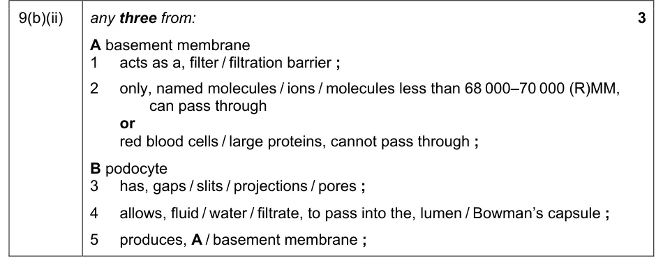
Paper 4 · May/June 2022 (42) · Q9(b)(ii) · 3 marks · Easy
Describe the roles of the basement membrane and the podocytes in the formation of the glomerular filtrate.
Show the mark scheme
Tap an image to open it full size.
From the examiners' report: Give one suitable example of what can or cannot pass through the basement membrane: students who put red blood cells in a long list of 'molecules' could not score. A molecular size needs qualifying, for example as relative molecular mass. Few saw that the pores let fluid move in bulk into the capsule, carrying dissolved substances with it.
Selective reabsorption in the proximal convoluted tubule (14.1.6 and 14.1.7)
Question 11
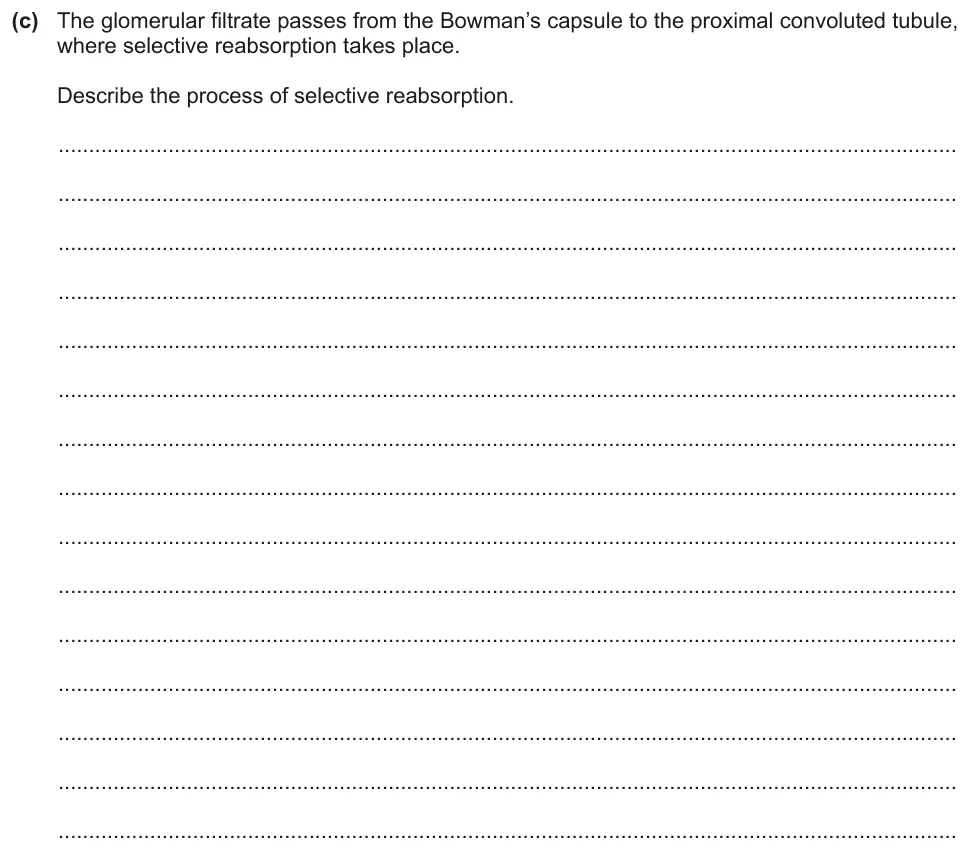
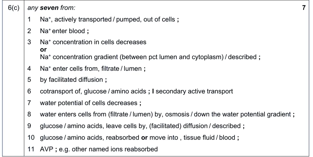
Paper 4 · May/June 2023 (42) · Q6(c) · 7 marks
Describe the process of selective reabsorption.
Show the mark scheme
Tap an image to open it full size.
From the examiners' report: Include the details: sodium ions are pumped out of the cells and then enter the cells again from the lumen or filtrate. Many wrongly said that the water potential of the blood decreases, rather than that of the cells. How the cells are adapted was not asked for and gained no credit.
Question 12
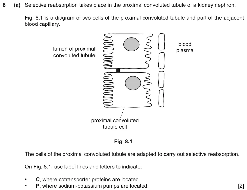
Paper 4 · October/November 2022 (42) · Q8(a) · 2 marks
Add label lines to show where cotransporter proteins and sodium-potassium pumps are located in proximal convoluted tubule cells.
Show the mark scheme

Tap an image to open it full size.
From the examiners' report: Some students made no attempt and many answers were imprecise. Credit was lost if the label line did not touch the membrane or went through it.
Question 13
Paper 4 · October/November 2022 (42) · Q8(b) · 5 marks
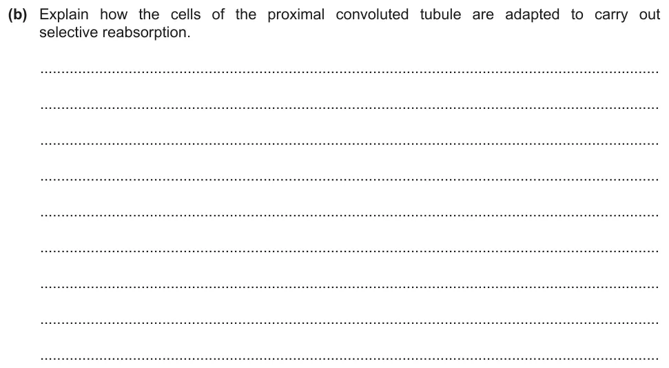
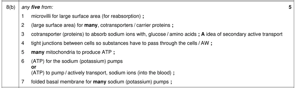
Explain how the cells of the proximal convoluted tubule are adapted to carry out selective reabsorption.
Show the mark scheme
Tap an image to open it full size.
From the examiners' report: Name each specific feature and say how it is used in selective reabsorption. Mitochondria were mentioned, but not that there are large numbers of them, and the quantity of cotransporters was rarely stated. Few used the diagram to help them.
ADH and osmoregulation (14.1.8)
Question 14
Paper 4 · October/November 2023 (42) · Q9(a) · 4 marks · Hard
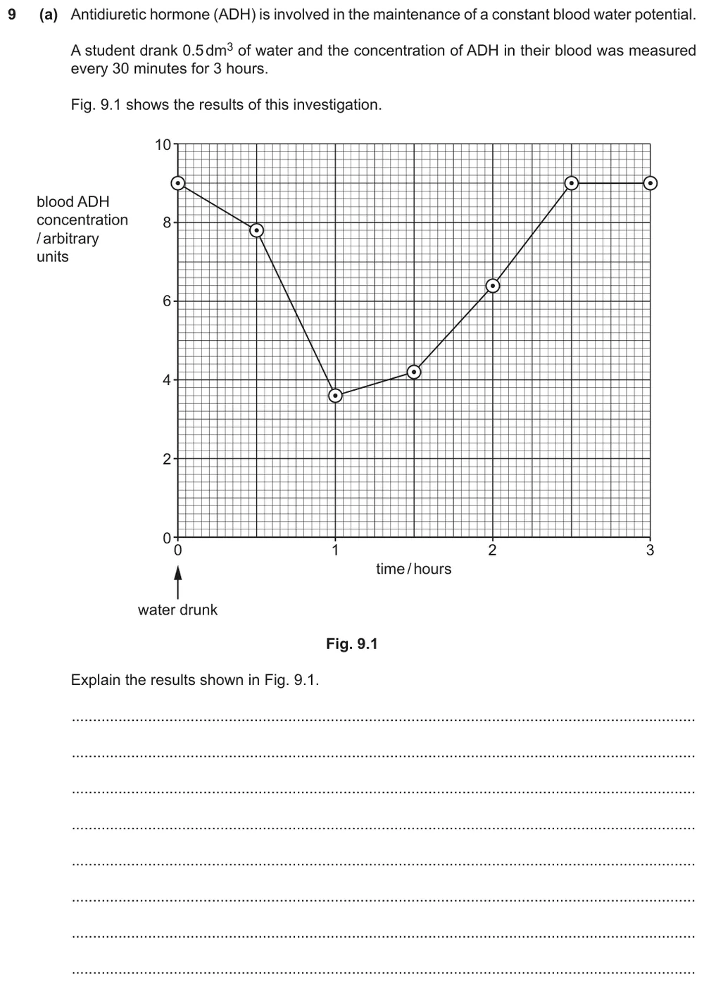
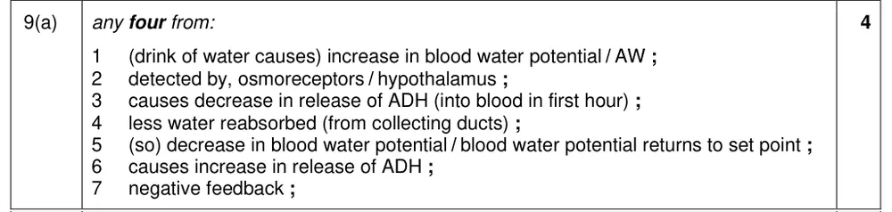
Explain the changes in blood ADH concentration after a student drinks water.
Show the mark scheme
Tap an image to open it full size.
From the examiners' report: Be precise: ADH is secreted, not just produced, and write water potential, not blood water concentration or level. ADH secretion decreases and then increases; it does not stop and start. Some only described the graph, and few said that less water is reabsorbed in the collecting duct.
Question 15
Paper 4 · February/March 2023 (42) · Q1(b) · 3 marks · Easy
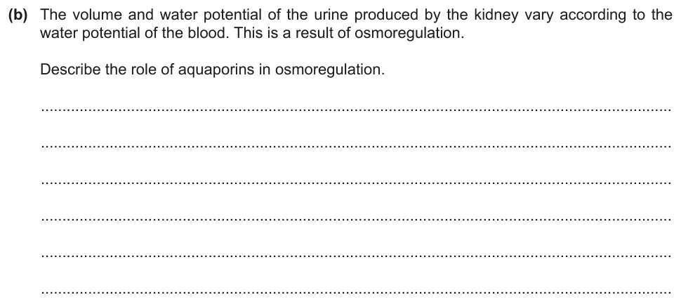
Describe the role of aquaporins in osmoregulation.
Show the mark scheme
Tap an image to open it full size.
From the examiners' report: Few said that aquaporins are channel proteins for water. Say where the water goes; 'leaving the collecting duct' or 'being absorbed' was too vague. Permeability was often linked to the collecting duct, its walls or its cells, without mentioning the cell surface membrane.
Question 16
Paper 4 · February/March 2023 (42) · Q1(c) · 3 marks
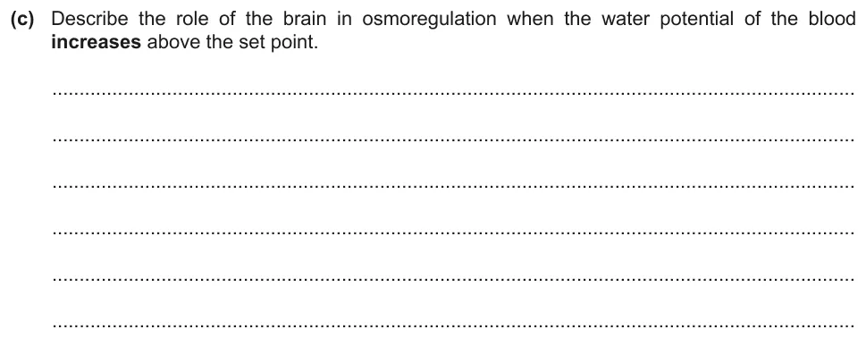
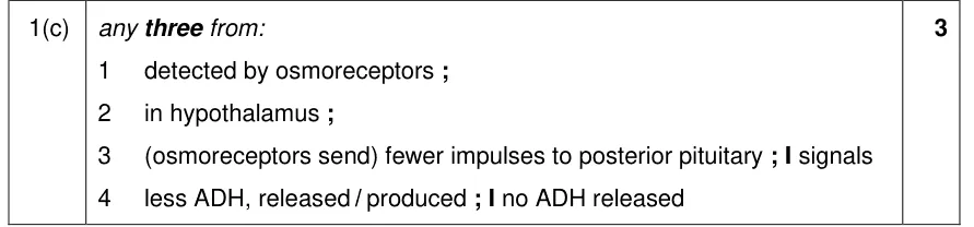
Describe the role of the brain in osmoregulation when the water potential of the blood increases above the set point.
Show the mark scheme
Tap an image to open it full size.
From the examiners' report: Write osmoreceptors, not 'osmoregulators'. Many wrongly said that more ADH would be secreted. Fewer impulses are sent to the posterior pituitary and less ADH is secreted: 'no impulses' and 'no ADH' are wrong. Write nerve impulses, not messages or signals.
Glucagon and cell signalling (14.1.9)
Question 17
Paper 4 · October/November 2023 (42) · Q6(a)(i) · 4 marks · Easy
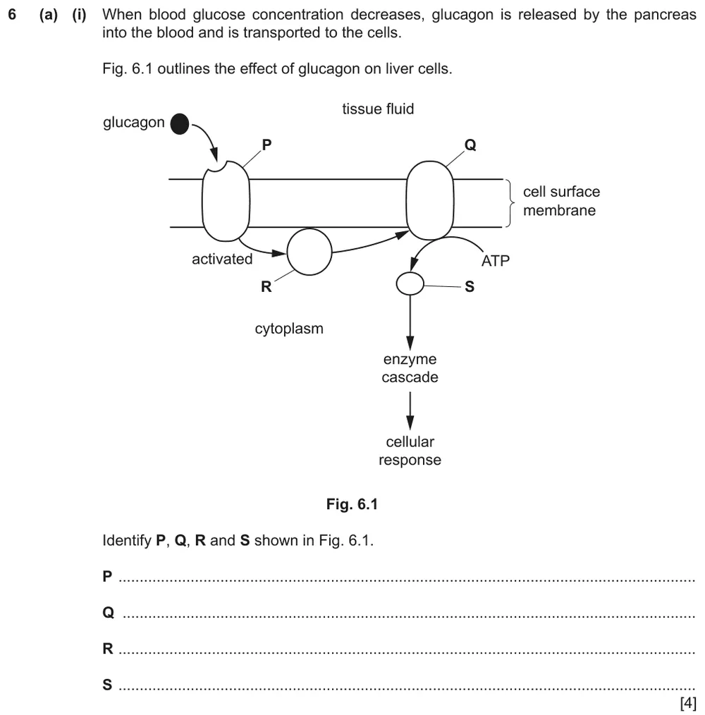
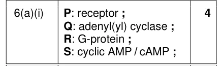
Identify four components of the glucagon signalling pathway in a liver cell.
Show the mark scheme
Tap an image to open it full size.
From the examiners' report: Most identified the receptor, although a few called it a receptor cell. G-protein and adenylyl cyclase were occasionally given the wrong way round.
Question 18
Paper 4 · February/March 2025 (42) · Q6(b) · 7 marks
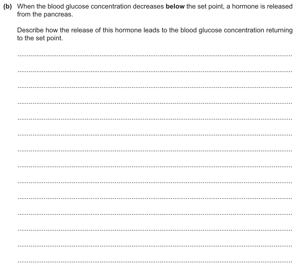
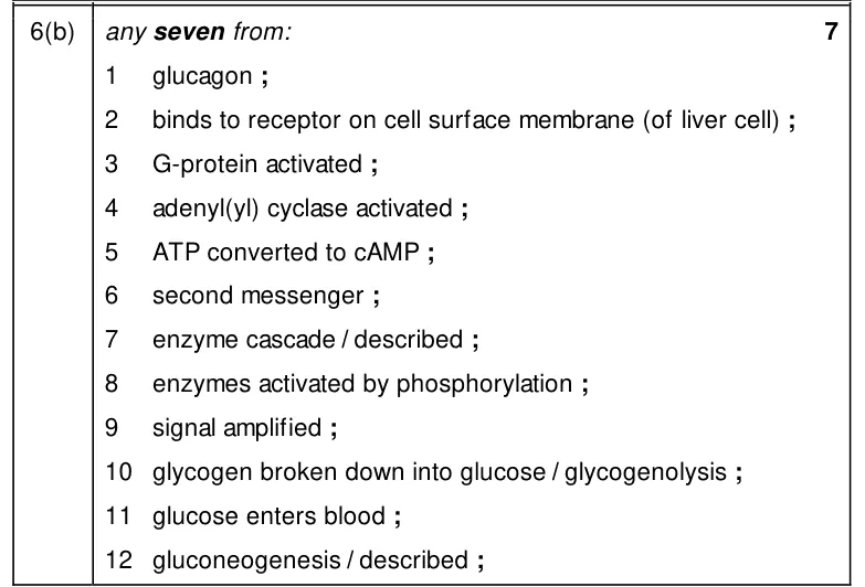
Describe how the hormone released when blood glucose concentration falls returns it to the set point.
Show the mark scheme
Tap an image to open it full size.
From the examiners' report: Strong answers started with glucagon binding to a receptor on the cell surface membrane and gave the events in the correct sequence. Weaker answers named the hormone and the breakdown of glycogen but left out other key points, or did not give a clear sequence.
Control of blood glucose concentration (14.1.10)
Question 19
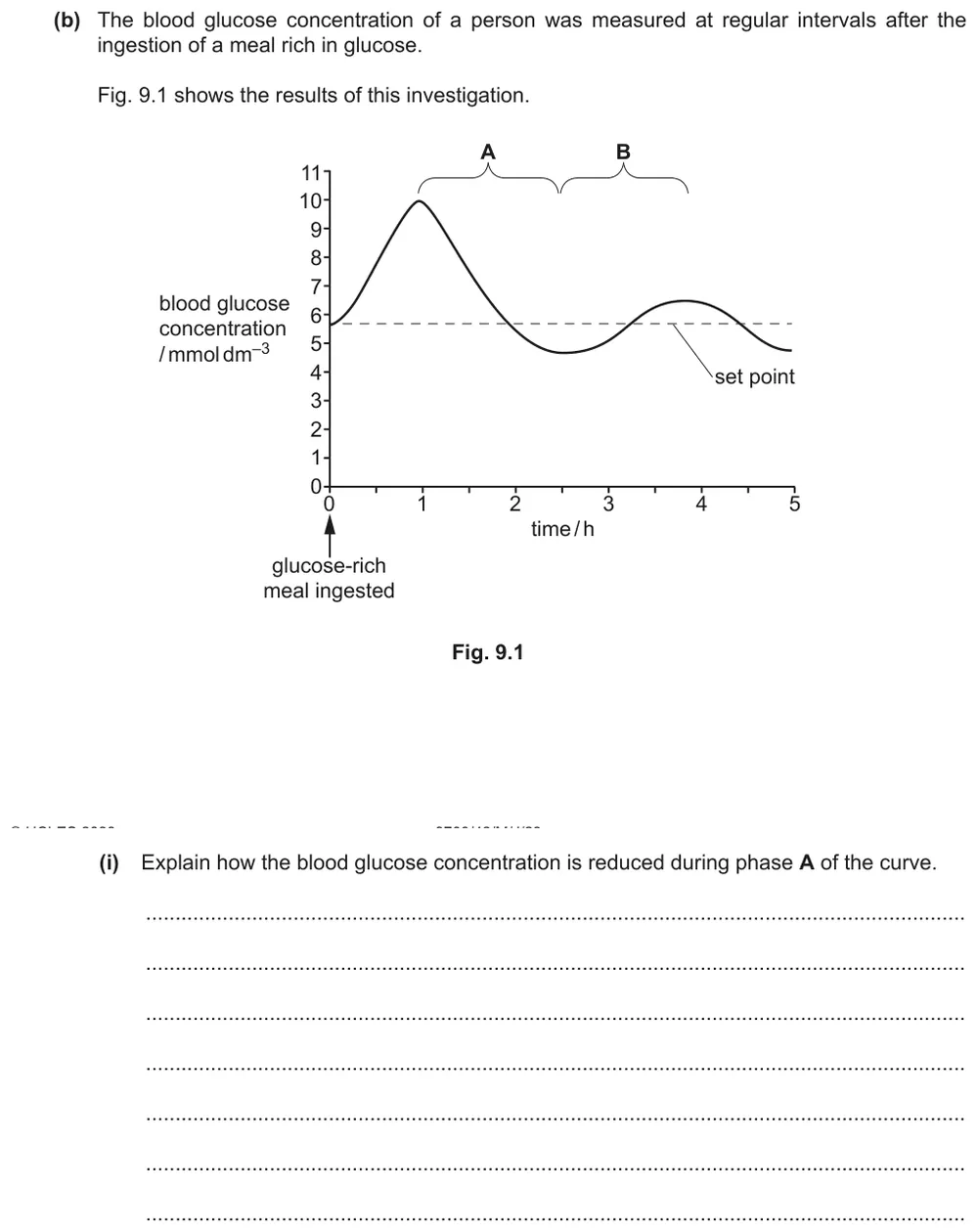
Paper 4 · May/June 2023 (42) · Q9(b)(i) · 3 marks
Explain how the blood glucose concentration is reduced after a glucose-rich meal.
Show the mark scheme

Tap an image to open it full size.
From the examiners' report: Most gave insulin secretion and glycogenesis. Some needed to be more precise: write an 'increase' in the uptake of glucose, and the cell 'surface' membrane.
Question 20
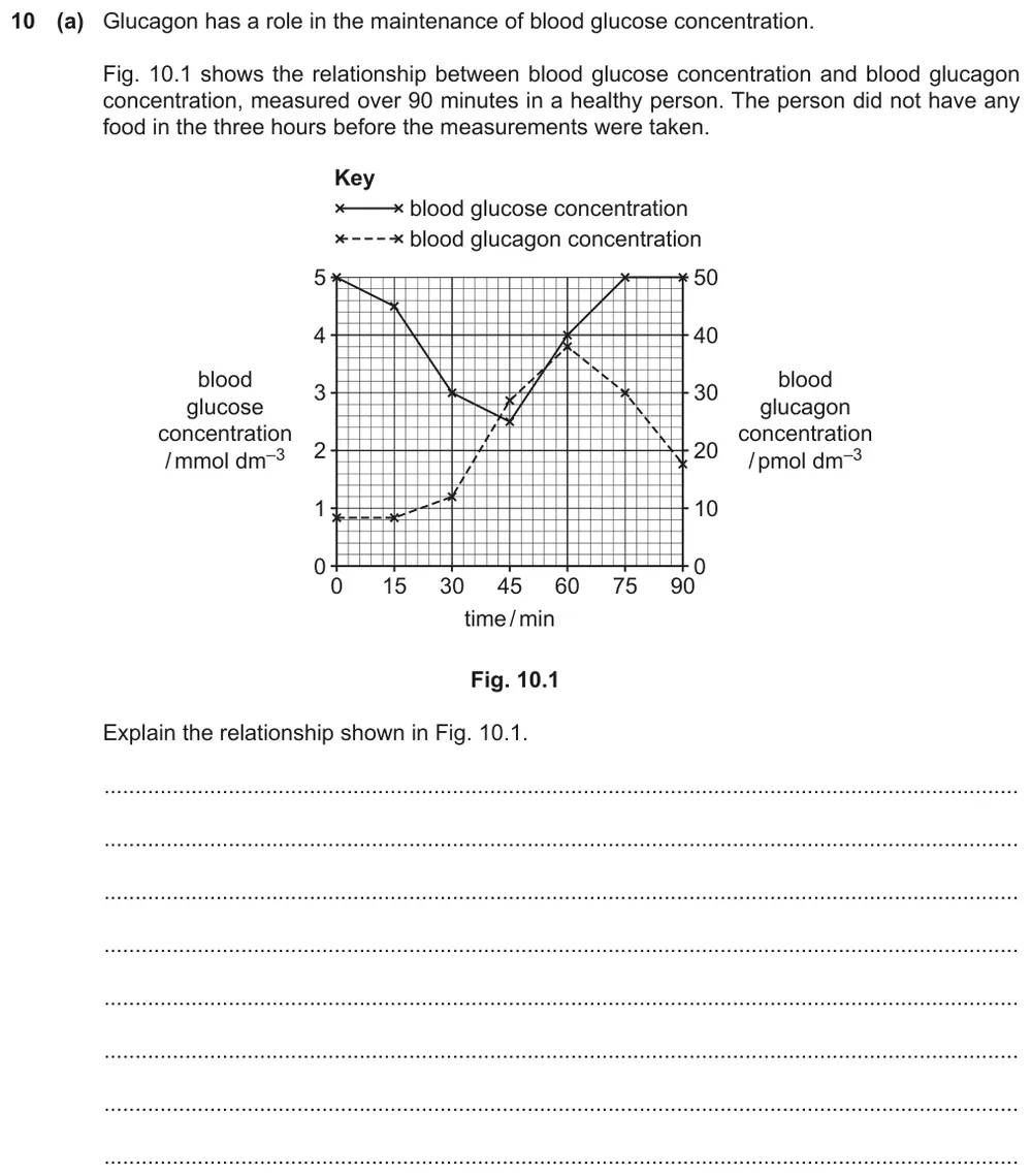
Paper 4 · October/November 2022 (42) · Q10(a) · 4 marks
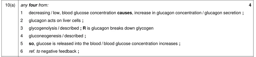
Explain the relationship between blood glucose concentration and blood glucagon concentration shown in the graph.
Show the mark scheme
Tap an image to open it full size.
From the examiners' report: Several students only described the graph instead of explaining the relationship. Weaker answers suggested that the breakdown of glucagon itself raises the blood glucose concentration. Few mentioned that the process is negative feedback.
Test strips and biosensors (14.1.11)
Question 21
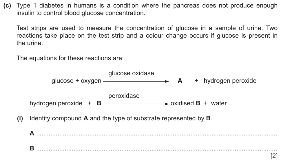
Paper 4 · May/June 2024 (42) · Q10(c)(i) · 2 marks
Identify compound A and the type of substrate represented by B in the two reactions on a glucose test strip.
Show the mark scheme

Tap an image to open it full size.
From the examiners' report: Quite well answered, although some students struggled with the spelling of gluconic acid.
Question 22
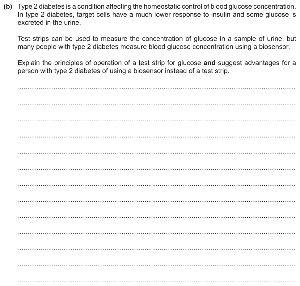
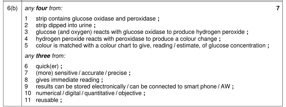
Paper 4 · October/November 2023 (42) · Q6(b) · 7 marks · Easy
Explain the principles of operation of a glucose test strip and suggest advantages of using a biosensor.
Show the mark scheme
Tap an image to open it full size.
From the examiners' report: Most named the two enzymes on the test strip and explained what they do. Some were imprecise about the colour change, which needs to be related to the concentration of glucose. Speed, accuracy and being reusable were the most common advantages given.
Where students lose marks (from Cambridge examiners' reports and mark schemes)
Each point below comes from the examiners' reports and mark schemes, and is followed by wording that gained marks.
Negative feedback is the mechanism. Students wrote 'homeostasis' when asked to name the mechanism that keeps blood glucose concentration around a set point.
✔ Gains credit: 'negative feedback'.
High pressure, not higher pressure. Students wrote that blood pressure is 'higher' or 'increased', wrote 'pressure' without saying which, or described diffusion.
✔ Gains credit: 'to generate a high blood pressure' or 'hydrostatic pressure', 'to force plasma', 'fluid' or 'water' 'through basement membrane' or 'into the Bowman’s capsule'.
Red blood cells are not molecules. Students gave a long list of 'molecules' that included red blood cells, or quoted a molecular size without saying what the figure was.
✔ Gains credit: the basement membrane 'acts as a filter'; 'red blood cells' and 'large proteins' 'cannot pass through'; the podocyte 'has gaps' or 'slits'.
The cells' water potential falls. Students said that the water potential of the blood decreases during selective reabsorption.
✔ Gains credit: 'Na+ actively transported out of cells', 'water potential of cells decreases', 'water enters cells from filtrate by osmosis'.
Many mitochondria, not just mitochondria. Students named features of proximal convoluted tubule cells without the detail or the use of each one.
✔ Gains credit: 'microvilli for large surface area', 'many mitochondria to produce ATP', 'folded basal membrane for many sodium (potassium) pumps'.
Less ADH, not no ADH. Students said that more ADH is secreted when blood water potential rises, or that no impulses are sent and no ADH is secreted.
✔ Gains credit: 'detected by osmoreceptors' 'in hypothalamus', 'fewer impulses to posterior pituitary', 'less ADH released'.
Say where the reabsorbed water goes. Students wrote that water leaves the collecting duct or is absorbed, and linked permeability to the duct or its walls.
✔ Gains credit: aquaporins are 'water channel (proteins)' that 'increase (cell surface) membrane permeability' 'of collecting duct (cells)' and 'allow water to be reabsorbed' or 'move into tissue fluid or blood'.
Glucagon is not broken down. Students described the graph, or suggested that the breakdown of glucagon itself raises the blood glucose concentration.
✔ Gains credit: 'glucagon acts on liver cells', 'glycogenolysis', 'so glucose is released into the blood', with a 'ref. to negative feedback'.
Download the questions
Download the PDF pack (22 questions, mark schemes at the end)
Keep practising
- Homeostasis in Plants (14.2): Cambridge (CIE) exam questions by topic
- Cambridge (CIE) A level Biology 9700 past papers, mark schemes and examiners' reports
- Cambridge (CIE) A level Biology grade thresholds
- Book a one-to-one CIE lesson or a free 15-minute discovery chat with Tom
- Register your interest in a CIE A level Biology group class
Questions, mark schemes and examiners' report extracts © Cambridge University Press & Assessment.